. 2024 Jul 31;14(15):1287.
doi: 10.3390/nano14151287.

Polyethylene Terephthalate Microplastics Generated from Disposable Water Bottles Induce Interferon Signaling Pathways in Mouse Lung Epithelial Cells

Affiliations

Polyethylene Terephthalate Microplastics Generated from Disposable Water Bottles Induce Interferon Signaling Pathways in Mouse Lung Epithelial Cells

Luna Rahman et al. Nanomaterials (Basel). .

Abstract

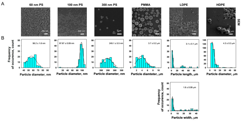
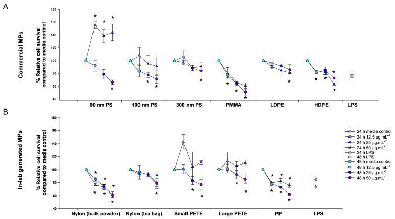
Microplastics (MPs) are present in ambient air in a respirable size fraction; however, their potential impact on human health via inhalation routes is not well documented. In the present study, methods for a lab-scale generation of MPs from regularly used and littered plastic articles were optimized. The toxicity of 11 different types of MPs, both commercially purchased and in-lab prepared MPs, was investigated in lung epithelial cells using cell viability, immune and inflammatory response, and genotoxicity endpoints. The underlying mechanisms were identified by microarray analysis. Although laborious, the laboratory-scale methods generated a sufficient quantity of well characterized MPs for toxicity testing. Of the 11 MPs tested, the small sized polyethylene terephthalate (PETE) MPs prepared from disposable water bottles induced the maximum toxicity. Specifically, the smaller size PETE MPs induced a robust activation of the interferon signaling pathway, implying that PETE MPs are perceived by cells by similar mechanisms as those employed to recognize pathogens. The PETE MPs of heterogenous size and shapes induced cell injury, triggering cell death, inflammatory cascade, and DNA damage, hallmark in vitro events indicative of potential in vivo tissue injury. The study establishes toxicity of specific types of plastic materials in micron and nano size.

Keywords: cytotoxicity; environmental exposure; genotoxicity; inhalation toxicology; micronuclei; microplastics; polyethylene terephthalate; transcriptomic.


Conflict of interest statement

The authors declare no conflicts of interest. The design of the study; the collection, analyses, or interpretation of data; the writing of the manuscript; or the decision to publish the results were not influenced by the funder.

Figures

Figure 1
Figure 1
A schematic showing methods for in-lab generation of MPs.
Figure 2
Figure 2
Raman characterization of commercial and in-lab generated MPs. The bright field images of MPs (A) and associated Raman spectrographs (B). The yellow arrows shows MPs in the nano size ranges.
Figure 3
Figure 3
Representative scanning electron micrographs showing the morphology of commercial MPs used in this study (A) and size distribution histograms (B). The diameter of the spherical particles or primary particle lengths and widths for non-spherical particles were determined using ImageJ. The mean primary particle length and width were obtained from the histogram plots obtained from distribution of particle lengths and widths using SigmaPlot 15.
Figure 4
Figure 4
Representative scanning electron micrographs showing the morphology of the lab-generated MPs (A) and size distribution histogram plots (B). The diameter of the spherical particles or primary particle lengths and widths for non-spherical particles were determined using ImageJ. The mean primary particle length and width were obtained from the histogram plot obtained from distribution of particle lengths and widths using SigmaPlot 15.
Figure 5
Figure 5
Relative cell survival post-24 h and 48 h exposure to 12.5, 25, and 50 µg mL−1 of commercial MPs or 1 µg mL−1 LPS compared with untreated controls (A) and post-exposure to 12.5, 25, and 50 µg·mL−1 of individual lab-generated MPs or 1 µg mL−1 LPS compared with untreated controls (B). The results are expressed as average % relative survival compared with the media controls and error bars depict standard errors. * Represents statistical significance. Statistical significance between the exposed samples and matched media control was determined (three biological replicates) by conducting a one-way ANOVA with Dunnett’s post hoc using p ≤ 0.05.
Figure 6
Figure 6
Analysis of pro-inflammatory proteins. IL-6 and IL-1 β expression in cell supernatant post-48 h exposure to 12.5, 25, and 50 µg·mL−1 of commercial MPs or 1 µg·mL−1 LPS via single cytokine ELISA (A) and IL-6 and IL-1 β expression levels in supernatant of cells post-48 h exposure to 12.5, 25, and 50 µg·mL−1 of lab-generated MPs or 1 µg mL−1 LPS via single cytokine ELISA (B). The results are expressed as average fold change compared with the media controls and error bars depict standard errors (three biological replicates, two technical replicates). * represents statistical significance. Statistical significance between the exposed samples and matched media control was determined by conducting a two-way ANOVA with a Dunnett’s post hoc using p ≤ 0.05.
Figure 7
Figure 7
Analysis of pro-inflammatory cytokines via 23-plex cytokine ELISA—cytokine expression in supernatant of cells exposed to 12.5, 25, and 50 µg·mL−1 of 100 nm PS MP, PETE MP, and PMMA MP or 1 µg·mL−1 LPS for 48 h. Statistical significance (three biological replicates, two technical replicates) was calculated using Student’s t-test (p ≤ 0.1 and fold change compared with media control ≥ 1.3).
Figure 8
Figure 8
Micronuclei (MN) induction in treated versus untreated controls following 40 h post-exposure to commercial MPs (A) and lab-generated MPs (B). Bar graphs are the fold change in micronuclei induction compared with matched controls. The z-axis (line graph) presents the relative cell survival compared with untreated controls at 40 h post-exposure to commercial MPs (A) and lab-generated MPs (B). The error bars depict standard error. * represents statistical significance in MN induction and # represents statistical significance in % cell survival. Statistical significance between the exposed samples and matched media control was determined by conducting a one-way ANOVA with a Tukey’s post hoc multiple comparisons test using p ≤ 0.05. All experiments were conducted with three biological replicates and three technical replicates. The dashed red line represents the condition for genotoxicity (40% of the relative survival and 2-fold change threshold).
Figure 9
Figure 9
Differentially expressed genes post-small PETE exposure. The total numbers of DEGs following 48 h exposure to 12.5, 25, and 50 µg·mL−1 concentrations of PETE: (A) Red: upregulated and Blue: downregulated; Venn diagram showing the common DEGs between different concentration groups; (B) the upward and downward arrows depict up- and downregulated genes, and the heatmap showing the top 10 upregulated and downregulated DEGs (C).
Figure 10
Figure 10
Pathway perturbation post-small PETE exposure. The total number of enriched canonical pathways (A); the enriched canonical pathways associated with immune and inflammation responses (B); and canonical pathways associated with cellular injury, apoptosis, cell cycle, and DNA replication (C).
Figure 11
Figure 11
Two-dimensional view and three-dimensional rendering showing the small PETE interacting with the actin cytoskeleton in FE1-mouse lung epithelial cells. The 3D views represent the area marked by the white box in the 2D view. The 2D view (left) represents the of overlay of immunofluorescence image with EDF image in the same field of view (left) and corresponding hyperspectral images of the same field of view with mapped MPs (pseudo-colored in cyan).

References

    1. Wright S.L., Thompson R.C., Galloway T.S. The physical impacts of microplastics on marine organisms: A review. Environ. Pollut. 2013;178:483–492. doi: 10.1016/j.envpol.2013.02.031. - DOI - PubMed
    1. Rochman C.M., Cook A., Koelmans A.A. Plastic debris and policy: Using current scientific understanding to invoke positive change. Environ. Toxicol. Chem. 2016;35:1617–1626. doi: 10.1002/etc.3408. - DOI - PubMed
    1. Gregory M.R. Plastic ‘scrubbers’ in hand cleansers: A further (and minor) source for marine pollution identified. Mar. Pollut. Bull. 1996;32:867–871. doi: 10.1016/S0025-326X(96)00047-1. - DOI
    1. Gewert B., Plassmann M.M., MacLeod M. Pathways for degradation of plastic polymers floating in the marine environment. Environ. Sci. Process. Impacts. 2015;17:1513–1521. doi: 10.1039/C5EM00207A. - DOI - PubMed
    1. Cao G., Cai Z. Getting Health Hazards of Inhaled Nano/Microplastics into Focus: Expectations and Challenges. Environ. Sci. Technol. 2023;57:3461–3463. doi: 10.1021/acs.est.3c00029. - DOI - PMC - PubMed