. 2024 Aug 9;81(1):336.
doi: 10.1007/s00018-024-05385-y.

Overexpression of long noncoding RNA DUXAP8 inhibits ER-phagy through activating AKT/mTOR signaling and contributes to preeclampsia

Affiliations

Overexpression of long noncoding RNA DUXAP8 inhibits ER-phagy through activating AKT/mTOR signaling and contributes to preeclampsia

Xiao-Hong Wei et al. Cell Mol Life Sci. .

Abstract

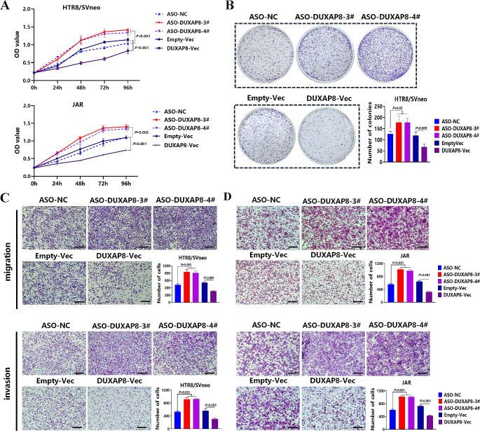
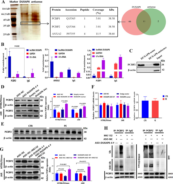
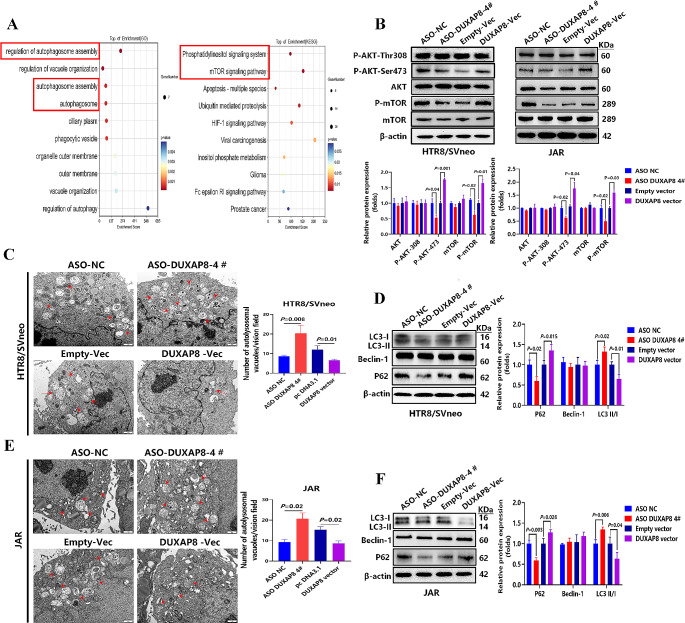
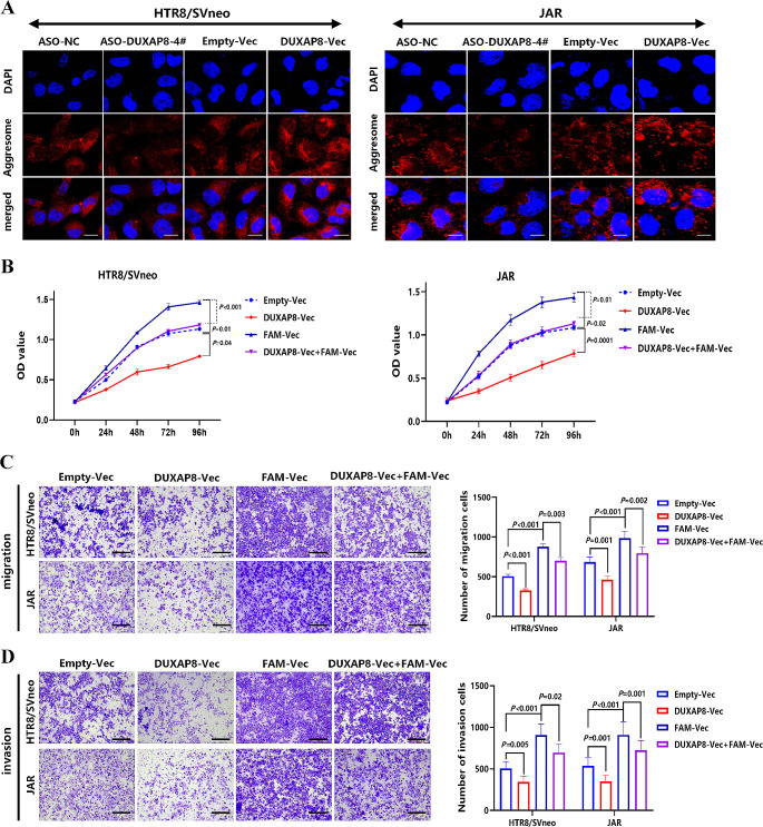
Preeclampsia (PE) is a life-threatening pregnancy-specific complication with controversial mechanisms and no effective treatment except delivery is available. Currently, increasing researchers suggested that PE shares pathophysiologic features with protein misfolding/aggregation disorders, such as Alzheimer disease (AD). Evidences have proposed defective autophagy as a potential source of protein aggregation in PE. Endoplasmic reticulum-selective autophagy (ER-phagy) plays a critical role in clearing misfolded proteins and maintaining ER homeostasis. However, its roles in the molecular pathology of PE remain unclear. We found that lncRNA DUXAP8 was upregulated in preeclamptic placentae and significantly correlated with clinical indicators. DUXAP8 specifically binds to PCBP2 and inhibits its ubiquitination-mediated degradation, and decreased levels of PCBP2 reversed the activation effect of DUXAP8 overexpression on AKT/mTOR signaling pathway. Function experiments showed that DUXAP8 overexpression inhibited trophoblastic proliferation, migration, and invasion of HTR-8/SVneo and JAR cells. Moreover, pathological accumulation of swollen and lytic ER (endoplasmic reticulum) was observed in DUXAP8-overexpressed HTR8/SVneo cells and PE placental villus trophoblast cells, which suggesting that ER clearance ability is impaired. Further studies found that DUXAP8 overexpression impaired ER-phagy and caused protein aggregation medicated by reduced FAM134B and LC3II expression (key proteins involved in ER-phagy) via activating AKT/mTOR signaling pathway. The increased level of FAM134B significantly reversed the inhibitory effect of DUXAP8 overexpression on the proliferation, migration, and invasion of trophoblasts. In vivo, DUXAP8 overexpression through tail vein injection of adenovirus induced PE-like phenotypes in pregnant rats accompanied with activated AKT/mTOR signaling, decreased expression of FAM134B and LC3-II proteins and increased protein aggregation in placental tissues. Our study reveals the important role of lncRNA DUXAP8 in regulating trophoblast biological behaviors through FAM134B-mediated ER-phagy, providing a new theoretical basis for understanding the pathogenesis of PE.

Keywords: DUXAP8; ER-phagy; FAM134B; Preeclampsia; Trophoblast cells.


Conflict of interest statement

None of the authors have any conflicts of interest to declare.

Figures

Fig. 1
Fig. 1
DUXAP8 expression is increased in preeclamptic placentae and primarily located in the nucleus of HTR-8/SVneo and JAR cells. A-B Heat map and volcano plot of the most differentially expressed lncRNAs in the placental tissues between the PE (n = 5, preeclampsia) and CTR (n = 5, normal control) group. C Validated expression of DUXAP8 in placentae of PE (n = 40) and CTR (n = 46) group was detected by RT-qPCR. Data are presented as the mean ± SEM, and statistical significance was evaluated using t-test for 2 comparisons. D The distribution and expression of DUXAP8 in the placental villi of CTR and PE group were detected by RNA-FISH (scar bar = 50 μm) (DAPI: nucleus; CK7: Cytokeratin 7, trophoblast marker protein). E-F Intracellular localization of DUXAP8 in HTR8/SVneo and JAR cells was observed by RNA-FISH (scar bar = 10 μm) (U6: nuclear marker; 18 S/β-actin: cytoplasmic marker) and cell fractionation assays (n = 3, Data are presented as the percentage)
Fig. 2
Fig. 2
Overexpression of DUXAP8 inhibits the proliferation, migration, and invasion of HTR-8/SVneo and JAR cells. A-B The effects of knockdown or overexpression of DUXAP8 on the proliferation of HTR-8/SVneo cells and JAR cells as detected by CCK-8 (n = 3) and colony formation assays (n = 3). C-D The effects of knockdown or overexpression of DUXAP8 on the migration and invasion ability of HTR-8/SVneo (C) and JAR cells (D) as detected by Transwell assay (without or with Matrigel) (n = 3, scar bar = 100 μm). Data are presented as the mean ± SD, statistical significance was evaluated using the t-test or Mann-Whitney U for 2 comparisons, one-way ANOVA and Tukey test or two-way ANOVA for multiple comparison vs. negative control
Fig. 3
Fig. 3
DUXAP8 specifically binds to the PCBP2 protein and affects its ubiquitin-mediated degradation. The sense strand DUX (+) and antisense strand DUX (-) of DUXAP8 were transfected into HTR-8/SVneo cells. A Combined with the Mass spectrometry analysis of RNA–protein binding solution, three proteins (PCBP2, ANXA2, and PCBP1) corresponding to the band size in the silver staining of binding solution in the pull-down experiment. B RNA-RIP and RT-qPCR were used to verify protein specifically bound to DUXAP8 in HTR-8/SVneo cells (n = 3). IgG: Negative control for RIP assay, GAPDH/U1sRNA: negative control for RT-qPCR analysis. C the expression of PCBP2 protein in the RNA–protein binding solution was detected by western blot. D-F The protein and mRNA expression of PCBP2 in HTR-8/SVneo and JAR cells after knockdown or overexpression of DUXAP8 (n = 3) and placental tissues of CTR and PE group (n = 12). CTR: normal control group; PE: Preeclampsia. G The expression level of PCBP2 protein between the ASO-DUXA8-4# group and ASO-DUXA8-4# +MG132 (proteasome inhibitor, 100nM) group (n = 3). H PCBP2 ubiquitination levels were verified by Co-immunoprecipitation (Co-IP) after downregulation of DUXAP8 in HTR-8/SVneo and JAR cells. IgG: Co-IP negative control. Data are presented as the mean ± SD/SE, statistical significance was evaluated using the t-test or Mann-Whitney U for 2 comparisons
Fig. 4
Fig. 4
DUXAP8 Overexpression increases the phosphorylation of the AKT/mTOR signaling pathway and decreases autophagy levels of trophoblast cells. A GO and KEGG analysis of differentially expressed genes identified by RNA sequencing. B the effects of knockdown or overexpression of DUXAP8 on the expression of key signaling proteins (AKT, mTOR, p-AKTSer473, p-AKTThr308, p-mTORS2248) in HTR-8/SVneo(n = 3) and JAR (n = 3) cells were determined by western blot. C-F The effects of knockdown or overexpression of DUXAP8 on the autolysosomal vacuoles (6000×, red arrows: autolysosomes) or autophagy-related proteins (LC3-II/LC3-I, P62, Beclin-1) in HTR-8/SVneo and JAR cells were separately examined by transmission electron microscopy (TEM) (n = 3) or western blot (n = 3).Data are all presented as the mean ± SD, statistical significance was evaluated using the t-test or Mann-Whitney U for 2 comparisons
Fig. 5
Fig. 5
DUXAP8 overexpression decreases FAM134B-mediated ER-phagy in trophoblast cells by activating AKT/mTOR signaling pathway. A Pathological changes of endoplasmic reticulum in villus trophoblastic cells of placentae from CTR and PE patients and HTR-8/SVneo cells with overexpressed DUXAP8 were observed by transmission electron microscopy (TEM). B Immunofluorescence analysis of the LC3 and FAM134B proteins in HTR-8/SVneo and JAR cells (Scale bar = 10 μm). C The effects of knockdown or overexpression of DUXAP8 on the expression of FAM134B were examined by western blot analysis (n = 3). D The expression of FAM134B protein in HTR-8/SVneo and JAR cells treated with ASO-DUXA8-4# and MHY1485 (mTOR agonist, 50 nM) was detected by western blot (n = 3). E-F After transfected with ASO-DUXA8-4# or ASO-NC in HTR-8/SVneo and JAR cells, immunofluorescence analysis of the co-localization of LC3 with WT or LIRmut HA-FAM134B (Scale bar = 5 μm) and Co-immunoprecipitation (Co-IP) assay was used to verify the interaction between WT or LIRmut HA-FAM134B and LC3. Data are all presented as the mean ± SD, statistical significance was evaluated using the t-test or Mann-Whitney U for 2 comparisons
Fig. 6
Fig. 6
Upregulation of FAM134B promotes the proliferation, migration, and invasion of HTR-8/SVneo and JAR cells. A The effects of knockdown or overexpression of DUXAP8 on intracellular protein aggregation in HTR-8/SVneo and JAR cells was detected by Protein Aggresome detection kit (Scale bar = 10 μm) (DAPI: nucleus; Aggresome: protein aggregates). B-D After co-transfecting with DUXAP8-Vec and FAM-Vec in HTR-8/SVneo (n = 3) and JAR (n = 3) cells, CCK-8 and Transwell assays (without or with Matrigel) were performed to detect proliferation, migration (scar bar = 100 μm), and invasion (scar bar = 100 μm) ability. Data are presented as the mean ± SD, statistical significance was evaluated using the t-test or Mann-Whitney U for 2 comparisons, one-way ANOVA and Tukey test or two-way ANOVA for multiple comparison
Fig. 7
Fig. 7
DUXAP8 overexpression induces preeclampsia-like phenotypes in pregnant rats. A Schematic diagram of animal experimental design. B The expression of DUXAP8 in placenta was detected by RT-qPCR (n = 6). C Systolic blood pressure (SBP) and Diastolic blood pressure (DBP) of pregnant rats were measured by tail-cuff system (GD9 and GD15) or carotid artery incubation (GD19) (n = 6); 24-hour proteinuria levels were detected by a protein assay kit(n = 6). D Placental weight of pregnant rats (n = 6). E Placental morphological features were identified by Masson’s trichrome staining(n = 6), and the histopathologic analysis of renal tissues was detected by periodic acid Schiff (n = 6). F The expression of key signaling proteins (AKT, mTOR, p-AKTSer473, p-AKTThr308, p-mTORS2248), FAM134B and LC3 proteins were detected by Western blot (n = 6). G The protein aggregation in placental trophoblast cells of pregnant rats was detected by Protein Aggresome detection kit (Scale bar = 50 μm) (DAPI: nucleus; Aggresome: protein aggregates). Data are presented as mean ± SD, statistical significance was evaluated using the t-test or Mann-Whitney for 2 comparisons and two-way ANOVA for multiple comparison

References

    1. Rana S, Lemoine E, Granger JP, Karumanchi SA (2019) Preeclampsia: Pathophysiology, Challenges, and Perspectives Circ Res, 2019. 124(7): pp. 1094–1112.10.1161/circresaha.118.313276 - PubMed
    1. Brown MA, Magee LA, Kenny LC, Karumanchi SA, McCarthy FP, Saito S et al (2018) Hypertensive Disorders of Pregnancy: ISSHP Classification, Diagnosis, and Management Recommendations for International Practice Hypertension, 2018. 72(1): pp. 24–43.10.1161/hypertensionaha.117.10803 - PubMed
    1. Mol BWJ, Roberts CT, Thangaratinam S, Magee LA, de Groot CJM, Hofmeyr GJ (2016) Pre-eclampsia The Lancet, 2016. 387(10022): pp. 999–1011.10.1016/S0140-6736(15)00070-7 - PubMed
    1. Hitti J, Sienas L, Walker S, Benedetti TJ, Easterling T (2018) Contribution of hypertension to severe maternal morbidity Am J Obstet Gynecol, 2018. 219(4): p. 40510.1016/j.ajog.2018.07.002 - PubMed
    1. Roberts JM (2014) Pathophysiology of ischemic placental disease Semin Perinatol, 2014. 38(3): pp. 139 – 4510.1053/j.semperi.2014.03.005 - PMC - PubMed

MeSH terms

LinkOut - more resources