Review
. 2024 Sep 1;5(9):1394-1401.
doi: 10.34067/KID.0000000000000531. Epub 2024 Aug 9.

Lupus Nephritis: Immune Cells and the Kidney Microenvironment

Affiliations
Review

Lupus Nephritis: Immune Cells and the Kidney Microenvironment

Irene Chernova. Kidney360. .

Abstract

Lupus nephritis (LN) is the most common major organ manifestation of the autoimmune disease SLE (lupus), with 10% of those afflicted progressing to ESKD. The kidney in LN is characterized by a significant immune infiltrate and proinflammatory cytokine milieu that affects intrinsic renal cells and is, in part, responsible for the tissue damage observed in LN. It is now increasingly appreciated that LN is not due to unidirectional immune cell activation with subsequent kidney damage. Rather, the kidney microenvironment influences the recruitment, survival, differentiation, and activation of immune cells, which, in turn, modify kidney cell function. This review covers how the biochemical environment of the kidney ( i.e ., low oxygen tension and hypertonicity) and unique kidney cell types affect the intrarenal immune cells in LN. The pathways used by intrinsic renal cells to interact with immune cells, such as antigen presentation and cytokine production, are discussed in detail. An understanding of these mechanisms can lead to the design of more kidney-targeted treatments and the avoidance of systemic immunosuppressive effects and may represent the next frontier of LN therapies.


Conflict of interest statement

Disclosure forms, as provided by each author, are available with the online version of the article at http://links.lww.com/KN9/A617.

Figures

Figure 1
Figure 1
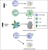
Pathways upregulated by infiltrating immune cells to adapt to the biochemical environment of the lupus kidney. In response to low oxygen tension, T cells and macrophages upregulate HIF-1α, resulting in decreased apoptosis and increased cytokine production, respectively. In response to hypertonicity, B cells upregulate Na+-K+-ATPase, resulting in improved survival in high-sodium conditions and B-cell accumulation in the LN kidney. Macrophages in LN upregulate TonEBP, a regulator of responses to hypertonic stress, leading to decreased efferocytosis (phagocytosis of apoptotic cells) and transcriptional upregulation of proinflammatory cytokines, although it is unknown whether hypertonicity is the trigger for increased TonEBP expression. HIF-1α, hypoxia-inducible factor 1α, LN, lupus nephritis; TonEBP, tonicity-responsive enhancer binding protein. Created with BioRender.com.
Figure 2
Figure 2
Intrinsic renal cell interactions with infiltrating immune cells. Selected contact-dependent and -independent effects of podocytes, endothelial cells, MCs, and RTECs on intrarenal immune components are summarized. Podocytes: cytokine secretion leads to glomerular inflammation, complement factor H secretion leads to IC deposition in GBM, and possible antigen presentation to T cells results in T-cell proliferation. GECs: cytokine secretion leads to neutrophil and macrophage recruitment; possible antigen presentation and interactions between endothelial glycocalyx hyaluronic acid with T-cell CD44 lead to enhanced T-cell proliferation/function. MCs: antigen presentation leads to T-cell proliferation and IFN-γ secretion. IL-6 production leads to glomerular inflammation, M1 macrophage polarization (in contrast to M2 macrophage polarization with other, undefined cytokines), and possibly MHC-II and ICAM-1 upregulation. RTECs: BAFF production leads to increased kidney inflammation, and reduced arginase 1 production in response to IL-23 in the lupus kidney leads to higher arginine levels and increased T-cell proliferation. RTECs express both stimulatory and inhibitory cell surface receptors, which, likely in combination with anti-inflammatory cytokines, lead to attenuated T- and B-cell proliferative responses. BAFF, B-cell activating factor; GBM, glomerular basement membrane; GEC, glomerular endothelial cell; IC, immune complex; ICAM-1, intracellular adhesion molecule 1; MC, mesangial cell; MHC, major histocompatibility complex; PDL-1, programmed death-ligand 1; RTEC, renal tubular epithelial cell; TCR, T-cell receptor; VCAM-1, vascular cell adhesion molecule 1. Created with BioRender.com.

References

    1. Kwon MS, Lim SW, Kwon HM. Hypertonic stress in the kidney: a necessary evil. Physiology (Bethesda). 2009;24:186–191. doi:10.1152/physiol.00005.2009 - DOI - PubMed
    1. Chen P-M Wilson PC Shyer JA, et al. . Kidney tissue hypoxia dictates T cell–mediated injury in murine lupus nephritis. Sci Transl Med. 2020;12(538):eaay1620. doi:10.1126/scitranslmed.aay1620 - DOI - PMC - PubMed
    1. Bhargava R, Li H, Tsokos GC. Pathogenesis of lupus nephritis: the contribution of immune and kidney resident cells. Curr Opin Rheumatol. 2023;35(2):107–116. doi:10.1097/BOR.0000000000000887 - DOI - PubMed
    1. Tsokos GC, Boulougoura A, Kasinath V, Endo Y, Abdi R, Li H. The immunoregulatory roles of non-haematopoietic cells in the kidney. Nat Rev Nephrol. 2024;20(4):206–217. doi:10.1038/s41581-023-00786-x - DOI - PMC - PubMed
    1. Sabiu G, Kasinath V, Jung S, Li X, Tsokos GC, Abdi R. Targeted nanotherapy for kidney diseases: a comprehensive review. Nephrol Dial Transplant. 2023;38(6):1385–1396. doi:10.1093/ndt/gfac233 - DOI - PMC - PubMed

LinkOut - more resources