. 2024 Sep 16;13(5):e240079.
doi: 10.1530/ETJ-24-0079. Print 2024 Oct 1.

Decreased sirtuin 4 levels promote cellular proliferation and invasion in papillary thyroid carcinoma

Affiliations

Decreased sirtuin 4 levels promote cellular proliferation and invasion in papillary thyroid carcinoma

Hyun-Jin Lee et al. Eur Thyroid J. .

Abstract

Objective: This study examined the effect of sirtuin 4 (SIRT4), a NAD+-dependent deacetylase, on the proliferation and progression of papillary thyroid carcinoma (PTC).

Methods: Data from The Cancer Genome Atlas (TCGA) were analyzed to identify SIRT4 expression in thyroid cancer. Subsequently, the correlation between SIRT4 expression and clinical characteristics was examined in 205 PTC tissue samples. In vitro assays using three human thyroid cancer cell lines (B-CPAP, TPC-1, and SNU-790) were conducted to assess the effects of regulated SIRT4 expression on cell growth, apoptosis, invasion, and migration. Furthermore, in vivo experiments were performed in a xenograft mouse model.

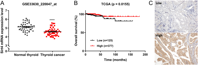
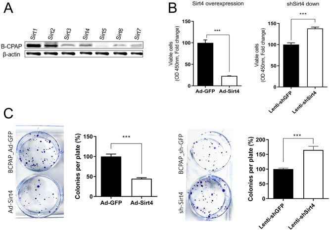
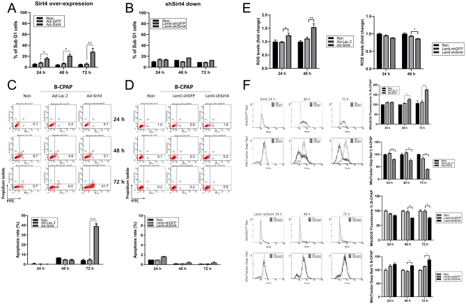
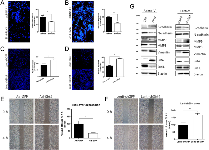
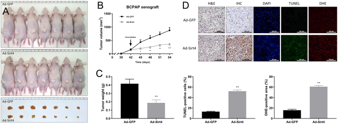
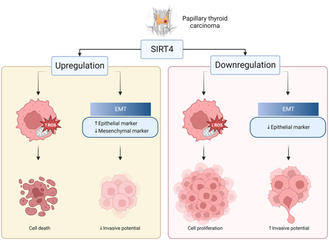
Results: Gene Expression Omnibus (GEO) and TCGA data indicated that SIRT4 expression is lower in thyroid cancer and SIRT4 downregulation is associated with poor overall survival. In PTC tissues, positive SIRT4 expression was associated with decreased extracapsular extension. In in vitro experiments using three human thyroid cancer cell lines, overexpression of SIRT4 decreased cell survival, clonogenic potential, and invasion and migratory capabilities, as well as inducing apoptosis and increasing reactive oxygen species levels. SIRT4 overexpression upregulated E-cadherin and downregulated N-cadherin, suggesting its potential involvement in the regulation of epithelial-mesenchymal transition. These findings were confirmed in vivo using a xenograft mouse model.

Conclusion: This study provides novel insight into the potential contribution of SIRT4 to the regulation of the pathological progression of PTC. The data suggest that SIRT4 plays a tumor-suppressive role in PTC by inhibiting growth, survival, and invasive potential. Future research should investigate the molecular mechanisms underlying these effects of SIRT4.

Keywords: SIRT4 protein; epithelial–mesenchymal transition; papillary thyroid cancer; reactive oxygen species; thyroid cancer.


Conflict of interest statement

The authors declare that they have no known competing financial interests or personal relationships that could have appeared to influence the work reported in this paper.

Figures

Figure 1
Figure 1
Clinical significance of SIRT4 expression in thyroid cancer. (A) Analysis of the GSE33630 database showed that the mRNA expression levels of SIRT4 are significantly lower in thyroid cancer tissues (n = 45) than in adjacent normal thyroid tissues (n= 60; P < 0.0001, t-test). (B) Overall survival was analyzed using a Kaplan–Meier (KM) plot based on the TCGA dataset, which included patients with high SIRT4 expression and those with low SIRT4 expression (P = 0.0155, log-rank test). (C) Representative IHC staining image of PTC tissues with different degrees of SIRT4 expression. Number of positive cells: (−) <10%; (+) >10%. ****P < 0.0001. PTC, papillary thyroid carcinoma; TCGA, The Cancer Genome Atlas.
Figure 2
Figure 2
SIRT4 inhibits B-CPAP cell proliferation. (A) Western blot analysis of the expression levels of sirtuins in B-CPAP cells. (B) CCK-8 assay of SIRT4-overexpressing and shSIRT4-transfected B-CPAP cells compared with the controls. B-CPAP cells were seeded into 24-well plates and treated with CCK-8 solution 72 h post seeding. SIRT4 significantly suppressed the viability of B-CPAP cells. (C) Clonogenic assay of SIRT4-overexpressing and shSIRT4-transfected B-CPAP cells compared with the controls. Surviving clones were decreased in SIRT4-overexpressing B-CPAP cells but increased in SIRT4-downregulated B-CPAP cells. *P < 0.05; **P < 0.01; ***P < 0.001. CCK-8, Cell Counting Kit-8.
Figure 3
Figure 3
SIRT4 regulates the cell cycle and promotes cell death by modulating ROS levels in B-CPAP cells. (A) Percentage of cells in sub-G1 phase in control and SIRT4-overexpressing B-CPAP cells. B-CPAP cells were treated for 24, 48, and 72 h and examined by flow cytometry. Histograms represent PI fluorescence intensity. SIRT4 overexpression significantly increased the number of sub-G1 cells. (B) Percentage of cells in sub-G1 phase in control and shSIRT4-transfected B-CPAP cells. (C) Apoptosis rate in control and SIRT4-overexpressing B-CPAP cells analyzed by flow cytometry using an Annexin V (FITC)-PI apoptosis detection kit. SIRT4 overexpression significantly increased the apoptosis rate at 72 h. (D) Apoptosis rate in control and SIRT4-downregulated B-CPAP cells. (E) Differences in total ROS levels upon regulation of SIRT4 expression. (F) SIRT4 upregulation significantly increased MitoSOX red levels and reduced the intensity of MTDR over time, and SIRT4 downregulation had the opposite effect. MTDR, MitoTracker Deep Red; ROS, reactive oxygen species.
Figure 4
Figure 4
SIRT4 regulates the invasion and migration of B-CPAP cells by modulating the expression of EMT-related proteins. (A) Relative invasion and (B) migration abilities of SIRT4-overexpressing B-CPAP cells compared with the controls analyzed using Transwell assays. Cells were incubated for 24 h, and invaded and migrated cells were counted under a fluorescence microscope at ×100 magnification in five random fields. (C) Relative invasion and (B) migration abilities of shSIRT4-transfected B-CPAP cells compared with the controls analyzed using Transwell assays. (E) Wound healing assays of SIRT4 overexpressing and (F) SIRT4 knockdown cells compared with the controls. Representative images at the indicated times are on the right. (G) Modulation of EMT-related proteins by SIRT4 expression. The expression of E-cadherin, N-cadherin, and other EMT markers was analyzed and quantified. *P < 0.05, **P < 0.01, ***P < 0.001. EMT, epithelial–mesenchymal transition.
Figure 5
Figure 5
SIRT4 inhibits tumor growth in a B-CPAP xenograft mouse model. (A) On day 42 after implanting tumor cells, mice received intratumoral injections of Ad-GFP or Ad-SIRT4. The tumor images show excised tumors from each group, with the top group representing the control group and the bottom group representing the SIRT4-expression group. (B) Tumor diameter was measured every 3 days using digital calipers and tumor volume was calculated. The diagram shows the tumor growth curve of the 14 mice at the indicated times after intratumoral injections of Ad-GFP or Ad-SIRT4. (C) Tumor weight was compared between the SIRT4-expression group and the control group. (D) SIRT4 overexpression induces apoptosis and superoxide production in mouse tumors. FFPE sections of tumors were co-stained with DAPI (0.5 µg/mL) to visualize nuclei, along with TdT enzyme (200 µg/mL) and DHE (10 μmol/L) to detect apoptosis and superoxide production, respectively. Apoptotic green fluorescence, oxidative red fluorescence, and nuclear blue fluorescence were analyzed under a fluorescence microscope at 400× magnification. The graph below shows the quantified signal of cells stained positively by TdT and DHE in five random fields. n = 8. *P < 0.05, **P < 0.01, ***P < 0.001. DAPI, 4′,6-diamidino-2-phenylindole; DHE, dihydroethidium; FFPE, formalin-fixed paraffin-embedded; TdT, terminal deoxynucleotidyl transferase; TUNEL, TdT-mediated dUTP nick end labeling.
Figure 6
Figure 6
Schematic diagram of the role of SIRT4 on papillary thyroid cancer cells. Figure created with BioRender.com.

References

    1. Agate L Lorusso L & Elisei R. New and old knowledge on differentiated thyroid cancer epidemiology and risk factors. Journal of Endocrinological Investigation 201235(Supplement) 3–9. - PubMed
    1. Hundahl SA Fleming ID Fremgen AM & Menck HR. A national cancer data base report on 53,856 cases of thyroid carcinoma treated in the U.S., 1985–1995 (see comments). Cancer 1998832638–2648. (https://doi.org/10.1002/(sici)1097-0142(19981215)83:12<2638::aid-cncr...) - PubMed
    1. Chen D, Tan Y, Li Z, Li W, Yu L, Chen W, Liu Y, Liu L, Guo L, Huang W, et al.Organoid cultures derived from patients with papillary thyroid cancer. Journal of Clinical Endocrinology and Metabolism 20211061410–1426. (10.1210/clinem/dgab020) - DOI - PubMed
    1. Toniato A Boschin I Casara D Mazzarotto R Rubello D & Pelizzo M. Papillary thyroid carcinoma: factors influencing recurrence and survival. Annals of Surgical Oncology 2008151518–1522. (10.1245/s10434-008-9859-4) - DOI - PubMed
    1. Ito Y Kudo T Kobayashi K Miya A Ichihara K & Miyauchi A. Prognostic factors for recurrence of papillary thyroid carcinoma in the lymph nodes, lung, and bone: analysis of 5,768 patients with average 10-year follow-up. World Journal of Surgery 2012361274–1278. (10.1007/s00268-012-1423-5) - DOI - PubMed

MeSH terms