. 2024;100(s1):S291-S304.
doi: 10.3233/JAD-240640.

Exposure to Lead in Drinking Water Causes Cognitive Impairment via an Alzheimer's Disease Gene-Dependent Mechanism in Adult Mice

Affiliations

Exposure to Lead in Drinking Water Causes Cognitive Impairment via an Alzheimer's Disease Gene-Dependent Mechanism in Adult Mice

Katharina Kohler et al. J Alzheimers Dis. 2024.

Abstract

Background: Exposure to lead (Pb) is a major public health problem that could occur through contaminated soil, air, food, or water, either during the course of everyday life, or while working in hazardous occupations. Although Pb has long been known as a neurodevelopmental toxicant in children, a recent and growing body of epidemiological research indicates that cumulative, low-level Pb exposure likely drives age-related neurologic dysfunction in adults. Environmental Pb exposure in adulthood has been linked to risk of late-onset Alzheimer's disease (AD) and dementia.

Objective: Although the biological mechanism underlying this link is unknown, it has been proposed that Pb exposure may increase the risk of AD via altering the expression of AD-related genes and, possibly, by activating the molecular pathways underlying AD-related pathology.

Methods: We investigated Pb exposure using a line of genetically modified mice with AD-causing knock-in mutations in the amyloid precursor protein and presenilin 1 (APPΔNL/ΔNL x PS1P264L/P264L) that had been crossed with Leprdb/db mice to impart vulnerability to vascular pathology.

Results: Our data show that although Pb exposure in adult mice impairs cognitive function, this effect is not related to either an increase in amyloid pathology or to changes in the expression of common AD-related genes. Pb exposure also caused a significant increase in blood pressure, a well known effect of Pb. Interestingly, although the increase in blood pressure was unrelated to genotype, only mice that carried AD-related mutations developed cognitive dysfunction, in spite of showing no significant change in cerebrovascular pathology.

Conclusions: These results raise the possibility that the increased risk of dementia associated with Pb exposure in adults may be tied to its subsequent interaction with either pre-existing or developing AD-related neuropathology.

Keywords: Aging; Alzheimer’s disease; amyloid; amyloid precursor protein; hypertension; presenilin 1; vascular contributions to cognitive impairment and dementia.


Conflict of interest statement

CONFLICTS OF INTEREST

M. Paul Murphy is an Editorial Board Member of this journal but was not involved in the peer-review process nor had access to any information regarding its peer-review. The remaining authors have no conflicts of interest to report.

Figures

Figure 1.
Figure 1.. Pb Exposure Resulted in Increased Blood Pressure.
(A) [Pb] by ICP-MS (n=6 / group; data shown are for 3 Lepr+/+, 6 Leprdb/+, 9 Leprdb/db, ½ M / F, divided equally between exposure groups; in this study, all mice were APP+/+ × PS1+/+, but we have not noted any genotype effects in this or other studies); as mice were not perfused, Pb detected in the brain may be due to residual blood (**). (B) ~3 months of Pb exposure caused an increase in blood pressure (Pb Treated/Control: 11/12; sex = 13F / 10M; genotype = 15 Lepr+/+, 8 Leprdb/db, 3 APP+/+ × PS1+/+, 20 APPΔNLh/ΔNLh × PS1P264L/P264L); we did not see differences by genotype, but we did see some evidence that the effect might be larger in males (sex*treatment interaction, p=0.02), possibly since the males had a lower baseline BP than the females (p<0.01; not shown). (C) Pb did not affect either fasting glucose levels, or affect changes in blood glucose following a bolus injection of dextrose (N = 44, ½ M / F; 23 Lepr+/+, 21 Leprdb/db; 17 APP+/+ × PS1+/+, 27 APPΔNLh/ΔNLh × PS1P264L/P264L). (D) Pb had no effect on body weight (note the slight decrease during the water maze testing period; baseline weights / group (g): control, 46.7 ± 0.8; Pb exposed, 45.1 ± 0.8).
Figure 2.
Figure 2.. Pb Exposure Impaired Cognitive Function, but Only in Mutant Mice.
(A) Pb had no effect on either swim speed (all genotypes combined) or on the ability to locate a flagged platform (separated by genotype) in the Morris water maze; N = 135 mice: WT (Lepr+/+ × APP+/+ x PS1+/+), n=27; db (Leprdb/db × APP+/+ x PS1+/+), n=32; AD (Lepr+/+ × APPΔNLh/ΔNLh × PS1P264L/P264L), n=46; db/AD (Leprdb/db × APPΔNLh/ΔNLh × PS1P264L/P264L), n=30; Pb treated, n = 69 / untreated, n = 66; sex balance = 69F / 66M. Note again the lack of genotype differences on the visual cued task. (B) Mice were given 4 trials / day over 5 days in the MWM; consistent with our earlier results in this line, the db/AD mice were the worst at this task (shown is the overall main effect of genotype, regardless of Pb exposure). (C) Pb exposed mice were impaired at learning the location of the hidden platform (p<0.003); however, this effect was driven almost entirely by mice with APP and PS1 mutations (p<0.005), as wild type animals were unaffected. The Pb effect was particularly pronounced on the last day of training (N.B.: days 2 and 3 have been left off of the genotype figure due to space constraints). (D) APP × PS1 mice also showed a deficit on a probe test of memory for the hidden platform location and, as expected, the db/AD mice performed worse than AD mice; in both cases, Pb exposure made the deficit worse. We did not detect a difference on training trial escape latency between Lepr+/+ and Leprdb/db mice. (E) Representative mouse swim patterns (shown are both male, Lepr+/+ × APPΔNLh/ΔNLh × PS1P264L/P264L). * = p<0.05, ** = p<0.01; relative to comparison group.
Figure 3.
Figure 3.. Pb Exposure Did Not Affect AD-related mRNA Expression or the Amount of Amyloid In the Brain.
(A) There were no effects (all adjusted p-values were n.s.) of Pb on major AD-related genes by qRT-PCR (Amyloid Precursor Protein, APP; Anterior Pharynx-Defective 1, APH1; β-Amyloid Precursor Protein Cleaving Enzyme, BACE (1 or 2); Endothelin Converting Enzyme, ECE (1 or 2); Insulin Degrading Enzyme, IDE; Neprilysin, NEP; Nicastrin, NIC; Presenilin, PS (1 or 2); Presenilin Enhancer 2, PEN2). Of these genes, only APP (F[2,13] = 6.61, p=0.010) and PS1 (F[2,13] = 5.71, p=0.017) showed a significant difference in the unadjusted p-values. RNA were extracted from one hemibrain; we did not evaluate expression by region. Data shown are for 7 Lepr+/+, 13 Leprdb/+, 10 Leprdb/db, 17 M / 13 F, divided between exposure groups as follows: Control (n=8), 0.004% Pb (n=13), 0.2% Pb (n = 9). All mice shown in this combined subgroup were APP+/+ x PS1+/+, and we have not observed any genotype differences. Note that this subgroup was exposed earlier, during weaning and pre-weaning period, in order to maximize chances to detect a difference in gene expression. (B) Aβ pathology was unchanged (human Aβ specific ELISA); WT (Lepr+/+ × APP+/+ x PS1+/+) and db (Leprdb/db × APP+/+ x PS1+/+) mice show minimal signal and are not shown.
Figure 4.
Figure 4.. Pb Exposure Did Not Affect Expression of AD-related Proteins
(A) RIPA-extracted brains from Pb exposed mice were separated by SDS-PAGE, and blotted for multiple AD-related proteins. Shown are Tau (detected using Tau46; Cell Signaling), and APP (detected using AbCT20. Equal protein loading was verified by probing for βActin (AC15, Sigma). A longer exposure was used to visualize CTFs (top); a lighter APP exposure is also shown (bottom) to better show intermouse variability. Note that KI mice can be clearly identified by an increased APP CTF signal, even though they do not over express APP. Representative blots are shown for a subset of mice; quantitation was performed using spot and western blots on all genotype, sex, and treatment combinations (n = 3 / subgroup; N = 48); we did not find any effects of Pb. (B) Pb exposure did not affect BACE1 (AD and WT are shown here, but other genotypes were also not different); equal protein loading shown through GAPDH levels.
Figure 5.
Figure 5.. Pb Exposure Had No Effect on Vascular Events.
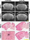
We did not detect any increase in the occurrence of microbleeds in the db/AD mice, either via MRI (top) or via examination by Prussian blue staining. We did detect some Prussian blue infarcts (an example is shown at higher magnification in the bottom left), but these were rare events. N = 36; 18 Lepr+/+, 18 Leprdb/db; 18 APP+/+ x PS1+/+, 18 APPΔNLh/ΔNLh × PS1P264L/P264L; 12 M / 24 F; 12 Pb exposed, 24 control).

References

    1. Bakulski KM, Rozek LS, Dolinoy DC, et al. Alzheimer’s disease and environmental exposure to lead: the epidemiologic evidence and potential role of epigenetics. Curr Alzheimer Res 2012; 9: 563–573. - PMC - PubMed
    1. Nabi M and Tabassum N. Role of Environmental Toxicants on Neurodegenerative Disorders. Front Toxicol 2022; 4: 837579. - PMC - PubMed
    1. Lu K, Liu T, Wu X, et al. Association between serum iron, blood lead, cadmium, mercury, selenium, manganese and low cognitive performance in old adults from National Health and Nutrition Examination Survey (NHANES): a cross-sectional study. Br J Nutr 2023; 130: 1743–1753. - PubMed
    1. Sanders T, Liu Y, Buchner V, et al. Neurotoxic effects and biomarkers of lead exposure: a review. Rev Environ Health 2009; 24: 15–45. - PMC - PubMed
    1. Li K, Wu J, Mei Y, et al. Metallomics analysis of metal exposure and cognitive function in older adults: A combined epidemiological and bioinformatics study. Chemosphere 2023; 341: 140049. - PubMed

LinkOut - more resources