. 2024 Aug 9;10(32):eadl5473.
doi: 10.1126/sciadv.adl5473. Epub 2024 Aug 9.

Spatial transcriptomic characterization of pathologic niches in IPF

Affiliations

Spatial transcriptomic characterization of pathologic niches in IPF

Christoph H Mayr et al. Sci Adv. .

Abstract

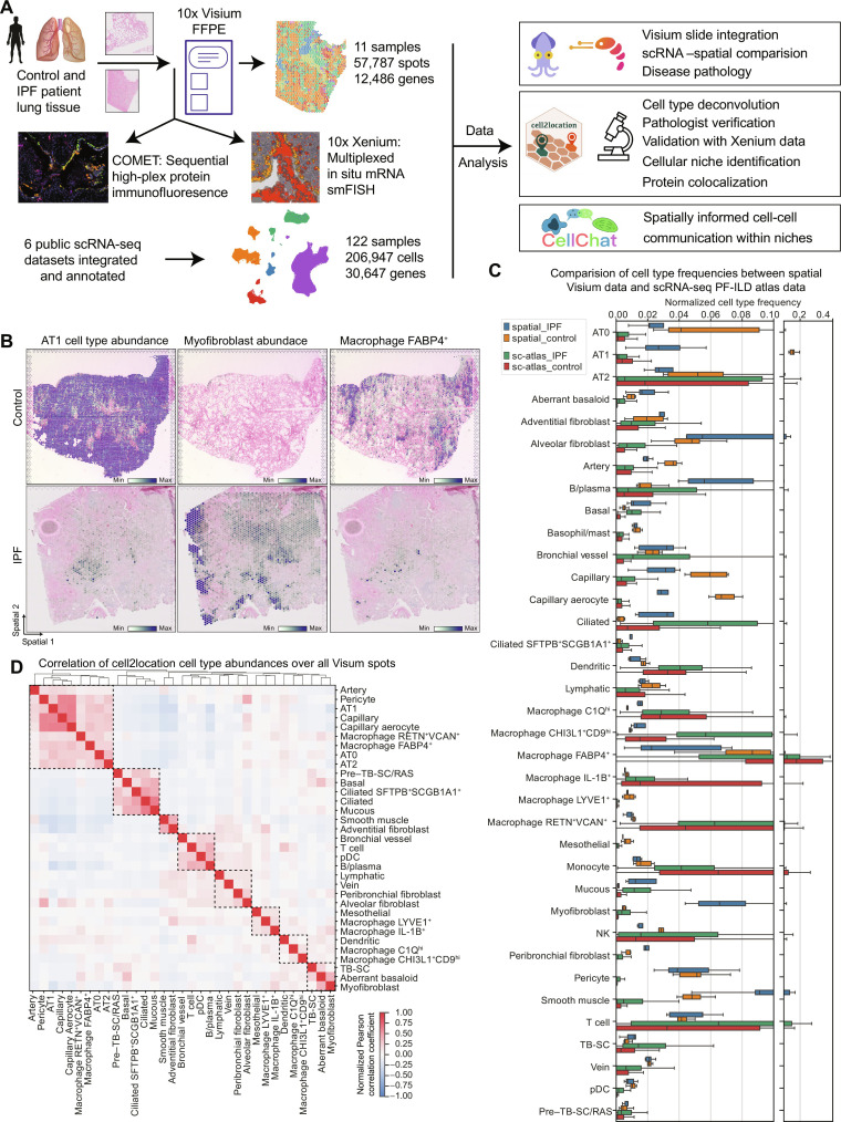
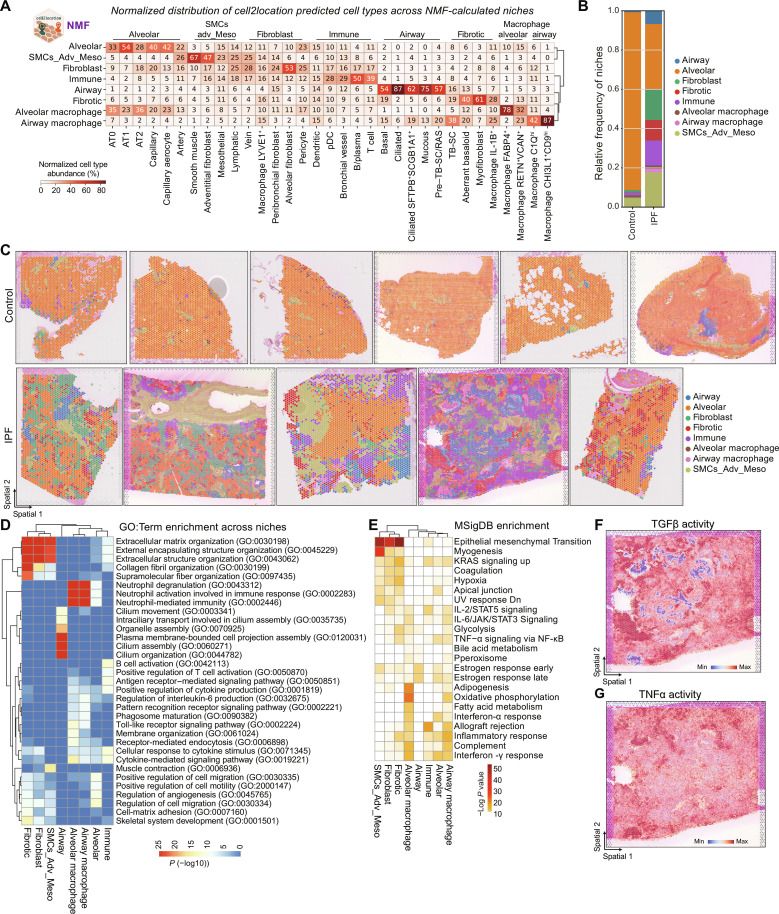
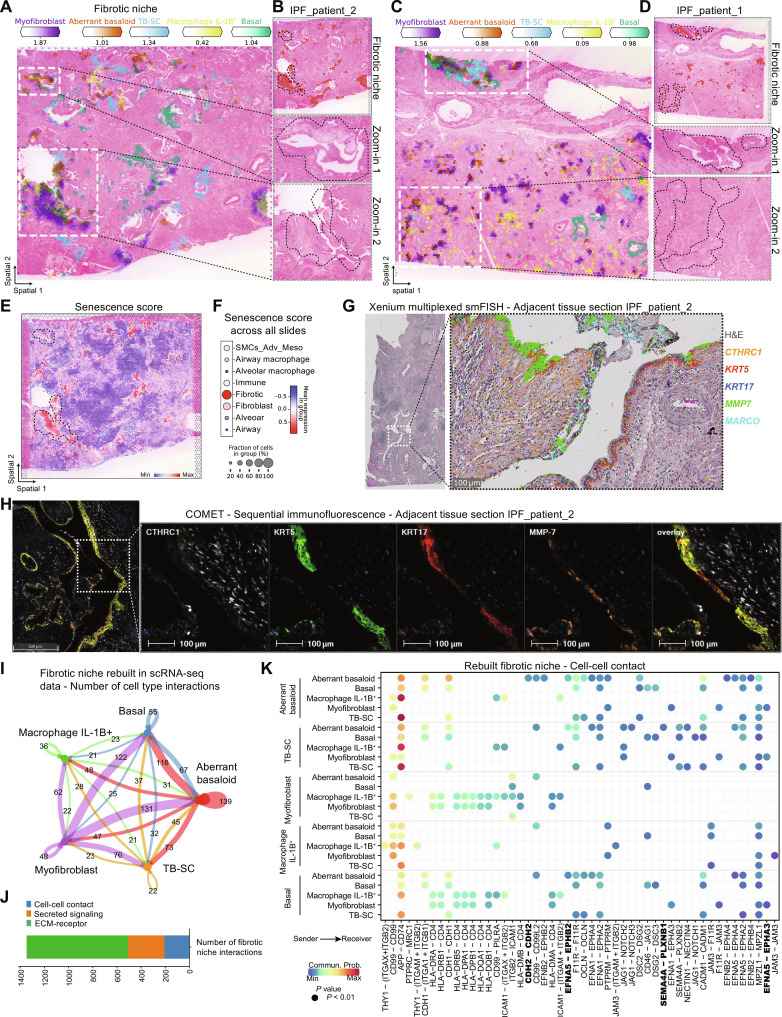
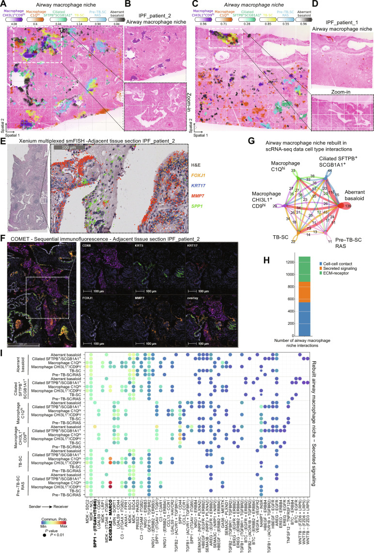
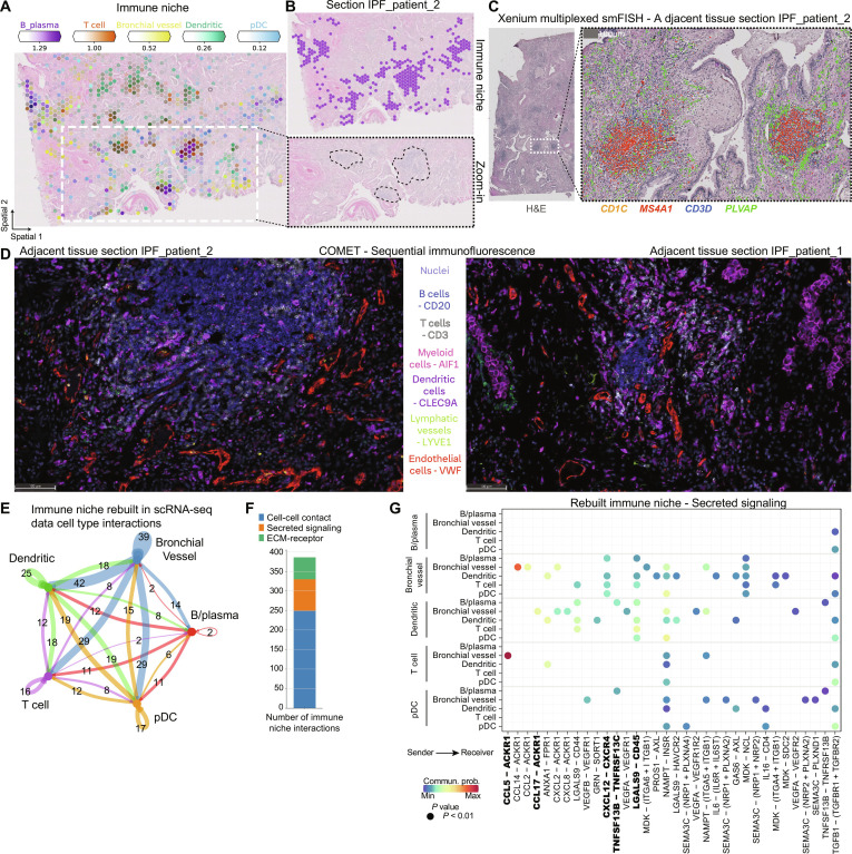
Despite advancements in antifibrotic therapy, idiopathic pulmonary fibrosis (IPF) remains a medical condition with unmet needs. Single-cell RNA sequencing (scRNA-seq) has enhanced our understanding of IPF but lacks the cellular tissue context and gene expression localization that spatial transcriptomics provides. To bridge this gap, we profiled IPF and control patient lung tissue using spatial transcriptomics, integrating the data with an IPF scRNA-seq atlas. We identified three disease-associated niches with unique cellular compositions and localizations. These include a fibrotic niche, consisting of myofibroblasts and aberrant basaloid cells, located around airways and adjacent to an airway macrophage niche in the lumen, containing SPP1+ macrophages. In addition, we identified an immune niche, characterized by distinct lymphoid cell foci in fibrotic tissue, surrounded by remodeled endothelial vessels. This spatial characterization of IPF niches will facilitate the identification of drug targets that disrupt disease-driving niches and aid in the development of disease relevant in vitro models.


Figures

Fig. 1.
Fig. 1.. Combining spatial transcriptomics and scRNA-seq places genes and cell types to their tissue location.
(A) Experimental design included spatial transcriptomics using Visium for FFPE and CytAssist (10x Genomics) on control and IPF patient samples (seven donors, 4× sections Visium CytAssist for FFPE, and 7× sections with Visium for FFPE; see fig. S1A for distribution of donors and sections). On two adjacent IPF and two control sections, as well as three additional IPF sections, we applied Xenium in situ (10x Genomics) for multiplexed in situ mRNA imaging similar to Single Molecule Fluorescence In Situ Hybridization (smFISH) and high-plex sequential protein immunofluorescence on the COMET platform. Six publicly available scRNA-seq datasets were integrated and annotated into a PF-ILD atlas. Visium and scRNA-seq data were combined for various analysis steps, including cell type deconvolution or cell-cell communication. (B) Spatial plots show cell2location mapping of estimated cell type abundance of AT1, FABP4+ alveolar macrophages, and myofibroblast cells on one control and one IPF tissue section. (C) Comparison of normalized cell type frequencies in spatial transcriptomic data and scRNA-seq PF-ILD atlas. (D) Pearson correlation of cell type abundances estimated with cell2location across all tissue sections identifies spatial colocated cell type modules.
Fig. 2.
Fig. 2.. Identification of three characteristic cellular niches in IPF lung tissue.
(A) Heatmap shows the normalized distribution of estimated cell type abundance in percentages across the niches, which were computed using the NMF method of the cell2location package. SMCs_Adv_Meso refers to smooth muscle, adventitial fibroblast, and mesothelial cells. (B) Relative frequency of niches across disease conditions is shown. Values are aggregated for four control and three IPF donors. (C) Spatial plots visualize the niche annotation across all samples and across conditions. (D) The heatmap shows top 3 enriched GO: terms per niche, colored by–log10 P value. (E) The heatmap shows top enriched Molecular Signatures Database (MSigDB) hallmark genes sets per niche. UV, ultraviolet; STAT5, signal transducers and activators of transcription 5; JAK, Janus kinase; NF-κB, nuclear factor κB; KRAS, Kirsten rat sarcoma virus; Dn, down-regulated. (F and G) Spatial plots exemplary show pathway activity for one IPF section.
Fig. 3.
Fig. 3.. The fibrotic niche localizes around airways.
(A to D) Spatial plots show, for two IPF tissue sections, (A and C) the abundance of fibrotic niche–associated cell types and (B and D) the fibrotic niche distribution and zoom into H&E-stained tissue for highlighted regions. (E and F) Senescence gene score (CDKN1A, CDKN1B, CDKN2B, TP53, SERPINE1, and GLB1) calculated using hotspot gene-module scoring (29) (E) on one IPF tissue section and (F) as dot plot against the other niches. (G) Xenium mRNA in situ hybridization data on an adjacent tissue section from (A). (H) Multiplexed protein immunofluorescence on an adjacent tissue section from (A). (I) Bubble plot summarizing the number of interactions within the rebuilt fibrotic niche in the scRNA-seq PF-ILD atlas data. (J) Number of interactions within the fibrotic niche per communication category. (K) Heatmap of statistically significant ligand-receptor pairs from the cell-cell contact category within the fibrotic niche.
Fig. 4.
Fig. 4.. SPP1+ macrophages are localized to fibrotic airway lumen.
(A to D) Spatial plots show, for two IPF tissue sections, (A and C) the abundance of airway macrophage niche associated cell types and (B and D) the niche distribution and zoom into H&E-stained tissue for highlighted regions. (E) Xenium mRNA in situ hybridization data on an adjacent tissue section from (A). (F) Multiplexed protein immunofluorescence on an adjacent tissue section from (A). (G) Bubble plot summarizing the number of interactions within the rebuilt airway macrophage niche in the scRNA-seq PF-ILD atlas data. (H) Number of interactions within the airway macrophage niche per communication category. (I) Heatmap of statistically significant ligand-receptor pairs from the secreted signaling category within the airway macrophage niche.
Fig. 5.
Fig. 5.. Immune cells from foci are recruited by IPF-specific bronchial vessels.
(A and B) Spatial plots show, for one IPF tissue section, (A) the abundance of immune niche–associated cell types and (B) the immune niche distribution and zoom into H&E-stained tissue for highlighted regions. (C) Xenium mRNA in situ hybridization data on an adjacent tissue section from (A). (D) Multiplexed protein immunofluorescence on an adjacent tissue section from (A). (E) Bubble plot summarizing the number of interactions between cell types within the rebuilt immune niche in the scRNA-seq PF-ILD atlas data. (F) Number of interactions within the immune niche per communication category. (G) Heatmap of statistically significant ligand-receptor pairs from the cell-cell contact category within the immune niche.

References

    1. Henderson N. C., Rieder F., Wynn T. A., Fibrosis: From mechanisms to medicines. Nature 587, 555–566 (2020). - PMC - PubMed
    1. Podolanczuk A. J., Thomson C. C., Remy-Jardin M., Richeldi L., Martinez F. J., Kolb M., Raghu G., Idiopathic pulmonary fibrosis: State of the art for 2023. Eur. Respir. J. 61, 2200957 (2023). - PubMed
    1. Raghu G., Chen S.-Y., Yeh W.-S., Maroni B., Li Q., Lee Y.-C., Collard H. R., Idiopathic pulmonary fibrosis in US medicare beneficiaries aged 65 years and older: Incidence, prevalence, and survival, 2001–11. Lancet Respir. Med. 2, 566–572 (2014). - PubMed
    1. Reyfman P. A., Walter J. M., Joshi N., Anekalla K. R., McQuattie-Pimentel A. C., Chiu S., Fernandez R., Akbarpour M., Chen C.-I., Ren Z., Verma R., Abdala-Valencia H., Nam K., Chi M., Han S., Gonzalez-Gonzalez F. J., Soberanes S., Watanabe S., Williams K. J. N., Flozak A. S., Nicholson T. T., Morgan V. K., Winter D. R., Hinchcliff M., Hrusch C. L., Guzy R. D., Bonham C. A., Sperling A. I., Bag R., Hamanaka R. B., Mutlu G. M., Yeldandi A. V., Marshall S. A., Shilatifard A., Amaral L. A. N., Perlman H., Sznajder J. I., Argento A. C., Gillespie C. T., Dematte J., Jain M., Singer B. D., Ridge K. M., Lam A. P., Bharat A., Bhorade S. M., Gottardi C. J., Budinger G. R. S., Misharin A. V., Single-cell transcriptomic analysis of human lung provides insights into the pathobiology of pulmonary fibrosis. Am. J. Respir. Crit. Care Med. 199, 1517–1536 (2018). - PMC - PubMed
    1. Morse C., Tabib T., Sembrat J., Buschur K. L., Bittar H. T., Valenzi E., Jiang Y., Kass D. J., Gibson K., Chen W., Mora A., Benos P. V., Rojas M., Lafyatis R., Proliferating SPP1/MERTK-expressing macrophages in idiopathic pulmonary fibrosis. Eur. Respir. J. 54, 1802441 (2019). - PMC - PubMed