. 2024 Aug 9;10(32):eado0636.
doi: 10.1126/sciadv.ado0636. Epub 2024 Aug 9.

Cyclin F-EXO1 axis controls cell cycle-dependent execution of double-strand break repair

Affiliations

Cyclin F-EXO1 axis controls cell cycle-dependent execution of double-strand break repair

Hongbin Yang et al. Sci Adv. .

Abstract

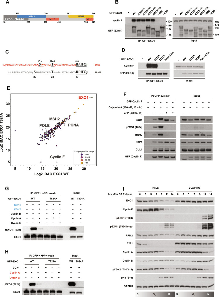
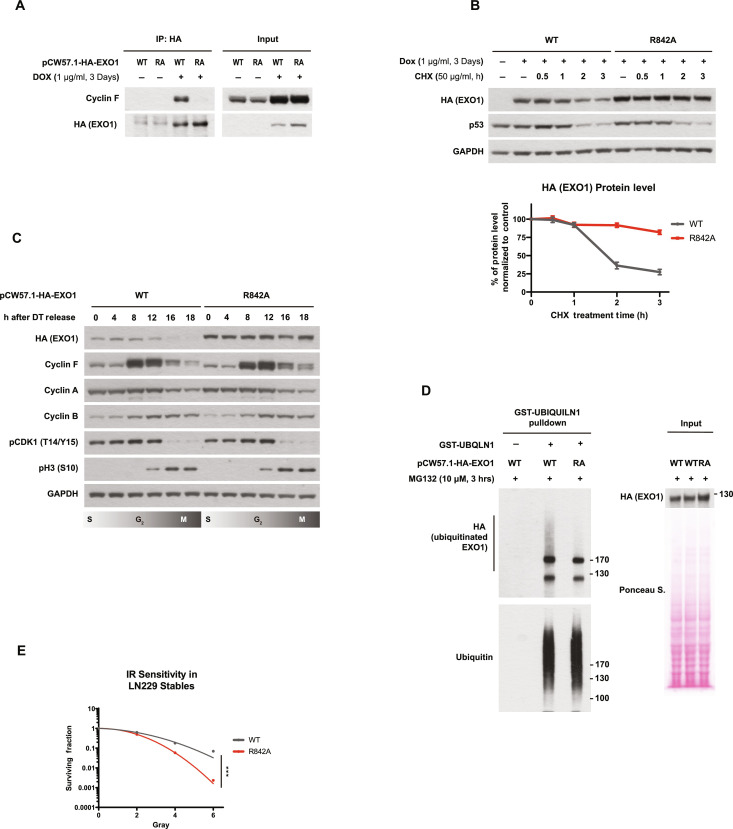
Ubiquitination is a crucial posttranslational modification required for the proper repair of DNA double-strand breaks (DSBs) induced by ionizing radiation (IR). DSBs are mainly repaired through homologous recombination (HR) when template DNA is present and nonhomologous end joining (NHEJ) in its absence. In addition, microhomology-mediated end joining (MMEJ) and single-strand annealing (SSA) provide backup DSBs repair pathways. However, the mechanisms controlling their use remain poorly understood. By using a high-resolution CRISPR screen of the ubiquitin system after IR, we systematically uncover genes required for cell survival and elucidate a critical role of the E3 ubiquitin ligase SCFcyclin F in cell cycle-dependent DSB repair. We show that SCFcyclin F-mediated EXO1 degradation prevents DNA end resection in mitosis, allowing MMEJ to take place. Moreover, we identify a conserved cyclin F recognition motif, distinct from the one used by other cyclins, with broad implications in cyclin specificity for cell cycle control.


Figures

Fig. 1.
Fig. 1.. CRISPR screen of the ubiquitin system identified genes required for cell survival after IR.
(A) Schematic representation of the CRISPR screen. Two days after transducing Cas9-expressing LN229 with the sgRNA library, sample T0 was collected as a reference sample of sgRNA expression. Cells were then selected with puromycin for 7 days before ionizing radiation (IR) treatment. Fourteen days after IR (4 Gy), cells were collected for genomic DNA extraction and PCR amplification of the sgRNA sequences. PCR product was sent for next-generation sequencing so that change in relative abundance of each sgRNA before and after IR could be assessed. (B) Identification of genes affecting IR sensitivity in LN229 by z ratio. Positive controls identified are highlighted in gray two hits from the screen are highlighted in red. See table S2 for the full list of genes. (C) Over-representation analysis (ORA) of statistically significant hits identified from the screen (absolute value of z ratio > 1.96). Pathways colored in dark red are highly confident (κ > 1). Pathways in red are confident (κ > 0.5). Pathways in pink are potential pathways (κ < 0.5). (D) Colony formation assay in CCNF K/O’s cells. HeLa parental cells or HeLa CCNF K/O cells were seeded for colony formation assay and challenged with the indicated dose of IR. Seven days after IR, cells were stained with crystal violet and counted. Error bars represent SDs of three biological replicates. Two-tailed unpaired t test was performed as statistical analysis. **P ≤ 0.01; ***P ≤ 0.001; ****P ≤ 0.0001. (E) Immunoblotting after IR treatment and recovery as indicated. DNA damage markers detected: pRPA32 S4/8 (ssDNA at DSB), pRPA32 S33 (single-strand breaks), and γH2Ax (DNA DSBs).
Fig. 2.
Fig. 2.. Cyclin F interacts and ubiquitinates EXO1.
(A) Volcano plot representing MS analysis of differential TurboID–cyclin F interacting partners in the presence versus absence of MLN4924. Proteins were isolated with streptavidin after labeling for 1 hour with biotin. (B) Immunoblotting after IP of GFP–cyclin F in HEK293T treated with MLN4924 as indicated. Input samples before IP are presented on the right. (C) Immunoblotting after treating cells with cycloheximide (CHX) for the indicated time (h = hour) (top). Relative quantification of EXO1 protein levels in HeLa parental cells or CCNF K/O cells after normalization with EXO1 levels at T0 for each cell line (bottom). Error bars: SDs of three biological replicates. (D) Immunoblotting after expression of GFP–cyclin F, Flag-EXO1, and HA-ubiquitin in HEK293T. Flag-EXO1 is isolated via Flag agarose beads pulldown before immunoblotting. Input samples before IP are presented on the right. (E) Immunoblotting after isolation of endogenous ubiquitinated proteins using recombinant GST-tagged UBA domain of UBQLN1 protein. Input samples before IP are on the right.
Fig. 3.
Fig. 3.. EXO1 accumulation mediates increased sensitivity to IR upon cyclin F depletion.
(A) HeLa parental cells, HeLa CCNF K/O, HeLa EXO1 K/O, and HeLa CCNF K/O EXO1 K/O as indicated were seeded for colony formation assay and challenged with the indicated doses of IR. Seven days after IR, cells were stained with crystal violet and counted. Error bars represent SDs of three biological replicates. Two-tailed unpaired t test was performed as statistical analysis. **P ≤ 0.01; ***P ≤ 0.001; ****P ≤ 0.0001. (B) Colony formation assay representative image of (A). (C). Immunoblotting in HeLa cells after IR treatment and recovery as indicated. DNA damage markers detected: pRPA32 S4/8 (ssDNA at DSB), pRPA32 S33 (single strand breaks), and γH2Ax (DNA DSBs). (D) Immunoblotting in LN229 cells after IR treatment and recovery as indicated. DNA damage markers detected as in (C). LN229 cells were transiently transfected with Cas9 protein and sgRNA, as indicated, 4 days before IR treatment.
Fig. 4.
Fig. 4.. EXO1 T824 phosphorylation by CDK1/cyclin A is required for the interaction with cyclin F and subsequent ubiquitination.
(A) Schematic representation of EXO1 depicting the domains and known interacting partners. (B) Immunoblotting after IP of GFP-EXO1 WT and the indicated EXO1 fragments. Input samples before IP are presented on the right. (C) The last 60 amino acids of EXO1 C terminus are highlighted and aligned with cyclin F interaction motif on RRM2. Amino acids conserved in both EXO1 and RRM2 are enlarged and labeled in black/gray. (D) Immunoblotting after IP of GFP-EXO1 WT and the indicated GFP-EXO1 mutants (S815A, T824A, and RAI/842-844/AAA). Input samples before IP are presented on the right. (E) Identification of differential interactors by MS after IP of Flag-EXO1 WT versus Flag-EXO1 T824A. Unique peptide ranges are labeled with the indicated colors. (F) Immunoblotting after IP of GFP–cyclin F plus treatment with calyculin A or λPP as indicated. λPP treatment was conducted on beads after IP. Input samples before IP are presented on the right. (G) Immunoblotting of in vitro phosphorylation assay using GFP-EXO1 WT or GFP-EXO1 T824A purified from HEK293T cells as substrates and the recombinant cyclin E/CDK2, cyclin A/CDK2, and cyclin A-B/CDK1 as indicated. Isolated GFP-EXO1 WT or GFP-EXO1 T824 mutant were dephosphorylated by λPP on beads before being used for in vitro phosphorylation. (H) Immunoblotting of in vitro phosphorylation assay using GFP-EXO1 WT or GFP-EXO1 T824A purified from HEK293T cells as substrates and recombinant cyclin A/CDK1 or cyclin B/CDK1 as indicated. (I) Immunoblotting of cell cycle synchronized HeLa cells via double thymidine (DT) block release.
Fig. 5.
Fig. 5.. Cyclin F interaction with EXO1 define a specific bivalent recognition domain (F-deg).
(A) Immunoblotting after IP of GFP-EXO1 WT, GFP-EXO1 T824A, GFP-EXO1 P825A, GFP-EXO1 R842A, GFP-EXO1 I844A, GFP-EXO1 F845A, and GFP-EXO1 Q846A mutants as indicated. Input samples before IP are presented at the bottom. (B) Immunoblotting after pulldown with streptavidin from cell extracts using as bait a biotinylated peptide encompassing EXO1 832-846 or a biotinylated peptide EXO1 832-846 where the RxIF residues were changed to alanine. (C) Docking of EXO1 peptide to cyclin F protein using COSMIC2. The peptide sequence from L823 to Q846 of EXO1 was used for the prediction. To mimic the phosphorylated status of EXO1, T824 was substituted to an E before docking. Cyclin F structure used in the docking is a predicted structure by AlphaFold Protein Structure Database. The iptm + ptm value is 0.7984765224933627. (D) Predicted aligned error (PAE) value plot of the prediction represented in (C). (E) Immunoblotting after IP of GFP–cyclin F WT or GFP–cyclin F Y387F mutant as indicated. Input samples before IP are presented on the right.
Fig. 6.
Fig. 6.. EXO1 R842A mutation phenocopies CCNF K/O.
(A) Immunoblotting after IP of stably integrated HA-EXO1 WT or HA-EXO1 R842A in LN229 using a doxycycline-inducible promoter. Cells were treated with doxycycline (1 μg/ml) for 3 days to induce expression. Input samples before IP are on the right. (B) Immunoblotting after treating cells described in (A) with CHX for the indicated time (top). Relative quantification of EXO1 protein levels in HA-EXO1 WT or HA-EXO1 R842A after normalization with EXO1 levels at T0 for each cell line (bottom). Error bars: SDs of three biological replicates. (C) Immunoblotting of cell cycle synchronized cells described in (A) via DT block release. (D) Immunoblotting after isolation of endogenous ubiquitinated proteins using recombinant GST-tagged UBA domain of UBQLN1 protein of cells indicated in (A). Input samples before IP are on the right. (E) LN229 cells expressing HA-EXO1 WT or HA-EXO1 R842A were seeded for colony formation assay and challenged with the indicated dose of IR. Seven days after IR, cells were stained with crystal violet and counted. Error bars represent SDs of three biological replicates. Two-tailed unpaired t test was performed as statistical analysis. Two-tailed unpaired t test. **P ≤ 0.01; ***P ≤ 0.001; ****P ≤ 0.0001.
Fig. 7.
Fig. 7.. EXO1 R842A mutant leads to hyper-resection and toxic single-strand annealing after IR.
(A) Representative images and quantification of native BrdU signal in IR-treated LN229 expressing HA-EXO1 WT or HA-EXO1R842A, as indicated. Error bars indicate SDs of three biological replicates. Two-tailed unpaired t test was performed as statistical analysis. ***P ≤ 0.001. (B) Representative images and quantification of the SMART assay in LN229 lines described in (A). >300 events were quantified. Error bars indicate SDs of three biological replicates. Mann-Whitney U test was used to derive significance. P = 0.0059, U = 17873.5, and Z = −2.7533. (C) Representative images and quantification of RPA32 positive anaphase ultrafine bridges (UFBs) in LN229 lines expressing HA-EXO1 WT or HA-EXO1R842A. Error bars indicate SDs of three biological replicates. Statistical analysis was performed using two-tailed unpaired t test. **P ≤ 0.01. (D) Quantification of chromosome aberrations in hTERT RPE-1 stably expressing HA-EXO1 or HA-EXO1 R842A mutant. A total of 100 metaphase events were analyzed for each condition. Percentages of mitotic events demonstrating 0, 1, 2, 3, 4, or >4 aberrations were calculated and plotted. (E) LN229 expressing HA-EXO1 WT or HA-EXO1 R842A transfected with Cas9 and sgPOLQ or control sgRNA were treated with IR and subjected to colony formation assay. Statistical analysis derived with two-tailed unpaired t test. (F) LN229 cells expressing HA-EXO1 WT or HA-EXO1 R842A transfected with Cas9 and sgRAD52 or control sgRNA were treated with IR and subjected to colony formation assay. Statistical analysis derived with two-tailed unpaired t test. (G) Quantification of MMEJ and SSA reporter assays as indicated. Repair efficiency was normalized against cells expressing control sgRNA. Statistical analysis was performed using two-tailed unpaired t test in three biological replicates. **P ≤ 0.01. (H) Schematic illustration of cyclin F–EXO1 axis controlling DSB repair pathway choice in mitosis.

References

    1. Jackson S. P., Bartek J., The DNA-damage response in human biology and disease. Nature 461, 1071–1078 (2009). - PMC - PubMed
    1. Schwertman P., Bekker-Jensen S., Mailand N., Regulation of DNA double-strand break repair by ubiquitin and ubiquitin-like modifiers. Nat. Rev. Mol. Cell Biol. 17, 379–394 (2016). - PubMed
    1. Chapman J. R., Taylor M. R., Boulton S. J., Playing the end game: DNA double-strand break repair pathway choice. Mol. Cell 47, 497–510 (2012). - PubMed
    1. Bennardo N., Cheng A., Huang N., Stark J. M., Alternative-NHEJ is a mechanistically distinct pathway of mammalian chromosome break repair. PLOS Genet. 4, e1000110 (2008). - PMC - PubMed
    1. Bhargava R., Onyango D. O., Stark J. M., Regulation of single-strand annealing and its role in genome maintenance. Trends Genet. 32, 566–575 (2016). - PMC - PubMed

Publication types

LinkOut - more resources