. 2024 Oct 8;8(19):5100-5111.
doi: 10.1182/bloodadvances.2023012161.

Altered enhancer-promoter interaction leads to MNX1 expression in pediatric acute myeloid leukemia with t(7;12)(q36;p13)

Affiliations

Altered enhancer-promoter interaction leads to MNX1 expression in pediatric acute myeloid leukemia with t(7;12)(q36;p13)

Dieter Weichenhan et al. Blood Adv. .

Abstract

Acute myeloid leukemia (AML) with the t(7;12)(q36;p13) translocation occurs only in very young children and has a poor clinical outcome. The expected oncofusion between break point partners (motor neuron and pancreas homeobox 1 [MNX1] and ETS variant transcription factor 6 [ETV6]) has only been reported in a subset of cases. However, a universal feature is the strong transcript and protein expression of MNX1, a homeobox transcription factor that is normally not expressed in hematopoietic cells. Here, we map the translocation break points on chromosomes 7 and 12 in affected patients to a region proximal to MNX1 and either introns 1 or 2 of ETV6. The frequency of MNX1 overexpression in pediatric AML is 2.4% and occurs predominantly in t(7;12)(q36;p13) AML. Chromatin interaction assays in a t(7;12)(q36;p13) induced pluripotent stem cell line model unravel an enhancer-hijacking event that explains MNX1 overexpression in hematopoietic cells. Our data suggest that enhancer hijacking may be a more widespread consequence of translocations in which no oncofusion product was identified, including t(1;3) or t(4;12) AML.


Conflict of interest statement

Conflict-of-interest disclosure: L.B. has received honoraria from AbbVie, Amgen, Astellas, Bristol Myers Squibb, Celgene, Daiichi Sankyo, Gilead, Hexal, Janssen, Jazz Pharmaceuticals, Menarini, Novartis, Pfizer, Roche, and Sanofi; and research support from Bayer and Jazz Pharmaceuticals. D.B.L. receives honoraria from Infectopharm GmbH. The remaining authors declare no competing financial interests.

Figures

None
Graphical abstract
Figure 1.
Figure 1.
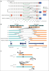
WGS analysis of t(7;12)(q36;p13) AML. (A) Copy numbers (blue, loss; red, gain), structural rearrangements (green bow connecting 2 chromosomes), and mutations in known AML driver genes for 6 t(7;12)(q36;p13) AML samples based on WGS. Samples T1, T2, T3, and T4 were profiled in this study, whereas TARGET-20-PARUNX and TARGET-20-PASIBG are from the TARGET-AML cohort 15. (B) Sketch of the rearranged chr7 and chr12 and zoom-in on the region around the break points. (C) Schematic overview of chr7 (turquoise), chr12 (orange), and derivative chromosomes der(12) and der(7) resulting from the reciprocal t(7;12) translocation involving MNX1 on chr7 and ETV6 on chr12. Red lines indicate positions of break/fusion points.
Figure 2.
Figure 2.
MNX1 expression in pediatric AML with t(7;12)(q36;p13) translocation. (A-B) MNX1 expression in 2 different pediatric AML cohorts. (A) Balgobind et al; 237 samples profiled with Affymetrix arrays. The mean expression level is shown with a dashed line, and the mean plus 3 standard deviations is shown with a horizontal line. (B) TARGET-AML; 1319 samples profiled with RNAseq (cutoff, 0.5 TPM). Samples with cytogenetically detected t(7;12)(q36;p13) translocation are shown in green and other samples in gray. exp, expression; TPM, transcripts per million.
Figure 3.
Figure 3.
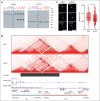
MNX1 protein expression and chromatin interaction of the MNX1 gene with the ETV6 region in ChiPSC22t(7;12) cells. (A) Western blot with an MNX1 antibody (left) and iPSC (blue) and HSPC (red) protein extracts from ChiPSC22WT and ChiPSC22t(7;12) sublines 14D7, 23G8, and 24C7. The MNX1 protein (asterisk) is only detected in HSPCs of ChiPSC22t(7;12) sublines 14D7, 23G8, and 24C7. The common band at ∼120 kD results from an unknown protein cross-reacting with the MNX1 antibody. To demonstrate loading of equal protein amounts, the unstripped blot was reincubated with an antibody against β-actin (right). (B) Chromatin interactions analyzed by Hi-C seq in the genomic region flanking the translocation break point in the ChiPSC22t(7;12) subline 24C7, either as iPSCs (top) or HSPCs (below). The neo-TAD is indicated by a black bar. ChIPseq data for CTCF and RAD21 in K562 were retrieved from the encode project (IDs ENCFF468HJA and ENCFF000YXZ). (C) Increased proximity between MNX1 and ETV6 in ChiPSC22t(7;12) subline 14D7–derived HSPCs compared with iPSCs. Representative STED images of FISH spots in 2 colors targeting MNX1 and ETV6 in iPSCs and HSPCs (left). Scale bars, 500 nm. 3D distances between the MNX1 and ETV6 signals (right). Red horizontal lines within boxes indicate medians; box limits indicate upper and lower quartiles. iPSCs, n = 154; HSPCs, n = 409, across 3 independent replicates. ∗P < .05, Wilcoxon rank-sum test.
Figure 4.
Figure 4.
Open chromatin and enhancer mark profiles in the ETV6 neo-TAD region of patient and cell line samples. Open chromatin profiles (ATAC) of patients with AML , T1 and T2, and of HSPCs from ChiPSC22WT and ChiPSC22t(7;12) sublines 14D7, 23G8, and 24C7 in the ETV6 neo-TAD region. HSPC-specific enhancer mark H3K27ac and H3K4me1 profiles and publicly available p300, H3K27ac, and H3K4me1 profiles from MOLM-1 and CD34+. Relevant common peak positions are highlighted by a gray shading. The chr12 break point (BP) position in T1 and T2 and in the ChiPSC22t(7;12) sublines are indicated.
Figure 5.
Figure 5.
Molecular validation of enhancer-promoter interaction in ChiPSC22t(7;12) upon differentiation. (A) Scheme of the enhancer deletion experiment performed to validate the interaction between the MNX1 promoter and enhancers distal to the BP. (B) Gene expression in HSPCs derived from ChiPSC22WT (n = 3), ChiPSC22t(7;12) (n = 4, from 2 independent cell lines), and ChiPSC22t(7;12)ΔEn (n = 3, from 2 independent cell lines) measured via qRT-PCR and shown as 2−ΔCt vs GUSB as endogenous reference. ∗∗∗∗P < .0001; ∗∗∗P < .001; ∗∗P < .01; ∗P < .05. ns, not significant.

References

    1. de Thé H, Pandolfi PP, Chen Z. Acute promyelocytic leukemia: a paradigm for oncoprotein-targeted cure. Cancer Cell. 2017;32(5):552–560. - PubMed
    1. Tosi S, Giudici G, Mosna G, et al. Identification of new partner chromosomes involved in fusions with the ETV6 (TEL) gene in hematologic malignancies. Genes Chromosomes Cancer. 1998;21(3):223–229. - PubMed
    1. Wlodarska I, La Starza R, Baens M, et al. Fluorescence in situ hybridization characterization of new translocations involving TEL (ETV6) in a wide spectrum of hematologic malignancies. Blood. 1998;91(4):1399–1406. - PubMed
    1. Espersen ADL, Noren-Nyström U, Abrahamsson J, et al. Acute myeloid leukemia (AML) with t(7;12)(q36;p13) is associated with infancy and trisomy 19: data from Nordic Society for Pediatric Hematology and Oncology (NOPHO-AML) and review of the literature. Genes Chromosomes Cancer. 2018;57(7):359–365. - PubMed
    1. von Bergh ARM, van Drunen E, van Wering ER, et al. High incidence of t(7;12)(q36;p13) in infant AML but not in infant ALL, with a dismal outcome and ectopic expression of HLXB9. Genes Chromosomes Cancer. 2006;45(8):731–739. - PubMed

Publication types

MeSH terms