Review
. 2024 Aug 8;187(16):4150-4175.
doi: 10.1016/j.cell.2024.05.059.

Guidelines for minimal information on cellular senescence experimentation in vivo

Affiliations
Review

Guidelines for minimal information on cellular senescence experimentation in vivo

Mikolaj Ogrodnik et al. Cell. .

Abstract

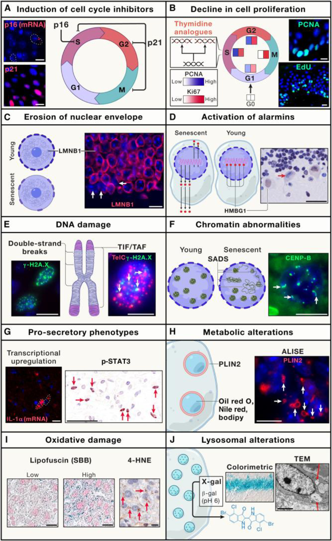
Cellular senescence is a cell fate triggered in response to stress and is characterized by stable cell-cycle arrest and a hypersecretory state. It has diverse biological roles, ranging from tissue repair to chronic disease. The development of new tools to study senescence in vivo has paved the way for uncovering its physiological and pathological roles and testing senescent cells as a therapeutic target. However, the lack of specific and broadly applicable markers makes it difficult to identify and characterize senescent cells in tissues and living organisms. To address this, we provide practical guidelines called "minimum information for cellular senescence experimentation in vivo" (MICSE). It presents an overview of senescence markers in rodent tissues, transgenic models, non-mammalian systems, human tissues, and tumors and their use in the identification and specification of senescent cells. These guidelines provide a uniform, state-of-the-art, and accessible toolset to improve our understanding of cellular senescence in vivo.

Keywords: aging; humans; in vivo; mouse; senescence; senotherapy.


Conflict of interest statement

Declaration of interests D.J.B. has potential financial interests related to this study. He is a co-inventor of patents held by the Mayo Clinic, patent applications licensed to or filed by Unity Biotechnology, and a Unity Biotechnology shareholder. Research in the Baker laboratory has been reviewed by the Mayo Clinic Conflict of Interest Review Board and is being conducted in compliance with Mayo Clinic Conflict of Interest policies. J. Gil has acted as a consultant for Unity Biotechnology, Geras Bio, Myricx Pharma Ltd., and Merck KGaA; owns equity in Geras Bio and share options in Myricx Pharma Ltd.; and is a named inventor in MRC and Imperial College patents related to senolytic therapies. J. Gil currently receives funding from Pfizer. Unity Biotechnology funded research on senolytics in J. Gil’s laboratory in the past. SenTraGor and GLF16 senescence detection compounds are under patent applications: EP3475287B1, and 20240100309 (Greek patent application) along with GB2406749.8 (UK patent application), respectively. J.M.S. is a co-inventor on patents held by Brown University on methods to inhibit retrotransposon activation in age-related diseases. He is the scientific co-founder of Transposon Therapeutics, chair of their scientific advisory board, and a consultant and holds stock options. He is also a consultant and holds equity in Atropos Therapeutics. Research in the Sedivy laboratory has been reviewed by the Brown University Conflict of Interest Review Board and is being conducted in compliance with Brown University Conflict of Interest policies. F.d.d.F. is an inventor on the patent applications PCT/EP2013/059753 and PCT/EP2016/068162. M.D. is co-inventor on patents held by the Buck Institute for Research on Aging. He is the scientific co-founder of Cleara Biotech and consultant for Oisin Biotechnologies. M.D.’s laboratory currently receives research funding from Ono Pharmaceuticals. J. Grillari is co-inventor on patents held by BOKU and is a co-founder and scientific advisor to TAmiRNA and Rockfish Bio.

Figures

Figure 1:
Figure 1:. Markers of cellular senescence in situ.
(A) Cell cycle inhibitors arrest cells in various phases: G1→ S (in the case of p16Ink4a) and G1 → S, S→ G2, and G2→ M (for p21Cip1/Waf). Images show RNA in situ hybridization (RNA-ISH) for p16 of an aged brain and IHF staining against p21 in the wounded epidermis. (B) Detection of cell proliferation can be performed using antibodies against Ki67 and PCNA, or by visualizing the incorporation of thymidine analogs into DNA. The images show EdU incorporation over 6 h on the left and PCNA staining on the right, both wounded skin samples. (C) Erosion of the nuclear envelope was visualized by detection of Lamin B1 (LMNB1). Images show neurons in the dentate gyrus of aged mice. White arrows show cells negative for LMNB1. (D) Senescent cells release Hmgb1 from chromatin, which translocates to the cytoplasm and outside the cell. The image shows the cerebellum of the aged mouse. The red arrow shows a Purkinje neuron negative for intranuclear Hmgb1. (E) Double-strand breaks (DSBs) can occur at telomeres (telomere-induced foci; TIF or Telomere-associated foci; TAF) or anywhere else on the chromosomes. Images show IHF staining against γ-H2A.X and immunoFISH co-staining for γ-H2A.X and TelC in the wounded skin. White arrows show colocalization of γ-H2A.X and TelC signals, indicating TAF. (F) Alteration of chromatin in senescent cells in situ can be visualized using FISH staining against Cenp-B sequences of peri-centromeric beta satellites. The image shows the hepatocytes in an aged liver. White arrows show senescence-associated satellite decondensation of satellites (SADS). (G) Senescence-associated secretory phenotype (SASP) can be visualized in situ by RNA-ISH (e.g., IL-1α in aged brain) or histologically by detecting the activation of pro-inflammatory pathways, for example, p-STAT3 (Y705) in wounded skin. White outlines show cells positive for IL-1α (left) and red arrows show cells positive for p-STAT3. (H) One facet of metabolic disruption in senescent cells involves an increased accumulation of lipid droplets (LDs) that can be visualized by staining against perilipin 2 (Plin2) or with dyes against neutral lipids such as Nile Red, Oil Red O, or BODIPY. The image shows LDs in cells surrounding the lateral ventricle of the aged brain. White arrows mark LDs. (I) Increased production of reactive oxygen species (ROS) by senescent cells can be detected by visualizing oxidative damage, such as lipofuscin and 4-hydroxynonenal (4-HNE). Exemplary images show Sudan Black B staining in aged livers and IHC for 4-HNE in aged livers. Red arrows indicate clusters of 4-HNE-positive granules. (J) Senescence-associated β-galactosidase (SA-β-gal) represents lysosomal activity and can be visualized in an enzymatic assay for β-galactosidase activity at acidic pH using colorimetric, fluorescent, or TEM-detectable reagents. Images show the hippocampal CA3 region from an aged brain and X-gal crystals visualized by TEM in a macrophage from an atherosclerotic plaque. Red arrows indicate the X-gal crystals. Scale bars: 10 μm for A-C, F, G (left), H, and I; 50 μm for D and G (right); 5 μm for E; 2 μm for J (right).
Figure 2:
Figure 2:. Heterogeneity of senescence markers in cell types and organs in situ.
Senescent cells found in different organs show heterogeneous phenotypes, and many have been reported to express only some markers of cellular senescence that are attributed to senescent cells in vitro. Similarities and differences between phenotypes associated with cellular senescence can be observed across mouse tissues, including the (A) brain, (B) skin, (C) lungs, (D) bone, (E) liver, (F) adipose tissue, and (G) muscle.
Figure 3:
Figure 3:. Approaches, limitations, and perspectives for research on cellular senescence in human samples.
(A) Examples of human material available for senescence research include cancer samples, postmortem organ pieces, biopsies, and medical fluids. (B) Limitations that challenge experimental approaches to work with human senescence. Senescent cells present a high level of heterogeneity across different types of cancer and across various tissue types. An additional layer of heterogeneity arises from the methodology used to obtain or process the samples prior to the detection of senescence markers. Finally, medical fluids can provide indirect information on senescence in organs; however, the methodology for making accurate predictions is still under development. (C) Perspectives for exploration of the properties and roles of senescent cells in human health and disease. With the recent advancements in single-cell and spatial omics, it is anticipated that these methods will provide accurate data on the characterization and prevalence of senescent cells in situ. Information derived from samples collected in a non-invasive manner (e.g., medical fluids) can be used as a biomarker to indirectly measure the quantity of senescent cells in peripheral organs.
Figure 4:
Figure 4:. Sequential approach to study senescence in vivo.
To accurately assess the relevance of cellular senescence to a specific phenotype, we suggest beginning with a general evaluation of the core markers for cellular senescence: p21 and/or p16 (STEP 1). Therefore, it is advisable to assess auxiliary markers of cellular senescence, including DNA damage, LMNB1, ALISE, oxidative damage, SASP, and SA-β-gal. The use of at least two auxiliary markers is recommended (STEP 2). While these initial steps are adequate for determining the presence or absence of senescence in a particular tissue or condition, to ascertain whether senescent cells impact a physiological process or pathology, we recommend reducing the quantity and/or activity of senescent cells using available transgenic models and/or drugs (STEP 3). Finally, to provide conclusive evidence that senescent cells influence a phenotype, we suggest confirming that a chosen drug/model has decreased the frequency of targeted senescent cells, and that this intervention has led to the expected alteration in the studied phenotype (STEP 4).

References

    1. Hayflick L, and Moorhead PS (1961). The serial cultivation of human diploid cell strains. Exp Cell Res 25, 585–621. 10.1016/0014-4827(61)90192-6. - DOI - PubMed
    1. Thery C, Witwer KW, Aikawa E, Alcaraz MJ, Anderson JD, Andriantsitohaina R, Antoniou A, Arab T, Archer F, Atkin-Smith GK, et al. (2018). Minimal information for studies of extracellular vesicles 2018 (MISEV2018): a position statement of the International Society for Extracellular Vesicles and update of the MISEV2014 guidelines. J Extracell Vesicles 7, 1535750. 10.1080/20013078.2018.1535750. - DOI - PMC - PubMed
    1. Brazma A, Hingamp P, Quackenbush J, Sherlock G, Spellman P, Stoeckert C, Aach J, Ansorge W, Ball CA, Causton HC, et al. (2001). Minimum information about a microarray experiment (MIAME)-toward standards for microarray data. Nat Genet 29, 365–371. 10.1038/ng1201-365. - DOI - PubMed
    1. Bringold F, and Serrano M (2000). Tumor suppressors and oncogenes in cellular senescence. Exp Gerontol 35, 317–329. 10.1016/s0531-5565(00)00083-8. - DOI - PubMed
    1. Kim WY, and Sharpless NE (2006). The regulation of INK4/ARF in cancer and aging. Cell 127, 265–275. 10.1016/j.cell.2006.10.003. - DOI - PubMed

LinkOut - more resources