. 2024 Sep;300(9):107653.
doi: 10.1016/j.jbc.2024.107653. Epub 2024 Aug 8.

The enzymatic oxygen sensor cysteamine dioxygenase binds its protein substrates through their N-termini

Affiliations

The enzymatic oxygen sensor cysteamine dioxygenase binds its protein substrates through their N-termini

Karishma Patel et al. J Biol Chem. 2024 Sep.

Abstract

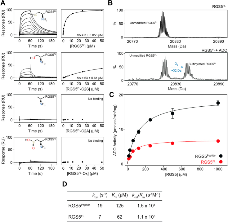
The non-heme iron-dependent dioxygenase 2-aminoethanethiol (aka cysteamine) dioxygenase (ADO) has recently been identified as an enzymatic oxygen sensor that coordinates cellular changes to hypoxia by regulating the stability of proteins bearing an N-terminal cysteine (Nt-cys) through the N-degron pathway. It catalyzes O2-dependent Nt-cys sulfinylation, which promotes proteasomal degradation of the target. Only a few ADO substrates have been verified, including regulators of G-protein signaling (RGS) 4 and 5, and the proinflammatory cytokine interleukin-32, all of which exhibit cell and/or tissue specific expression patterns. ADO, in contrast, is ubiquitously expressed, suggesting it can regulate the stability of additional Nt-cys proteins in an O2-dependent manner. However, the role of individual chemical groups, active site metal, amino acid composition, and globular structure on protein substrate association remains elusive. To help identify new targets and examine the underlying biochemistry of the system, we conducted a series of biophysical experiments to investigate the binding requirements of established ADO substrates RGS5 and interleukin-32. We demonstrate, using surface plasmon response and enzyme assays, that a free, unmodified Nt-thiol and Nt-amine are vital for substrate engagement through active site metal coordination, with residues next to Nt-cys moderately impacting association and catalytic efficiency. Additionally, we show, through 1H-15N heteronuclear single quantum coherence nuclear magnetic resonance titrations, that the globular portion of RGS5 has limited impact on ADO association, with interactions restricted to the N-terminus. This work establishes key features involved in ADO substrate binding, which will help identify new protein targets and, subsequently, elucidate its role in hypoxic adaptation.

Keywords: ADO; N-degron pathway; enzyme kinetics; hypoxia; nuclear magnetic resonance; oxygen-sensing; posttranslational modification; protein degradation; surface plasmon resonance.


Conflict of interest statement

Conflict of interest The authors declare that they have no conflicts of interest with the contents of this article.

Figures

Figure 1
Figure 1
ADO requires an unmodified Nt-cys to interact with RGS5 peptides.A, Left: Representative SPR sensorgrams for the titrations of modified Nt-cys RGS5 peptides with ADO. Right: fits of the equilibrium responses from the sensorgrams in the left panels to a 1:1 binding model. The identity, chemical structures of the modification state of the Nt-cys of each peptide, and Kᴅ values are shown (Kᴅ given as the geometric mean of a minimum of three independent SPR measurements). B, LC-MS spectra showing the RGS5 peptide species detected following a 45 s incubation of the modified Nt-cys RGS5 peptides (100 μM) with ADO (0.1 μM) at 37 °C (1-h incubations were also preformed, Fig. S4). The expected +32 da mass shift resulting from sulfinylation of the native RGS5 peptide in the presence of ADO is detected. The remaining RGS5 peptides do not get sulfinylated by ADO. The positions of the peptide species expected upon oxidation (or unmodified RGS5 in the case of the IL32-Ox peptide) by ADO are indicated. ADO, 2-aminoethanethiol dioxygenase; Nt-cys, N-terminal cysteine; SPR, surface plasmon resonance.
Figure 2
Figure 2
ADO requires an active site metal for substrate binding.A, Representative SPR sensorgrams for the titration of the native RGS5 peptide with ADO before treatment (top), postmetal removal using 10 mM 1, 10-phenanthroline and 100 mM EDTA (middle), and postreconstitution with iron using 0.1 mM FeSO4 (supplemented with 12.5 mM sodium ascorbate) (bottom). B, Fits of the equilibrium responses from the sensorgrams in (A) to a 1:1 binding model. The Kᴅ values presented are calculated from the specific SPR data presented in this figure. C, the iron occupancy of the native ADO and ADO-H193D determined by ICP-MS. The occupancy is provided as a percent based on amount of iron detected in the sample relative to the concentration of ADO present. D, Left: Representative SPR sensorgram for the titration of native RGS5 peptide with ADO-H193. Right: Fits of the equilibrium responses from the sensorgrams in the left panels to a 1:1 binding model. The Kᴅ value is shown (Kᴅ given as the geometric mean of a minimum of three independent SPR measurements). E, The metal (Fe2+, Co2, and Zn2+) occupancies of native ADO (left), ADO-Co2+ (middle), and ADO-Zn2+ (right) determined by ICP-MS. The occupancies are provided as percentages based on amount of the metals detected in the sample relative to the concentration of ADO present. F, Left: Representative SPR sensorgram for the titration of the native RGS5 peptide with ADO-Zn2+. Right: Fits of the equilibrium responses from the sensorgrams in the left panels to a 1:1 binding model. The Kᴅ value is shown (Kᴅ given as the geometric mean of a minimum of three independent SPR measurements). G, Representative single cycle kinetic (SCK) SPR sensorgram of the native RGS5 peptide with ADO-Co2+. The sensorgram is shown in red and the fit to the data is shown in black. The concentrations of RGS5 used in the titration and the Kᴅ value is shown (Kᴅ given as the geometric mean of a minimum of three independent SPR measurements). H, Time course showing the enzymatic turnover of the RGS5 peptide (100 μM) by native ADO, ADO-Co2+, and ADO-Zn2+ (0.1 μM enzyme) at 37 °C. The average of three independent experiments are shown (error bars show the standard error). O/N indicates overnight. ADO, 2-aminoethanethiol dioxygenase; ICP, inductively coupled plasma; MS, mass spectrometry; Nt-cys, N-terminal cysteine; SPR, surface plasmon resonance.
Figure 3
Figure 3
SPR and enzyme kinetic analyses of the RGS5 alanine mutational scan.A, Fold difference in Kᴅ relative to the native RGS5 peptide. The substitutions that generate a greater than 1.5-fold reduction in Kᴅ are colored in purple. The peptide sequences, with the mutation indicated in bold red lettering, and Kᴅ values for the interactions with ADO are provided (Kᴅ given as the geometric mean of a minimum of three independent SPR measurements). B, Left: Representative SPR sensorgram for the titration of native RGS5, RGS5-K3A, and RGS5-G4A with ADO. Right: Fits of the equilibrium responses from the sensorgrams in the left panels to a 1:1 binding model. The Kᴅ value is shown (Kᴅ given as the geometric mean of a minimum of three independent SPR measurements). Data for the native RGS5 peptide are provided for reference (first presented in Fig. 1A). C, The specific activity of ADO (0.05 μM) calculated by measuring the rate of native RGS5, RGS5-K3A, RGS5-G4A, and RGS5-L5A oxidation by LC-MS (100 μM peptide, 45 s incubation at 37 °C). D, Michaelis–Menten kinetic plots for native RGS5, RGS5-K3A, and RGS5-G4A performed in aerobic conditions at 37 °C. The average of three independent experiments are shown (error bars show the standard error). E, Table of the reaction kinetics for ADO catalysis of native RGS5, RGS5-K3A, and RGS5-G4A calculated from the data presented in D. ADO, 2-aminoethanethiol dioxygenase; SPR, surface plasmon resonance.
Figure 4
Figure 4
SPR binding and kinetic studies of the interaction between ADO and full-length RGS5.A, Left: Representative SPR sensorgrams for the titrations of modified Nt-cys RGS5 full-length proteins with ADO. Right: Fits of the equilibrium responses from the sensorgrams in the left panels to a 1:1 binding model. The identity, chemical structures of the modification state of the Nt-cys of each protein, and Kᴅ values are shown (Kᴅ given as the geometric mean of a minimum of three independent SPR measurements). B, LC-MS spectra showing the RGS5 protein species detected following a 45 s incubation of full-length RGS5 (100 μM) with and without ADO (0.1 μM) at 25 °C. The expected +32 Da mass shift resulting from sulfinylation of RGS5 in the presence of ADO is detected. C, Michaelis–Menten kinetic plots for native RGS5 peptide (RGS5Peptide) and full-length protein (RGS5FL) performed in aerobic conditions at 25 °C. The average of three independent experiments are shown (error bars show the standard error). D, Table of the reaction kinetics for ADO catalysis of RGS5Peptide and RGS5FL calculated from the data presented in (C). ADO, 2-aminoethanethiol dioxygenase; Nt-cys, N-terminal cysteine; SPR, surface plasmon resonance.
Figure 5
Figure 5
NMR studies of the interaction between ADO and full-length RGS5. For all titrations, RGS5 was used at a concentration of 50 μM and the assignments for some signals are indicated. A, 15N-HSQC spectra of RGS5FL-C2S alone (red) and in the presence of increasing concentrations of ADO (up to 800 μM, blue). B, 15N-HSQC spectra of RGS5FL alone (red) and in the presence of increasing concentrations of ADO-Co2+ (up to 300 μM, blue). The directions of chemical shift change for selected peaks are indicated by arrows. C, Top: Quantitation of signal intensity post titration of 400 μM ADO into RGS5FL-C2S. Bottom: The residues that display the greatest reduction in signal intensity (∼top 10%) are mapped onto the AlphaFold model of RGS5 (gray) in red. Unassigned residues and proline residues (which do not have signals in 15N-HSQC spectra) are depicted in dark gray. ADO, 2-aminoethanethiol dioxygenase; HSQC, heteronuclear single-quantum coherence; Nt-cys, N-terminal cysteine; SPR, surface plasmon resonance.

Similar articles

Cited by

References

    1. Jaakkola P., Mole D.R., Tian Y.M., Wilson M.I., Gielbert J., Gaskell S.J., et al. Targeting of HIF-α to the von Hippel-Lindau ubiquitylation complex by O2-regulated prolyl hydroxylation. Science. 2001;292:468–472. - PubMed
    1. Ivan M., Kondo K., Yang H., Kim W., Valiando J., Ohh M., et al. HIFα targeted for VHL-mediated destruction by proline hydroxylation: implications for O2 sensing. Science. 2001;292:464–468. - PubMed
    1. Fiorini G., Schofield C.J. Biochemistry of the hypoxia-inducible factor hydroxylases. Curr. Opin. Chem. Biol. 2024;79 - PubMed
    1. Masson N., Keeley T.P., Giuntoli B., White M.D., Lavilla Puerta M., Perata P., et al. Conserved N-terminal cysteine dioxygenases transduce responses to hypoxia in animals and plants. Science. 2019;364:65–69. - PMC - PubMed
    1. Chakraborty A.A., Laukka T., Myllykoski M., Ringel A.E., Booker M.A., Tolstorukov M.Y., et al. Histone demethylase KDM6A directly senses oxygen to control chromatin and cell fate. Science. 2019;363:1217–1222. - PMC - PubMed

LinkOut - more resources