. 2024 Sep;300(9):107644.
doi: 10.1016/j.jbc.2024.107644. Epub 2024 Aug 8.

Cdc14 phosphatases use an intramolecular pseudosubstrate motif to stimulate and regulate catalysis

Affiliations

Cdc14 phosphatases use an intramolecular pseudosubstrate motif to stimulate and regulate catalysis

Kedric L Milholland et al. J Biol Chem. 2024 Sep.

Abstract

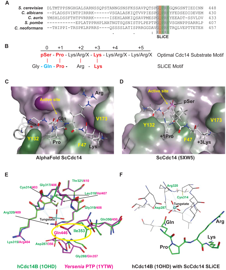
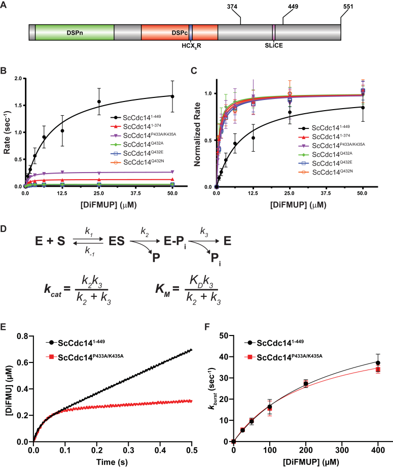
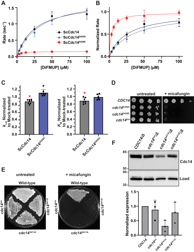
Cdc14 phosphatases are related structurally and mechanistically to protein tyrosine phosphatases (PTPs) but evolved a unique specificity for phosphoSer-Pro-X-Lys/Arg sites primarily deposited by cyclin-dependent kinases. This specialization is widely conserved in eukaryotes. The evolutionary reconfiguration of the Cdc14 active site to selectively accommodate phosphoSer-Pro likely required modification to the canonical PTP catalytic cycle. While studying Saccharomyces cerevisiae Cdc14, we discovered a short sequence in the disordered C terminus, distal to the catalytic domain, which mimics an optimal substrate. Kinetic analyses demonstrated this pseudosubstrate binds the active site and strongly stimulates rate-limiting phosphoenzyme hydrolysis, and we named it "substrate-like catalytic enhancer" (SLiCE). The SLiCE motif is found in all Dikarya fungal Cdc14 orthologs and contains an invariant glutamine, which we propose is positioned via substrate-like contacts to assist orientation of the hydrolytic water, similar to a conserved active site glutamine in other PTPs that Cdc14 lacks. AlphaFold2 predictions revealed vertebrate Cdc14 orthologs contain a conserved C-terminal alpha helix bound to the active site. Although apparently unrelated to the fungal sequence, this motif also makes substrate-like contacts and has an invariant glutamine in the catalytic pocket. Altering these residues in human Cdc14A and Cdc14B demonstrated that it functions by the same mechanism as the fungal motif. However, the fungal and vertebrate SLiCE motifs were not functionally interchangeable, illuminating potential active site differences during catalysis. Finally, we show that the fungal SLiCE motif is a target for phosphoregulation of Cdc14 activity. Our study uncovered evolution of an unusual stimulatory pseudosubstrate motif in Cdc14 phosphatases.

Keywords: Cdc14; enzyme catalysis; enzyme kinetics; enzyme mechanism; enzyme regulation; phosphatase; phosphorylation; pseudosubstrate; structural model.


Conflict of interest statement

Conflicts of interest The authors declare that they have no conflicts of interest with the contents of this article.

Figures

Figure 1
Figure 1
The highly conserved fungal SLiCE motif is predicted to bind the Cdc14 active site.A, multiple sequence alignment showing a portion of the C-terminal tail of Cdc14 orthologs from several model and pathogen fungal species generated with Clustal Omega. The invariant SLiCE motif is highlighted. Asterisks indicate identical amino acids in all aligned sequences. Orange and green highlighting indicates the Gln and Pro + Lys residues subjected to mutagenesis, respectively. B, comparison of the known optimal Cdc14 substrate motif with the fungal SLiCE motif. Red text indicates amino acids essential for efficient substrate recognition by Cdc14 enzymes (35, 36). Blue indicates the Gln predicted to facilitate coordination of nucleophilic water for phosphoenzyme hydrolysis. CD, AlphaFold2 structural prediction of ScCdc14 showing the active site region with bound SLiCE motif compared to same view of the ScCdc14 crystal structure bound to an optimal phosphopeptide substrate (RCSB: 5XW5). The catalytic domain is depicted in surface representation (green = nonpolar, purple = polar) and the SLiCE motif and phosphopeptide substrate in stick mode. The active site and several amino acids important for substrate binding are labeled in yellow for orientation. SLiCE motif residues and key substrate recognition positions are labeled in black. E, structures of hCdc14B (RCSB: 1OHD) and Yersinia PTP (RCSB: 1YTW), each bound to the PTP inhibitor tungstate, were superposed in Molecular Operating Environment software. The active site region is shown (tungstate from 1OHD was removed for simplicity) with hCdc14B backbone and residue labels in green and Yersinia PTP in magenta. The critical water-coordinating Gln of Yersinia PTP that is absent from Cdc14 enzymes is highlighted with yellow oval. F, the AlphaFold2 ScCdc14 structure was superposed on hCdc14B (1OHD) in Molecular Operating Environment. Relative position of the ScCdc14 SLiCE motif Gln to the bound tungstate in the hCdc14B active site is shown. SLiCE sequence is the thick green stick model with black residue labels. Critical catalytic residues of hCdc14B are labeled in green. SLiCE, substrate-like catalytic enhancer; PTP, protein tyrosine phosphatase.
Figure 2
Figure 2
The SLiCE motif enhances the rate-limiting Cdc14 catalytic step.A, domain map of ScCdc14. DSPn and DSPc are the N- and C-terminal dual-specificity phosphatase domains, respectively. HCX5R is the signature PTP phosphate-binding loop motif with catalytic Cys. Sites of truncations and the invariant fungal SLiCE motif are indicated. B, initial velocities as a function of increasing DiFMUP concentration were measured under steady-state conditions for the indicated ScCdc14 variants. All point mutations were made in ScCdc141-449. Data are average values from at least three independent measurements. Error bars are SDs. Lines are best fits for the Michaelis–Menten equation. Resulting kinetic parameters kcat and KM are listed in Table S1. C, to better visualize differences in KM, all Vmax values from data in panel A were normalized to 1. D, top: the general PTP-catalyzed reaction scheme with associated rate constants (50), where E is enzyme, S is phosphorylated substrate, P is dephosphorylated product, Pi is inorganic phosphate, and E-Pi is the covalent phosphoenzyme intermediate. For simplicity, the reverse catalytic steps are omitted and considered negligible under our experimental conditions. Bottom: definitions of kcat and KM for enzymes following the two step PTP reaction mechanism (47). E, a representative presteady-state reaction progress graph for reaction of the indicated ScCdc14 enzyme variants with 200 μM DiFMUP, measured in a stopped flow spectrofluorometer. Data were fit with Equation 1 (see Experimental procedures) to generate rates of the burst phase (kburst) of the reaction. F, kburst was plotted as a function of substrate concentration from presteady-state kinetic measurements with the substrate DiFMUP and the indicated ScCdc14 enzyme variants. Data are averages of three independent measurements and error bars are SDs. Data were fit in GraphPad Prism with Equation 2 (Experimental procedures). DiFMUP, 6,8-difluoro-4-methylumbelliferyl phosphate; PTP, protein tyrosine phosphatase; SLiCE, substrate-like catalytic enhancer.
Figure 3
Figure 3
The SLiCE motif functionally interacts with the Cdc14 active site.A, the ability of synthetic peptides containing WT or mutant SLiCE sequence to stimulate reaction rate of the ScCdc141-374 catalytic domain (lacking the SLiCE motif) was measured under the same steady-state conditions used in Figure 2 with near-saturating DiFMUP concentration (60 μM). WT SLiCE peptide = QTSPGQPRKGQN; P433A/K435A peptide = QTSPGQARAGQN. Rates are normalized values relative to reactions with no added peptide. Data are averages of at least three independent measurements and error bars are SDs. ∗ = p value <0.05 in two-way ANOVA with Tukey post hoc test comparing the WT and P433A/K435A peptides at each concentration; ns = not significant (p > 0.05). B, same as panel A with ScCdc141-449 containing the native SLiCE motif. All p values were >0.05. CD, steady-state kinetic analyses with the indicated ScCdc14 enzyme variants toward an optimal phosphopeptide substrate derived from the Saccharomyces cerevisiae Acm1 protein (sequence = MI(pS)PSKKRTIL, where pS is phosphoSer). Data are averages of three independent measurements and error bars are SDs. Lines were generated by fitting data with a modified Michaelis–Menten equation containing a substrate inhibition term in Prism. DiFMUP, 6,8-difluoro-4-methylumbelliferyl phosphate; SLiCE, substrate-like catalytic enhancer.
Figure 4
Figure 4
The SLiCE motif does not affect high affinity substrate binding but helps discriminate against poor substrates in vitro.A, binding of a phosphopeptide substrate analog containing a noncleavable pCF2Ser residue to the indicated ScCdc14 enzyme variants was measured by microscale thermophoresis. Data are the average of three independent measurements and error bars are SDs. Data were fit with a standard dose-response function in GraphPad Prism to extract the equilibrium dissociation constant (KD). B, catalytic efficiencies (kcat/KM) of the indicated Cdc14 enzyme variants were measured on a pooled collection of phosphopeptides of varying sequence using LC-MS detection. The chart at top illustrates the sequence variations represented in the pool, starting with an optimal substrate sequence (HT(pS)PIKSIG) derived from the Saccharomyces cerevisiae Yen1 protein (36). Each variant has one position known to influence substrate recognition altered to a different amino acid. pS, pT, and pY represent phosphorylated Ser, Thr, and Tyr amino acids. kcat/KM values in the bar graph at bottom are averages of at least four independent measurements with SD error bars. ∗ = p value <0.05 after analysis by multiple unpaired t test with individual variance in Prism comparing mean kcat/KM of ScCdc141-449 and ScCdc14P433A/K435A for each phosphopeptide substrate (otherwise p > 0.05). SLiCE, substrate-like catalytic enhancer.
Figure 5
Figure 5
Vertebrate Cdc14 enzymes have a distinct, but mechanistically similar, SLiCE motif.A, similar to Figure 1A, multiple sequence alignment showing a portion of the C-terminal tail of Cdc14A orthologs from representative vertebrate species generated in Clustal Omega. SLiCE motif is boxed in gray. The Gln predicted to help coordinate the nucleophilic water is highlighted in orange and residues predicted to contribute significantly to active site binding are highlighted in green. BC, active site region of AlphaFold2 structure predictions for hCdc14A and hCdc14B. The conserved catalytic domain is depicted in surface rendering. The putative SLiCE motif is depicted with stick mode and residues predicted to be important for SLiCE function are labeled. DE, steady-state kinetic analyses with hCdc14A enzyme variants toward DiFMUP as in Figure 2, B and C. Inset in panel D shows the full curve for WT hCdc14A. In E, Vmax values from data in panel D were normalized to 1 to show differences in KM. FG, presteady-state kinetic analysis of the indicated hCdc14A variants with 100 μM DiFMUP as in Figure 2, E and F. DiFMUP, 6,8-difluoro-4-methylumbelliferyl phosphate; SLiCE, substrate-like catalytic enhancer.
Figure 6
Figure 6
The human SLiCE motif stimulates the hCdc14A catalytic domain in trans but is not functionally interchangeable with the fungal SLiCE motif.A, the ability of synthetic peptides containing WT or mutant human SLiCE sequence to stimulate reaction rate of the hCdc14A3A mutant lacking a functional SLiCE motif was measured under steady-state conditions with near-saturating DiFMUP concentration (300 μM) as in Figure 3A. WT SLiCE peptide = TQGDKLRALKSQR; Q395A peptide = TAGDKLRALKSQR; 3A peptide = TQGDKAAALASQR. B, same as panel A with WT hCdc14A that contains a functional SLiCE motif. In both A and B, ∗ = p value <0.05 in two-way ANOVA with Tukey post hoc test comparing stimulation by WT peptide to Q395A and 3A peptides at each concentration; otherwise p > 0.05. C, dephosphorylation of phosphopeptide derivatives of the Saccharomyces cerevisiae (yeast) and human Cdc14A SLiCE motif sequences by ScCdc141-449 were compared to that of an optimal Acm1pS3 phosphopeptide substrate. The human SLiCE peptide sequence was T(pS)GDKLRALKSQR, and the yeast SLiCE peptide sequence was QTSPG(pS)PRKGQN where pS is phosphoserine. All reactions contained 200 μM peptide substrate and were run for 30 min. Enzyme concentrations varied: 1 μM for reactions with the human SLiCE substrate, 50 nM for ScCdc141-449 with the yeast SLiCE substrate, and 25 nM for ScCdc141-449 with the Acm1pS3 substrate. Data are means of six measurements and error bars are SDs. D, same as panel C with hCdc14A enzyme at 1 μM for human SLiCE substrate and 100 nM for yeast SLiCE and Acm1pS3 substrates. E, peptide stimulation assays using 10 μM DiFMUP as substrate were conducted as in Figures 3A and 6A with ScCdc141-374 (50 nM) and 2 mM each stimulating peptide. F, same as panel E with hCdc14A3A (10 nM) using 100 μM DiFMUP. In E and F, differences between the unnormalized means of the three datasets were assessed by one way ANOVA with Tukey post hoc test. ∗ = p value <0.05 comparing human or yeast SLiCE peptide stimulation to the unstimulated value; otherwise p > 0.05. DiFMUP, 6,8-difluoro-4-methylumbelliferyl phosphate; SLiCE, substrate-like catalytic enhancer.
Figure 7
Figure 7
The fungal SLiCE motif is regulated by phosphorylation.AB, steady-state kinetic analyses with ScCdc14 enzyme variants containing Ala or Glu substitutions in the S429 Cdk phosphorylation site, as in Figure 2, A and B. C, kcat and KM measurements of ScCdc14 and the ScCdc14S429A phosphosite mutant after 16 h treatment with affinity-purified Cdk1 (Clb2-Cdc28) and ATP. kcat and KM were normalized to values obtained from mock-treated enzymes (see Experimental procedures). Values are means of at least six independent measurements and error bars are SDs. Unpaired t-tests assuming equal variance (two-tailed) were used to determine statistical significance of mock-normalized kcat and KM measurement differences between Cdk1-treated ScCdc14 and ScCdc14S429A; ∗ = p value < 0.05. D, agar plate spotting assay with identical serial dilutions of liquid cultures of the indicated Saccharomyces cerevisiae strain genotypes on YPD or YPD + 20 ng/ml micafungin. Plates were incubated at 30 °C for 48 h (untreated) or 72 h (+micafungin) prior to imaging. E, agar plate patch assay with the indicated Candida albicans strains on YPD or YPD + 50 ng/ml micafungin. Plates were incubated at 30 °C for 72 h prior to imaging. Images in panels D and E are representative of multiple experiments performed with three independent isolates of the Ala and Glu mutant strains. F, top: immunoblotting of Cdc14-3xHA in cell extracts from log phase YPD liquid cultures of the indicated strains probed with anti-HA and anti-PSTAIR (load control) antibodies. Numbers at left are size markers, in kDa. Bottom: quantitation of blots from three independent experiments. Bars are means with SDs. Expression levels are normalized first to the load control and then to the WT CDC14 strains. Means were compared by one way ANOVA with Tukey post hoc test. All p values were >0.05. Cdk, cyclin-dependent kinase; HA, hemagluttinin; SLiCE, substrate-like catalytic enhancer; YPD, yeast extract/peptone/dextrose.

Similar articles

References

    1. Culotti J., Hartwell L.H. Genetic control of the cell division cycle in yeast. Exp. Cell Res. 1971;67:389–401. - PubMed
    1. Taylor G.S., Liu Y., Baskerville C., Charbonneau H. The activity of Cdc14p, an oligomeric dual specificity protein phosphatase from Saccharomyces cerevisiae, is required for cell cycle progression. J. Biol. Chem. 1997;272:24054–24063. - PubMed
    1. Visintin R., Craig K., Hwang E.S., Prinz S., Tyers M., Amon A. The phosphatase Cdc14 triggers mitotic exit by reversal of cdk-dependent phosphorylation. Mol. Cell. 1998;2:709–718. - PubMed
    1. Manzano-López J., Monje-Casas F. The multiple roles of the Cdc14 phosphatase in cell cycle control. Int. J. Mol. Sci. 2020;21:709. - PMC - PubMed
    1. Mocciaro A., Schiebel E. Cdc14: a highly conserved family of phosphatases with non-conserved functions? J. Cell Sci. 2010;123:2867–2876. - PubMed

MeSH terms

Substances

LinkOut - more resources