. 2024 Aug 10;15(1):6843.
doi: 10.1038/s41467-024-50788-x.

Enhancing homology-directed repair efficiency with HDR-boosting modular ssDNA donor

Affiliations

Enhancing homology-directed repair efficiency with HDR-boosting modular ssDNA donor

Ying-Ying Jin et al. Nat Commun. .

Abstract

Despite the potential of small molecules and recombinant proteins to enhance the efficiency of homology-directed repair (HDR), single-stranded DNA (ssDNA) donors, as currently designed and chemically modified, remain suboptimal for precise gene editing. Here, we screen the biased ssDNA binding sequences of DNA repair-related proteins and engineer RAD51-preferred sequences into HDR-boosting modules for ssDNA donors. Donors with these modules exhibit an augmented affinity for RAD51, thereby enhancing HDR efficiency across various genomic loci and cell types when cooperated with Cas9, nCas9, and Cas12a. By combining with an inhibitor of non-homologous end joining (NHEJ) or the HDRobust strategy, these modular ssDNA donors achieve up to 90.03% (median 74.81%) HDR efficiency. The HDR-boosting modules targeting an endogenous protein enable a chemical modification-free strategy to improve the efficacy of ssDNA donors for precise gene editing.


Conflict of interest statement

D.L., Y.J., P.Z., and H.C. have submitted two patent applications to the China National Intellectual Property Administration pertaining to the HDR-boosting modules and the combination of HDR-boosting modular ssDNA donor and M3814 inhibitor aspects of this work (application number 2023108476157 and 2024107260446). The remaining authors declare no competing interests.

Figures
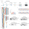
Fig. 1
Fig. 1. DSB repair-related proteins bind ODNs in a sequence-biased manner.
a Schematic overview of ODIP-Seq for capturing preference binding sequences of the protein of interest. The ODN pool was incubated with the cell lysate for 12 h, and the ODN-protein complexes were immunoprecipitated using the corresponding primary antibody. The ODNs were then recovered from the beads using the proteinase K-phenol-chloroform method, and a library was prepared for next-generation sequencing. ODIP, oligodeoxynucleotide immunoprecipitation. b Heatmap of relative TPM of precipitated ODNs with RAD51, RAD50, Ku80 and CtIP. Relative TPM for each ODN was calculated using the mean of two independent replicates. c Violin plots showing the overall relative TPM distribution of precipitated ODNs with the indicated DNA repair proteins. The white dot represents the median. The box spans from the 25th percentile (first quartile) to the 75th percentile (third quartile). The whiskers extend to the smallest and largest data points within 1.5 times the interquartile range (IQR). d, e Characterization of top-ranked ODNs bound to the RAD51 (d) or Ku80 (e) protein in Fig. 1b using the WebLogo 3 website. f, g Evaluation of the binding activity of RAD51 protein with top- and bottom-ranked SSO by biotin-ODN pulldown assay (f) and ODIP assay (g). The digits denote the number of SSOs. h, i Evaluation of the binding activity of Ku80 protein with top- and bottom-ranked SSO using biotin-ODN pulldown assay (h) and ODIP assay (i). The digits denote the number of SSOs. Data are representative of 3 independent experiments (fi). The sequences of all SSOs used are shown in Supplementary Data 3. Source data are provided as a Source Data file. Figure a Created with BioRender.com released under a Creative Commons Attribution-NonCommercial-NoDerivs 4.0 International license.
Fig. 2
Fig. 2. Addition of RAD51-preferred ODNs increases ssDNA donor-mediated HDR efficiency at the BFP locus.
a Schematic representation for gene editing in the BFP reporter cell lines. Single-copy-integrated BFP cell lines were electroporated with Cas9 RNP together with modular or canonical ssDNA donors. Then fluorescence conversion percentage was measured using flow cytometry. b, c HDR efficiencies of ssDNA donors connected with RAD51- or Ku80-preferred ODNs in HEK 293T-BFP cells (b) and K562-BFP cells (c). d HDR efficiencies of ssDNA donors at different concentration gradients in HEK 293T-BFP cells. e The effect of the incorporation position on the HDR improvement of HDR-boosting modules. f HDR efficiencies of ssDNA donors incorporated with the indicated mutant SSO14 modules. g Schematic representation for gene editing with ssDNA donors and independent HDR-boosting modules. h HDR efficiencies of ssDNA donors in HEK 293T-BFP cells electroporated with CRISPR-Cas9 RNP, canonical ssDNA donor, and independent HDR-boosting module. i Evaluation of the binding activity of RAD51 with ssDNA donors using the ODIP assay. Donor mix, an equal parts mix of the three ssDNA donors mentioned in the image. j Western blot analysis of the knockdown efficiencies of three siRNAs targeting RAD51 in HEK 293T cells 48 h post-transfection. siRAD51-mix, an equal parts mix of the three siRNAs of RAD51 mentioned in the image. k, l Effects of RAD51 knockdown on HDR efficiencies of ssDNA donors at the BFP site in HEK 293T-BFP cells (k) and endogenous FANCF site in HEK 293T cells (l). For all HDR efficiency-assessing experiments unless otherwise specified, 18 pmol Cas9 nuclease, 22 pmol gRNA and 6 pmol ssDNA donors corresponded to 2 × 10⁵ cells. HDR efficiency was measured three days after electroporation. Data are representative of 3 independent experiments (i, j). Values and error bars reflect mean ± SD of n = 3 (b, df, h, k) independent electroporation replicates. Values reflect n = 2 (c, l) independent electroporation replicates. The sequences of all gRNAs, ssDNA donors and siRNAs used are shown in Supplementary Data 1, 2, and 6. Source data are provided as a Source Data file. Figure (a, g) Created with BioRender.com released under a Creative Commons Attribution-NonCommercial-NoDerivs 4.0 International license.
Fig. 3
Fig. 3. HDR-boosting modules achieve efficient precise gene editing at endogenous genomic loci in multiple human cell types.
ad HDR efficiencies of HDR-boosting modular ssDNA donors and canonical ssDNA donors at the specified gene loci for EMX1, DNMT1, CXCR4, RUNX1, RNF2, and FANCF in HEK 293T cells (a), HeLa cells (b), U2OS cells (c), and K562 cells (d). e HDR efficiencies yielded with different concentrations of HDR-boosting modular ssDNA donors and canonical ssDNA donors at the FANCF site in HeLa cells. The digits denote the amount of ssDNA donors corresponding to 1 x 106 cells. f HDR efficiencies of ssDNA donors interrogating two endogenous gene sites simultaneously in HeLa cells. g HDR efficiencies of HDR-boosting modular ssDNA donors and canonical ssDNA donors at the FANCF site in hPB CD34+ cells. For all HDR efficiency-assessing experiments unless otherwise specified, 18 pmol Cas9 nuclease, 22 pmol gRNA and 6 pmol ssDNA donors corresponded to 2 × 105 cells. HDR efficiency was measured by NGS three days after electroporation. HDR efficiencies reflect the sequencing reads that contain the intended edit and do not contain indels among all treated cells (ag). Values reflect n = 2 (ag) independent electroporation replicates. The sequences of all gRNAs and ssDNA donors used are shown in Supplementary Data 1 and 2. Source data are provided as a Source Data file.
Fig. 4
Fig. 4. Genome-wide profile of donor integration and chromosomal translocation.
a Brief workflow of Tn5-based high-throughput genome-wide sequencing for integration of ssDNA donor or translocation at the DSB site. Genomic DNA was tagmented with Tn5, and genomic regions containing the inserted donor sequences and the translocated regions were amplified using the indicated primer pairs designed for integration and translocation, respectively. After nest-PCR, amplicons were barcoded and then sequenced. b Circos plots of genome-wide off-target integrations of donors and translocation junctions in edited cells. Off-target integrations and translocation junctions were binned to 0.1 Mb regions and plotted on a normalized scale (black bars). Green arrow, ssDNA donor on-target site. Blue arrow, bait primer targeting site. c The overall off-target integration and translocation rate were calculated as the percentage of off-target reads in HDR reads and the percentage of translocation reads in non-translocation (bait region) reads, respectively. Values reflect n = 2 (c) independent electroporation replicates. The sequences of all gRNAs, ssDNA donors and primers used are shown in Supplementary Data 1, 2 and 4. Source data are provided as a Source Data file.
Fig. 5
Fig. 5. HDR-boosting modules function in other types of precise gene editing.
a Schematic representation for DNA knock-in at the endogenous genomic loci cooperating with Cas9. b FLAG or loxP sequence insertion efficiency mediated by the indicated ssDNA donors at the FANCF gene locus in HeLa cells. 18 pmol Cas9 nuclease, 22 pmol gRNA and ssDNA donors with the indicated amount (per million cells) corresponded to 2 × 105 cells. c Schematic illustrating DNA double nicks using a pair of sgRNAs guiding Cas9 nickases (nCas9). The D10A mutation renders Cas9 able to cleave only the strand complementary to the sgRNA; the H840A mutation renders Cas9 able to cleave only the non-complementary strand. A pair of sgRNA-nCas9 complexes can nick both strands simultaneously. d HDR efficiencies in paired nCas9-mediated precise editing system using ssDNA donors tethered with HDR-boosting module. In this electroporation, 36 pmol nCas9 nuclease, 22 pmol gRNA, 22 pmol AS-gRNA and the indicated amount of ssDNA donor (per million cells) corresponded to 2 × 105 cells. e Schematic representation for gene editing using the Cas12a nuclease which generates sticky-end DSB. f HDR efficiencies of HDR-boosting modular ssDNA donors and canonical ssDNA donors cooperated with Cas12a at the RNF2 site in HEK 293T cells. 32 pmol Cas12a nuclease, 37.5 pmol gRNA and 6 pmol ssDNA donors corresponded to 2 × 10⁵ cells. For all HDR efficiency-assessing experiments, HDR efficiency was measured three days after electroporation. Values and error bars reflect mean ± SD of n = 3 (d) independent electroporation replicates. Values reflect n = 2 (b, f) independent electroporation replicates. The sequences of all gRNAs and ssDNA donors used are shown in Supplementary Data 1 and 2. Source data are provided as a Source Data file. Figure (a, c, e) Created with BioRender.com released under a Creative Commons Attribution-NonCommercial-NoDerivs 4.0 International license.
Fig. 6
Fig. 6. Enhanced potency of HDR-boosting modular donors with HDRobust or M3814.
a Genome editing efficiencies at four endogenous loci in HEK 293T cells introduced by the indicated donors, along with transient end-joining inhibition by HDRobust (M3814 + POLQ siRNA mix) or M3814. b Genome editing efficiencies at five endogenous loci in K562 cells introduced by the indicated donors, with or without treatment by M3814. Electroporation was carried out using a 20 µl mixture, containing 2 × 105 cells, 50.4 pmol Cas9 nuclease, 64 pmol gRNA and 40 pmol ssDNA donors. When applicable, we added POLQ siRNA mix containing 32 pmol of POLQ siRNA predesigned pool (siRNAs 485, 1390, 1397 and 2460) and 64 pmol of POLQ siRNA 765. For transient NHEJ inhibition, 2 µM M3814 was added for two days after electroporation, and editing efficiency was measured five days after electroporation. Values reflect n = 2 (a, b) independent electroporation replicates. The sequences of all gRNAs, ssDNA donors and POLQ siRNA used are shown in Supplementary Data 1, 2 and 6. Source data are provided as a Source Data file.

References

    1. Awwad, S. W., Serrano-Benitez, A., Thomas, J. C., Gupta, V. & Jackson, S. P. Revolutionizing DNA repair research and cancer therapy with CRISPR–Cas screens. Nat. Rev. Mol. Cell Biol.24, 477–494 (2023). 10.1038/s41580-022-00571-x - DOI - PubMed
    1. Scully, R., Panday, A., Elango, R. & Willis, N. A. DNA double-strand break repair-pathway choice in somatic mammalian cells. Nat. Rev. Mol. Cell Biol.20, 698–714 (2019). 10.1038/s41580-019-0152-0 - DOI - PMC - PubMed
    1. Sfeir, A. & Symington, L. S. Microhomology-mediated end joining: A back-up survival mechanism or dedicated pathway? Trends Biochem. Sci.40, 701–714 (2015). 10.1016/j.tibs.2015.08.006 - DOI - PMC - PubMed
    1. Bhargava, R., Onyango, D. O. & Stark, J. M. Regulation of single-strand annealing and its role in genome maintenance. Trends Genet32, 566–575 (2016). 10.1016/j.tig.2016.06.007 - DOI - PMC - PubMed
    1. Yeh, C. D., Richardson, C. D. & Corn, J. E. Advances in genome editing through control of DNA repair pathways. Nat. Cell Biol.21, 1468–1478 (2019). 10.1038/s41556-019-0425-z - DOI - PubMed

MeSH terms

LinkOut - more resources