. 2024 Aug 9;15(1):6637.
doi: 10.1038/s41467-024-50930-9.

Inherited defects of piRNA biogenesis cause transposon de-repression, impaired spermatogenesis, and human male infertility

Affiliations

Inherited defects of piRNA biogenesis cause transposon de-repression, impaired spermatogenesis, and human male infertility

Birgit Stallmeyer et al. Nat Commun. .

Abstract

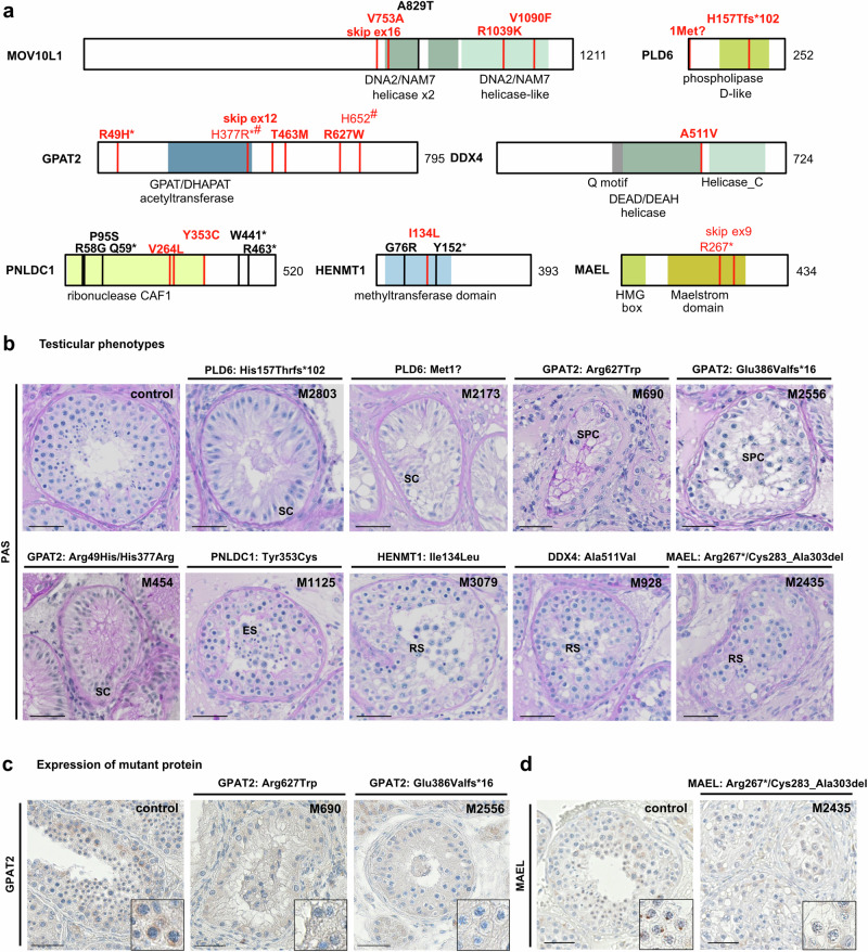
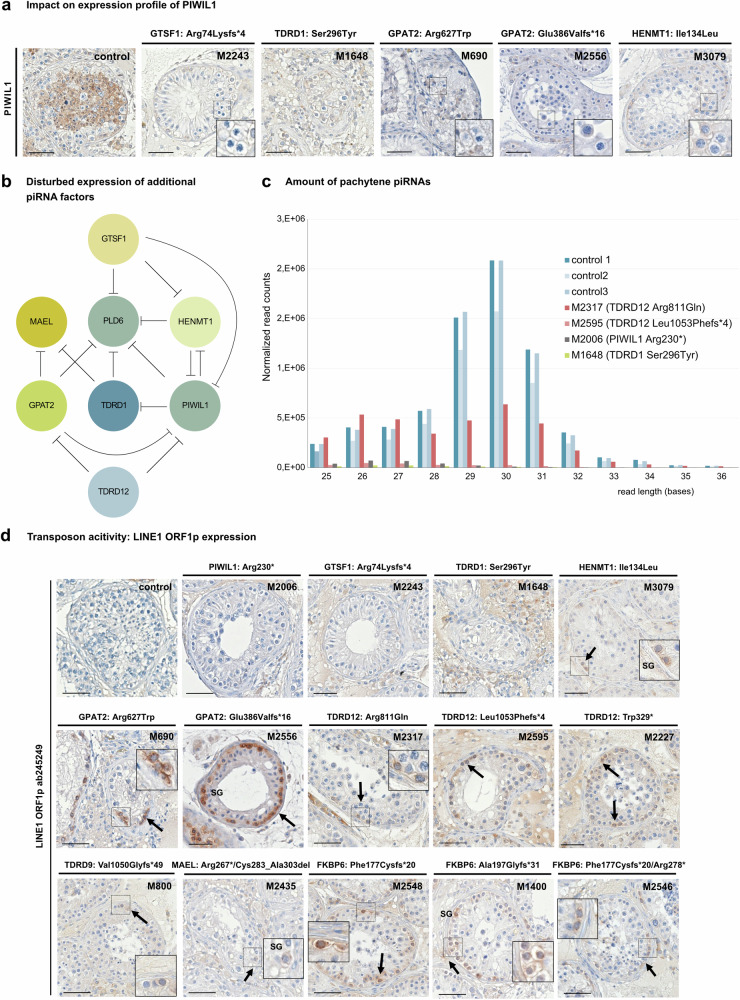
piRNAs are crucial for transposon silencing, germ cell maturation, and fertility in male mice. Here, we report on the genetic landscape of piRNA dysfunction in humans and present 39 infertile men carrying biallelic variants in 14 different piRNA pathway genes, including PIWIL1, GTSF1, GPAT2, MAEL, TDRD1, and DDX4. In some affected men, the testicular phenotypes differ from those of the respective knockout mice and range from complete germ cell loss to the production of a few morphologically abnormal sperm. A reduced number of pachytene piRNAs was detected in the testicular tissue of variant carriers, demonstrating impaired piRNA biogenesis. Furthermore, LINE1 expression in spermatogonia links impaired piRNA biogenesis to transposon de-silencing and serves to classify variants as functionally relevant. These results establish the disrupted piRNA pathway as a major cause of human spermatogenic failure and provide insights into transposon silencing in human male germ cells.


Conflict of interest statement

The authors declare no competing interests.

Figures

Fig. 1
Fig. 1. Genetic landscape of piRNA biogenesis-related male infertility.
a Workflow of scrutinizing biological processes related to genetically determined reduced sperm count and male infertility by Gene Ontology (GO) analysis. Pie chart shows first hierarchy of the two-tiered hierarchy. b Side-ways bar chart showing -log10(P-value of Bonferroni-adjusted Fisher’s Exact test) of individual GO terms. Number of piRNA pathway factors associated with GO term shown to the right of each bar and GO terms associated exclusively with piRNA pathway factors highlighted in blue. c Schematic overview on mammalian piRNA biogenesis related sub-pathways with proteins factors known to be involved from mice knockout studies. Proteins in which encoded biallelic high-impact variants were identified in infertile men are underlined. a, c Created with BioRender.com released under a Creative Commons Attribution-NonCommercial-NoDerivs 4.0 International license (https://creativecommons.org/licenses/by-nc-nd/4.0/deed.en).
Fig. 2
Fig. 2. Homozygous variants identified in genes of the piRISC complex and associated testicular phenotypes.
a Schematic representation and AlphaFold2 structure predictions of PIWIL1, PIWIL2, and GTSF1. The schematic depicts both novel (red) and known (black) homozygous variants, with amino acids affected by new variants highlighted in the protein structure in red. N N-terminal structured domain, PAZ Piwi/Argonaute/Zwille domain, PIWI Piwi-like domain; L1 linker domain 1, L2 linker domain 2, MID middle domain, ZF zinc finger domain. b Periodic acid-Schiff (PAS) staining of testicular tissue of men with normal spermatogenesis (control), M2006 [PIWIL1, p.(Arg230*)] and M2243 [GTSF1, p.(Arg74Lysfs*4)]. Representative tubules showing the most advanced stage of spermatogenesis observed in three independent sections are shown. Immunohistochemical staining (IHC) for round spermatid marker protein Cyclic AMP Element Modulator (CREM) and spermatocyte marker protein γH2AX. In M2006, round spermatids were detected as most advanced germ cells, whereas in M2243, in addition to spermatogonia, rarely pachytene spermatocytes with γH2AX positive sex bodies but no haploid germ cells were observed. c IHC staining for PIWIL1 and GTSF1 in controls and variant carriers demonstrating absence of PIWIL1 in M2006 due to homozygous stop-gain variant p.(Arg230*) and absence of GTSF1 in M2243 with homozygous frameshift variant p.(Arg74Lysfs*4). Representative tubules showing the staining pattern observed in independent sections (control: N = 3, proband: N = 2) are shown. Scale bar = 50 µm. SC Sertoli cell, SG spermatogonia, SPC spermatocyte, RS round spermatid.
Fig. 3
Fig. 3. Biallelic variants identified in human piRNA biogenesis-associated genes.
a Localization of variants in schematic of MOV10L1, PLD6, GPAT2, PNLDC1, MAEL, DDX4, and HENMT1 structure with protein domains colored and newly identified biallelic variants (red, bold for homozygous) as well as previously described homozygous variants (black) indicated. Pairs of compound heterozygous variants are indicated by identical symbols (*,#) in superscript. Helicase domains (green): DEAD/DEAH, Helicase_C, DNA2/NAM7; CAF1 chromatin assembly factor 1 domain (yellow); GPAT/DHAPAT acetyltransferase and methyltransferase domains (blue). b Periodic acid-Schiff (PAS) staining of representative testicular tissue of variant carriers demonstrating SCO in M2803 [PLD6, p.(His157Thrfs*102)], M2173 [PLD6, p.(Met1?)] and M454 [GPAT2, p.(His377Arg)/(Arg49His)] and presence of haploid germ cells (round/elongated spermatids) in M1125 [PNLDC1, p.(Tyr353Cys)], M3079 [HENMT1, p.(Ile134Leu)], M928 [DDX4, p.(Ala511Val)], and M2435 [MAEL, p.(Arg267*)/ p.(Cys283_Ala303del)]. Representative tubules showing the most advanced stage of spermatogenesis observed in three independent sections are shown. c Immunohistochemical (IHC) staining for GPAT2 in testicular tissue with full spermatogenesis (control) and GPAT2 variant carriers with meiotic arrest, [M690, p.(Arg627Trp), [M2556, p.(Glu386Valfs*16)]. In control tissue, GPAT2 is expressed in perinuclear structures in spermatocytes and this staining pattern is absent in M690 and M2556. Representative tubules showing the staining pattern observed in independent sections (control: N = 3, proband: N = 2) are shown. d IHC for MAEL in testicular tissue with full spermatogenesis (control) and M2435 with compound heterozygous presence of two MAEL LoF variants p.(Arg267*)/p.(Cys283_Ala303del). In control tissue, MAEL is expressed in perinuclear structures in spermatocytes and distinct condensed structures in round spermatids and this staining pattern is absent in the variant carrier. Representative tubules showing the staining pattern observed in independent sections (control: N = 3, proband: N = 2) are shown. Scale bar = 50 µm. SC Sertoli cell, SPC spermatocyte, RS round spermatid, ES elongated spermatid.
Fig. 4
Fig. 4. Homozygous high-impact variants identified in human genes of the tudor-domain containing gene family (TDRDs).
a Schematic representation and AlphaFold2 structure predictions of TDRDs. The schematic depicts both novel (red) and known (black) homozygous variants, with amino acids affected by new variants highlighted in the protein structures in red. MYND-ZF MYND-type zinc finger domain (gray), Tudor tudor domain (blue), helicase domains DEAD/DEAH (green), helicase-associated 2 (light green). b Periodic acid-Schiff (PAS) staining of testicular tissue of variant carriers demonstrating meiotic arrest in M1648 [TDRD1, p.(Ser296Tyr)], presence of elongated spermatids in M800 [TDRD9, p.(Val1050Glyfs*49)], SCO in M1642 [TDRD12, p.(Asn198Ser)] and round spermatid arrest in M2227 [TDRD12, p.(Trp329*)]. Representative tubules showing the most advanced stage of spermatogenesis observed in three independent sections are shown. Scale bar = 50 µm. SC Sertoli cell, SG spermatogonia, SPC spermatocyte, RS round spermatid.
Fig. 5
Fig. 5. Functional impact of disturbed piRNA biogenesis.
a Immunohistochemical staining demonstrating diminished expression of PIWIL1 in variant carriers M2243 [GTSF1, p.(Arg74Lysfs*4)], M1648 [TDRD1, p.(Ser296Tyr)], M690 [GPAT2, p.(Arg627Trp)], M2556, [GPAT2, p.(Glu386Valfs*16)] and M3079 [HENMT1, p.(Ile134Leu)]. Representative tubules showing the staining pattern observed in independent sections (control: N = 3, proband: N = 2) are shown. b Schematic depicting impact of loss of piRNA biogenesis protein function on expression of additional piRNA factors. Circles represent piRNA protein and inhibiting effects of loss-of-protein functions on the expression of further piRNA proteins are indicated. c Effect of biallelic variants in genes of the piRNA pathway on biogenesis of pachytene piRNAs. RNA isolated from snap frozen testicular tissue of M2006 [PIWIL1 p.(Arg230*)], M1648 [TDRD1 p.(Ser296Tyr)], M2317 [TDRD12 p.(Arg811Gln)] and M2595 [TDRD12 p.(Leu1053Phefs*4)] revealed a major loss of pachytene piRNAs with expected lengths of 26–31 bases when compared with controls with complete spermatogenesis (ctrl1-3; P = 0.000007). Shapiro-Wilk test was used to estimate the normality of the data. Since Shapiro-Wilk test indicated abnormal data distribution in both control and case groups, two-sided Mann-Whitney U test was used for comparing the expression changes of piRNAs with different length (26–31 nt) between both groups. Source data are provided as a Source Data file. d Immunohistochemical staining for LINE1 transposon specific protein LINE1 ORF1p in testicular tissue. LINE1 ORF1p was not detected in testicular tissue of controls with full spermatogenesis and PIWIL1, GTSF1, and TDRD1 variant carriers. In contrast, all three TDRD12 variant carriers, both GPAT2 variant carriers, and all three FKBP6 variant carriers revealed a concordant distinct and specific LINE1 ORF1p staining in spermatogonia. A similar effect was also seen in testicular tissue of MAEL, HENMT1, and TDRD9 variant carriers. Representative tubules showing the staining pattern observed in independent sections (control: N = 3, proband: N = 2) are shown. Scale bar = 50 µm. SG spermatogonia. b Created with BioRender.com released under a Creative Commons Attribution-NonCommercial-NoDerivs 4.0 International license (https://creativecommons.org/licenses/by-nc-nd/4.0/deed.en).
Fig. 6
Fig. 6. Comparison of piRNA factor-related male reproductive phenotypes.
a For each piRNA factor gene described in this study, the reproductive phenotype of the male knockout mice is compared with the phenotypes observed in novel and known human infertile biallelic variant carriers. Of note, for several genes, testicular phenotypes of human variant carriers differ from those described for the respective knockout mice. b Venn diagram depicting overlap between reproductive phenotypes of infertile men affected by biallelic variants in the same gene. LoF variants are indicated in red, missense variants are indicated in black. For several genes, (PIWIL2, GTSF1, MOV10L1, PNLDC1, TDRD9) a phenotypic overlap could be observed for missense and LoF variant carriers. LoF variant carriers are in most cases not affected by a more severe phenotype than missense variant carriers. SCO Sertoli cell-only, SpgA spermatogonia arrest, MeiA meiotic arrest, RsA round spermatid arrest, ES+ elongated spermatids present in testicular tissue.

Comment in

References

    1. Wang, X., Ramat, A., Simonelig, M. & Liu, M.-F. Emerging roles and functional mechanisms of PIWI-interacting RNAs. Nat. Rev. Mol. Cell Biol.24, 123–141 (2023). 10.1038/s41580-022-00528-0 - DOI - PubMed
    1. Ozata, D. M., Gainetdinov, I., Zoch, A., O’Carroll, D. & Zamore, P. D. PIWI-interacting RNAs: small RNAs with big functions. Nat. Rev. Genet.20, 89–108 (2019). 10.1038/s41576-018-0073-3 - DOI - PubMed
    1. Girard, A., Sachidanandam, R., Hannon, G. J. & Carmell, M. A. A germline-specific class of small RNAs binds mammalian Piwi proteins. Nature442, 199–202 (2006). 10.1038/nature04917 - DOI - PubMed
    1. Aravin, A. et al. A novel class of small RNAs bind to MILI protein in mouse testes. Nature442, 203–207 (2006). 10.1038/nature04916 - DOI - PubMed
    1. Özata, D. M. et al. Evolutionarily conserved pachytene piRNA loci are highly divergent among modern humans. Nat. Ecol. Evol.4, 156–168 (2020). 10.1038/s41559-019-1065-1 - DOI - PMC - PubMed

MeSH terms

LinkOut - more resources