. 2024 Aug 9;15(8):580.
doi: 10.1038/s41419-024-06940-y.

Integrative single-cell RNA-seq and spatial transcriptomics analyses reveal diverse apoptosis-related gene expression profiles in EGFR-mutated lung cancer

Affiliations

Integrative single-cell RNA-seq and spatial transcriptomics analyses reveal diverse apoptosis-related gene expression profiles in EGFR-mutated lung cancer

Motohiro Izumi et al. Cell Death Dis. .

Abstract

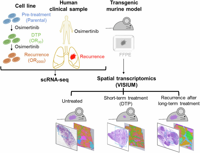
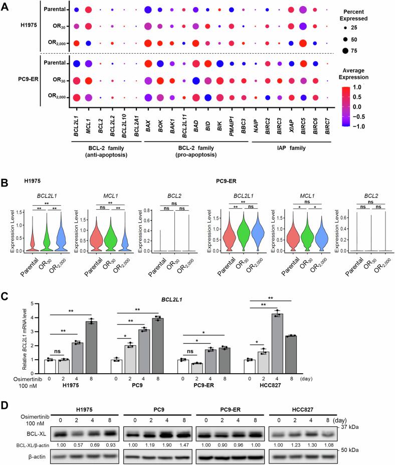
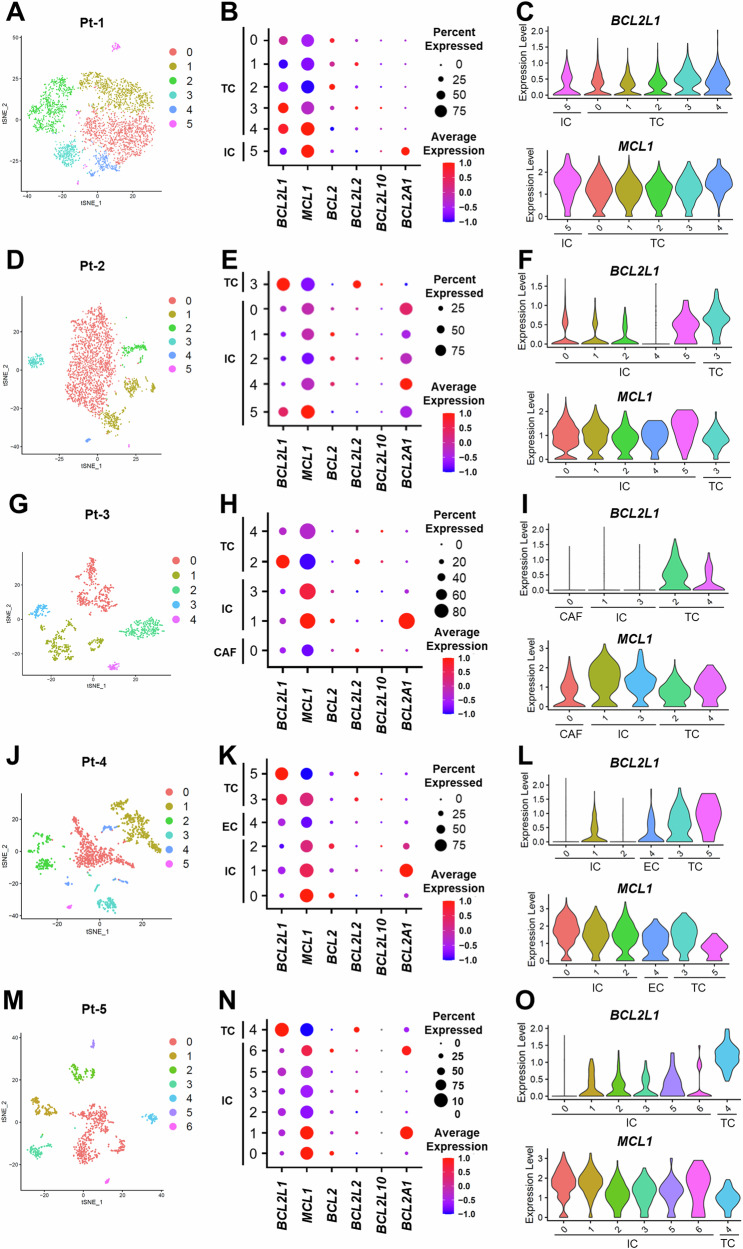
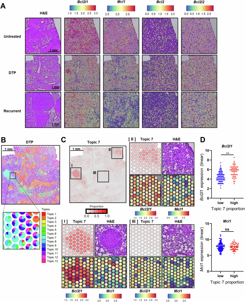
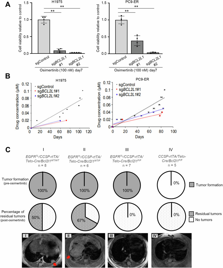
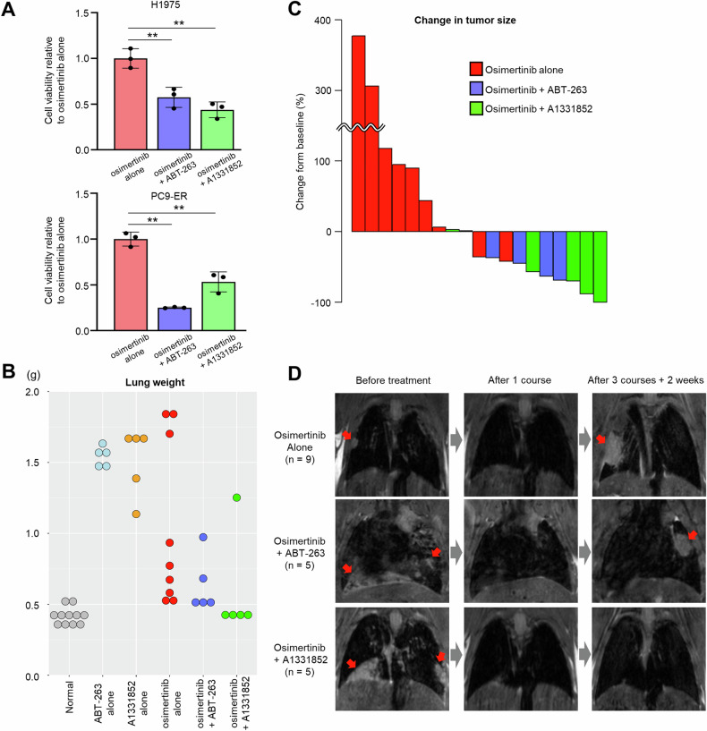
In EGFR-mutated lung cancer, the duration of response to tyrosine kinase inhibitors (TKIs) is limited by the development of acquired drug resistance. Despite the crucial role played by apoptosis-related genes in tumor cell survival, how their expression changes as resistance to EGFR-TKIs emerges remains unclear. Here, we conduct a comprehensive analysis of apoptosis-related genes, including BCL-2 and IAP family members, using single-cell RNA sequence (scRNA-seq) and spatial transcriptomics (ST). scRNA-seq of EGFR-mutated lung cancer cell lines captures changes in apoptosis-related gene expression following EGFR-TKI treatment, most notably BCL2L1 upregulation. scRNA-seq of EGFR-mutated lung cancer patient samples also reveals high BCL2L1 expression, specifically in tumor cells, while MCL1 expression is lower in tumors compared to non-tumor cells. ST analysis of specimens from transgenic mice with EGFR-driven lung cancer indicates spatial heterogeneity of tumors and corroborates scRNA-seq findings. Genetic ablation and pharmacological inhibition of BCL2L1/BCL-XL overcome or delay EGFR-TKI resistance. Overall, our findings indicate that BCL2L1/BCL-XL expression is important for tumor cell survival as EGFR-TKI resistance emerges.


Conflict of interest statement

The authors declare the following financial interests/personal relationships which may be considered potential competing interests: H.U. reports grants or contracts from Takeda, Boehringer Ingelheim, and Taiho; personal fees (honoraria) from Novartis, Taiho, Chugai, MSD, AstraZeneca, Daiichisankyo. S.S.K. reports research support from Boehringer Ingelheim, MiRXES, Johnson&Johnson, and Taiho Therapeutics, as well as personal fees (honoraria) from AstraZeneca, Boehringer Ingelheim, Bristol Meyers Squibb, Chugai Pharmaceutical, and Takeda Pharmaceuticals, plus royalties from Life Technologies; all interests are outside the submitted work. D.C. reports receiving consulting fees and honoraria from Takeda/Millennium Pharmaceuticals, AstraZeneca, Pfizer, Blueprint Medicines, and Janssen; institutional research support from Takeda/Millennium Pharmaceuticals, AstraZeneca, Pfizer, Merck Sharp and Dohme, Merrimack Pharmaceuticals, Bristol Myers Squibb, Clovis Oncology, Spectrum Pharmaceuticals, Tesaro, Taiho Pharmaceutical Company, and Daiichi Sankyo; and consulting fees from Teladoc and Grand Rounds by Included Health plus royalties from Life Technologies; all interests are outside the submitted work. The remaining authors declare no competing interests.

Figures

Fig. 1
Fig. 1. Overview of study.
To assess apoptosis-related gene expression in tumors following EGFR-TKI treatment, we established cell line models (top, left) representing untreated, drug-tolerant persister (DTP), and acquired resistance states using EGFR-mutated lung cancer lines and subjected them to single-cell RNA sequencing (scRNA-seq). To validate those results and compare gene expression profiles between tumor and normal cells, we conducted scRNA-seq on samples collected from patients with EGFR-mutated lung cancer pre-treatment or after the development of EGFR-TKI resistance (top, middle). To confirm scRNA-seq findings and assess spatial heterogeneity, we prepared formalin-fixed paraffin-embedded (FFPE) lung tissue sections from transgenic mice (top, right) harboring EGFR-driven lung cancer representative of three distinct states: untreated, short-term drug treatment reflecting the DTP state, and tumor recurrence after long-term treatment. Spatial transcriptomics was subsequently conducted.
Fig. 2
Fig. 2. Osimertinib induces BCL2L1 expression in EGFR-mutated lung cancer cell lines.
A Dotplots based on scRNA-seq showing apoptosis-related gene expression in H1975 (top) and PC9-ER (bottom) cells. B Violin plots based on scRNA-seq showing BCL2L1, MCL1, and BCL-2 expression in H1975 and PC9-ER cells. p-values were calculated using Kruskal–Wallis one-way ANOVA. C Relative BCL2L1 transcript levels in indicated EGFR-mutant cell lines based on real-time PCR. Each line was treated with 100 nM osimertinib and evaluated at 0, 2, 4, and 8 days. Data are means ± SD of triplicates from one experiment and are representative of three independent experiments. A Repeated measures one-way ANOVA with Dunnett’s test was used to compare treatment groups to a single control group. D Immunoblot analysis showing changes in BCL-XL expression in EGFR-mutant lung cancer lines treated with 100 nM osimertinib for 0, 2, 4, and 8 days. The BCL-XL/actin ratio was calculated using ImageJ software. All blots show representative images of three independent experiments. Asterisks indicate p-values as follows: *p < 0.05; **p < 0.005; ns, not significant. Data are presented as the mean ± SD.
Fig. 3
Fig. 3. Apoptosis-related gene expression in clinical samples.
A, D, G, J, M t-SNE plots showing clusters based on scRNA-seq in indicated patients. B, E, H, K, N Dot plots showing differences in expression of anti-apoptotic genes in tumor versus non-tumor cells. C, F, I, L, O Violin plots showing BCL2L1 (upper) and MCL1 (lower) expression in indicated cell types, indicating that BCL2L1 is specifically and highly expressed in tumor cells. TC tumor cell, IC immune cell, EC endothelial cell, CAF cancer-associated fibroblast.
Fig. 4
Fig. 4. Visualization of inter- and intra-tumor heterogeneity using spatial transcriptomics.
A H&E staining and Bcl2l1, Mcl1, Bcl2, and Bcl2l2 expression in three lung-specific EGFR-L858R-T790M (EGFRTL/CCSP-rtTA) transgenic mouse samples representing untreated (top), DTP (middle), and tumor-recurrent (bottom) states. B Deconvolved cell-type proportions for the DTP dataset, represented as pie charts for each pixel. C Pixel proportions showing the distribution of topic 7. Three areas with high (I), medium (II), and low (III) proportions of topic 7 are extracted. Topic 7 proportion, H&E staining, and Bcl2l1 and Mcl1 expression levels are visualized in each area. D Spots were divided into low (≥25%, <50%) and high (≥50%) groups based on the size of the Topic 7 proportion. Bcl2l1 and Mcl1 expression levels were compared between these two groups. p-values were calculated using the unpaired two-tailed Welch’s t-test. Asterisks indicate p-values as follows: **p < 0.005. ns not significant, H&E hematoxylin and eosin, DTP drug-tolerant persister.
Fig. 5
Fig. 5. BCL-XL deletion in lung cancer blocks the emergence of drug-tolerant cells and counters osimertinib resistance in vitro and in vivo.
A The number of cells remaining on day 7 after treatment with 100 nM osimertinib in H1975 (left) and PC9-ER (right) cells transduced with indicated sgRNAs. Data are the mean ± SD of triplicates from one experiment and are representative of three independent experiments. p-values were calculated using one-way ANOVA followed by the Tukey–Kramer multiple-comparison test. Asterisks indicate p-values as follows: **p < 0.005. B Comparison of the time course of acquired osimertinib resistance in H1975 (left) and PC9-ER (right) cells transduced with indicated sgRNAs. Cells were chronically exposed to gradually increasing osimertinib doses. C (top) The proportion of indicated transgenic mice exhibiting tumor formation after doxycycline administration; (middle) the percentage of mice exhibiting residual tumors after completion of osimertinib treatment; and (bottom) representative MRI images after completion of osimertinib treatment. Red arrows indicate lung tumors.
Fig. 6
Fig. 6. Combining osimertinib with pharmacological BCL-XL inhibition suppresses osimertinib resistance.
A The number of cells remaining after 7 days of treatment with 100 nM osimertinib plus either 1 µM ABT-263 or 1 µM A1331852 in H1975 (top) and PC9-ER (bottom) cells. Data are the mean ± SD of triplicates from one experiment and are representative of three independent experiments. p-values were calculated using one-way ANOVA followed by the Tukey–Kramer multiple-comparison test. Asterisks indicate p-values as follows: **p < 0.005. B Lung weight at the end of the experiment. Normal lungs were derived from CCSP-rtTA transgenic mice (no EGFRTL expression). C Waterfall plot showing changes in tumor size from baseline (before treatment) to the experimental endpoint. The bars shown represent a single mouse. D Representative MRI images of mouse lung before and after drug treatment. Red arrows indicate lung tumors. Drug administration protocols are provided in the Methods section.

References

    1. Siegel RL, Miller KD, Wagle NS, Jemal A. Cancer statistics, 2023. CA Cancer J Clin. 2023;73:17–48. 10.3322/caac.21763 - DOI - PubMed
    1. Gazdar AF. Activating and resistance mutations of EGFR in non-small-cell lung cancer: role in clinical response to EGFR tyrosine kinase inhibitors. Oncogene. 2009;28:S24–31. 10.1038/onc.2009.198 - DOI - PMC - PubMed
    1. Soria JC, Ohe Y, Vansteenkiste J, Reungwetwattana T, Chewaskulyong B, Lee KH, et al. Osimertinib in untreated EGFR-mutated advanced non-small-cell lung cancer. N Engl J Med. 2018;378:113–25. 10.1056/NEJMoa1713137 - DOI - PubMed
    1. Ramalingam SS, Vansteenkiste J, Planchard D, Cho BC, Gray JE, Ohe Y, et al. Overall survival with osimertinib in untreated, EGFR-mutated advanced NSCLC. N Engl J Med. 2020;382:41–50. 10.1056/NEJMoa1913662 - DOI - PubMed
    1. Sharma SV, Lee DY, Li B, Quinlan MP, Takahashi F, Maheswaran S, et al. A chromatin-mediated reversible drug-tolerant state in cancer cell subpopulations. Cell. 2010;141:69–80. 10.1016/j.cell.2010.02.027 - DOI - PMC - PubMed

MeSH terms