. 2024 Aug 9;15(1):6804.
doi: 10.1038/s41467-024-50710-5.

High-throughput identification of functional regulatory SNPs in systemic lupus erythematosus

Affiliations

High-throughput identification of functional regulatory SNPs in systemic lupus erythematosus

Qiang Wang et al. Nat Commun. .

Abstract

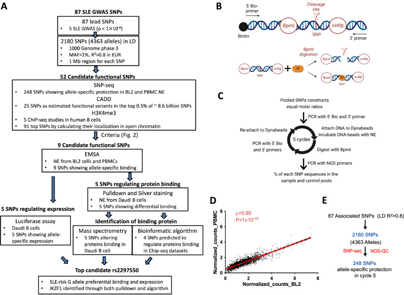
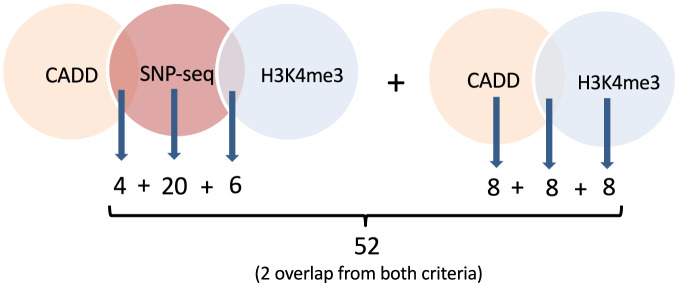
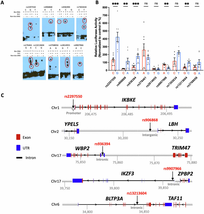
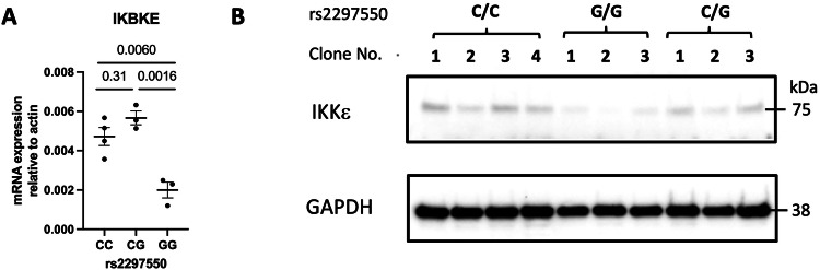
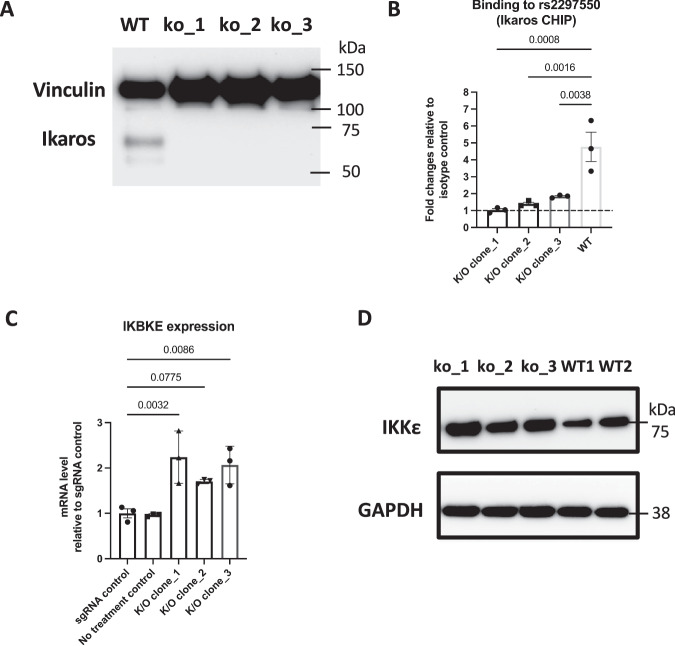
Genome-wide association studies implicate multiple loci in risk for systemic lupus erythematosus (SLE), but few contain exonic variants, rendering systematic identification of non-coding variants essential to decoding SLE genetics. We utilized SNP-seq and bioinformatic enrichment to interrogate 2180 single-nucleotide polymorphisms (SNPs) from 87 SLE risk loci for potential binding of transcription factors and related proteins from B cells. 52 SNPs that passed initial screening were tested by electrophoretic mobility shift and luciferase reporter assays. To validate the approach, we studied rs2297550 in detail, finding that the risk allele enhanced binding to the transcription factor Ikaros (encoded by IKZF1), thereby modulating expression of IKBKE. Correspondingly, primary cells from genotyped healthy donors bearing the risk allele expressed higher levels of the interferon / NF-κB regulator IKKε. Together, these findings define a set of likely functional non-coding lupus risk variants and identify a regulatory pathway involving rs2297550, Ikaros, and IKKε implicated by human genetics in risk for SLE.


Conflict of interest statement

The authors declare no competing interests.

Figures

Fig. 1
Fig. 1. Screening candidate regulatory variants for SLE by SNP-seq.
A Flow chart illustrating the identification of functional candidate SNPs from SLE GWAS data. B SNP-seq: To generate the SNP-seq construct (top), a 31 bp sequence centered on the SNP is positioned between two type IIS restriction enzyme (IIS RE) binding sites. SNPs that fail to bind regulatory proteins such as transcription factors (TF) are negatively selected (bottom), allowing enrichment of protected constructs by PCR. The whole construct can be amplified using primers as per Supplementary Data 9. Bio biotin. Figure 1B was created with BioRender.com released under a Creative Commons Attribution-NonCommerical-NoDerivs 4.0 International license. C The experimental procedure for SNP-seq; NE nuclear extract, Bio biotin. D Spearman’s correlation with a two-tailed test of SNP-seq allele counts normalized to control between PBMC and BL2 samples (ρ = 0.89, P < 1 × 10−15). E We selected 248 SNPs that passed next-generation sequencing quality control (NGS-QC) and demonstrated progressive allele-specific protection (Suppl. Fig. 1C). Source data are provided as a Source Data file.
Fig. 2
Fig. 2. Prioritization of candidate SNPs by SNP-seq and bioinformatic enrichment.
SNPs selected for experimental screening were the top 20 of 248 emerging form SNP-seq, the top eight emerging from H3K4me3 score, the top eight emerging from CADD, and 18 SNPs identified by at least two analyses: six SNP-seq+H3K4me3, four SNP-seq+CADD, and eight SNPs H3K4me3 + CADD.
Fig. 3
Fig. 3. EMSA and luciferase reporter assessment of 52 candidate regulatory SNPs.
A Seven biallelic and two triallelic (rs2297550 and rs936394) SNPs showed consistent allele-imbalanced binding for nuclear by EMSA. Allele-specific gel shift/binding marked with red circles. Experiments were repeated three times using distinct nuclear extracts. Representative blots from PBMCs are shown here; BL2 data and negative SNPs are provided in Supplementary Fig. 4 and Supplementary Data 6. B Luciferase reporter assay between the reference (red) and alternative (blue) alleles of nine candidate regulatory SNPs from A in Daudi B cells (mean ± s.d, n = 8 biological replicates, Mann–Whitney two-tailed U-test with two-stage step-up correction for multiple hypothesis testing: rs2297550 p = 0.00047, rs906868 p = 0.0013, rs936394 p = 0.00047, rs9907966 p = 0.0071, rs13213604 p = 0.0085). C Diagram displaying the position of 5 SNPs that showed consistent significant differences between alleles through EMSA and luciferase reporter assay; rs2297550 in promoter of IKBKE, rs906868 in intergenic region between YPEL5 and LBH, rs936394 in intron of WBP2, rs9907966 in intron of IKZF3, rs13213604 in intron of BLTP3A. The unit of chromosome position is in kilobases (Kb). A diagram of gene features including exon, intron, and UTR was generated using GSDS 2.0 (http://gsds.gao-lab.org/index.php). Source data are provided as a Source Data file.
Fig. 4
Fig. 4. Characterization of functional SNP rs2297550 by CRISPR-mediated editing in Daudi cells.
A Using base-edited clones (4C/C, 3C/G, and 3G/G), mRNA levels of IKBKE and β-actin were measured by RT-qPCR, and B protein levels of IKKϵ (80 KDa) and GAPDH (37 KDa) were measured by Western blotting, performed once (mean ± s.d, P values from one-way ANOVA corrected by Tukey’s multiple comparisons). Source data are provided as a Source Data file.
Fig. 5
Fig. 5. SNP rs2297550 associated with IKKϵ expression in primary PBMCs.
IKKϵ expression was determined by flow cytometry for 35 healthy subjects with C/C (n = 12), C/G (n = 11), and G/G (n = 12) genotype at SNP rs2297550. The mean fluorescence intensity (MFI) of IKKϵ expression in (AF) resting and (GL) 4 h LPS-stimulated PBMCs, including CD19+ B cells, CD3+CD4+ T cells, CD3+CD8+ T cells, CD3-CD56+ NK cells, and CD14+ monocytes (mean ± s.d, P values from one-way ANOVA corrected by Dunnett’s multiple comparisons test). Source data are provided as a Source Data file.
Fig. 6
Fig. 6. rs2297550 risk allele G bind Ikaros.
A Oligonucleotide pulldown western blot assay testing Ikaros binding to rs2297550-G; binding is eliminated in the presence of a 30-fold excess of non-biotinylated rs2297550-C or G competitor; positive control, nuclear extract. Pulldown western blot was performed once. B EMSA supershift assay showed that anti-Ikaros caused fading of the shifted band (oligo rs2297550-G, Daudi nuclear extract), whereas isotype control did not; anti-E2F4 also exhibited specificity. EMSA supershift was repeated twice. All blots are provided in the Source Data. C ChIP-qPCR using anti-Ikaros confirms binding to rs2297550 (Daudi cells, mean ± s.d, three biological replicates). D Allele discrimination ChIP-qPCR shows Ikaros preferentially binds to G allele over C allele of rs2297550 in CRISPR-HDR edited C/G Daudi cells (mean ± s.d, three biological replicates). All studies were done in unstimulated cells. Statistical analysis for C and D: paired t-test with two tails, without correction. Source data are provided as a Source Data file.
Fig. 7
Fig. 7. Ikaros binding to rs2297550 regulates IKBKE/IKKϵ expression.
CRISPR-Cas9 Ikaros knockout Daudi clones show A decreased Ikaros protein expression by western blotting, B loss of Ikaros binding to rs2297550 by ChIP-qPCR (mean ± s.d, three biological replicates), and C, D increased IKBKE mRNA by RT-qPCR (mean ± s.d, three biological replicates) and increased IKKϵ protein expression by western blotting. Statistical analysis panels B and C: one-way ANOVA corrected by Dunnett’s multiple comparisons test. Western blotting (A, D) was performed once. Source data are provided as a Source Data file.
Fig. 8
Fig. 8. Ikaros modulation of IKBKE expression via lupus risk SNP.
The lupus risk SNP rs2297550 allelically modulates the binding of the transcription factor Ikaros (encoded by IKZF1) to regulate the expression of IKBKE, encoding IKKε, a protein that modulates multiple processes implicated in SLE pathogenesis. Note that Ikaros binding can either amplify or repress IKBKE depending on context; amplification, shown here, reflects the direction of effect observed in genotyped healthy donors. The role of mechanisms downstream of IKKε remains to be defined. Figure 8 was created with BioRender.com released under a Creative Commons Attribution-NonCommerical-NoDerivs 4.0 International license.

Update of

References

    1. Deapen, D. et al. A revised estimate of twin concordance in systemic lupus erythematosus. Arthritis Rheum.35, 311–318 (1992). 10.1002/art.1780350310 - DOI - PubMed
    1. Block, S. R. et al. Studies of twins with systemic lupus erythematosus. A review of the literature and presentation of 12 additional sets. Am. J. Med.59, 533–552 (1975). 10.1016/0002-9343(75)90261-2 - DOI - PubMed
    1. Alarcon-Segovia, D. et al. Familial aggregation of systemic lupus erythematosus, rheumatoid arthritis, and other autoimmune diseases in 1,177 lupus patients from the GLADEL cohort. Arthritis Rheum.52, 1138–1147 (2005). 10.1002/art.20999 - DOI - PubMed
    1. Vinuesa, C. G., Shen, N. & Ware, T. Genetics of SLE: mechanistic insights from monogenic disease and disease-associated variants. Nat. Rev. Nephrol. 19, 558–572 (2023). - PubMed
    1. Caielli, S., Wan, Z. & Pascual, V. Systemic lupus erythematosus pathogenesis: interferon and beyond. Annu Rev. Immunol.41, 533–560 (2023). 10.1146/annurev-immunol-101921-042422 - DOI - PubMed

MeSH terms

LinkOut - more resources