. 2024 Aug 9;9(1):143.
doi: 10.1038/s41541-024-00930-z.

Inactivated rabies-based Lassa fever virus vaccine candidate LASSARAB protects nonhuman primates from lethal disease

Affiliations

Inactivated rabies-based Lassa fever virus vaccine candidate LASSARAB protects nonhuman primates from lethal disease

Gabrielle Scher et al. NPJ Vaccines. .

Abstract

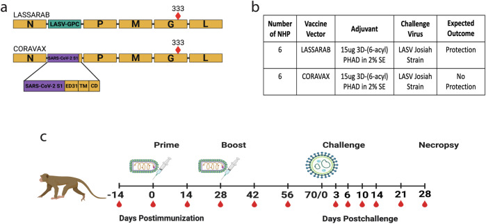
Lassa fever virus (LASV), a member of the Arenavirus family, is the etiological agent of Lassa fever, a severe hemorrhagic disease that causes considerable morbidity and mortality in the endemic areas of West Africa. LASV is a rodent-borne CDC Tier One biological threat agent and is on the World Health Organization's (WHO) Priority Pathogen list. Currently, no FDA-licensed vaccines or specific therapeutics are available. Here, we describe the efficacy of a deactivated rabies virus (RABV)-based vaccine encoding the glycoprotein precursor (GPC) of LASV (LASSARAB). Nonhuman primates (NHPs) were administered a two-dose regimen of LASSARAB or an irrelevant RABV-based vaccine to serve as a negative control. NHPs immunized with LASSARAB developed strong humoral responses to LASV-GPC. Upon challenge, NHPs vaccinated with LASSARAB survived to the study endpoint, whereas NHPs in the control group did not. This study demonstrates that LASSARAB is a worthy candidate for continued development.


Conflict of interest statement

The authors declare no competing interests.

Figures

Fig. 1
Fig. 1. Experimental design.
a Schematic of vaccine constructs with all RABV and foreign proteins indicated. Red diamond indicates the attenuating mutation at amino acid 333 of RABV-G. N nucleoprotein, P phosphoprotein, M matrix protein, G glycoprotein, L RNA-dependent RNA polymerase, LASV-GPC Lassa virus glycoprotein precursor, SARS-CoV-2 S1 severe acute respiratory syndrome coronavirus 2 spike protein subunit 1, ED31 31 amino acids of the ectodomain, TM transmembrane domain, CD cytoplasmic domain. b Table outlining the vaccine groups and expected experimental outcomes. c Experimental timeline. Red droplets indicate blood draws, rhabdovirus and syringe indicate immunization, and the arenavirus represents the LASV challenge. Figure made with BioRender.com.
Fig. 2
Fig. 2. Humoral responses to LASSARAB.
Antibody responses against LASV-GPC and RABV-G were measured through enzyme-linked immunosorbent assay (ELISA). a Antibody EC50 titers over time for LASV-GPC and RABV-G. b Antibody endpoint titers over time for LASV-GPC and RABV-G. Samples from each NHP were run in triplicate, and error bars are the mean with standard deviation (SD). The Mann-Whitney nonparametric T-test was used to determine statistical differences between groups at each time point. Where significance is not noted, samples have no significant difference. ****<0.0001; ***0.0002; **0.0021; *0.0332; P > 0.05 ns, not significant. LOD limit of detection.
Fig. 3
Fig. 3. Antibody functionality.
Assays measuring neutralizing and non-neutralizing antibody functions. a Pseudotype virus neutralization assay (VNA) for LASV with sera from immunized NHPs and human monoclonal anti-LASV-GPC antibody 37.7H as a positive control. Error bars represent standard deviation. b Antibody-dependent cellular cytotoxicity (ADCC) assay for LASV with sera from immunized NHPs. Error bars represent the standard error of the mean. c Rapid fluorescent focus inhibition test (RFFIT) to measure neutralizing antibodies against RABV (strain CVS-11). Neutralizing titers are represented as international units per mL (IU/mL). 0.5 IU/mL, the WHO threshold suggestive of protection, is indicated by the dotted line. NC negative control, ns not significant.
Fig. 4
Fig. 4. NHP survival and clinical signs.
Clinical measurements of LASV disease throughout the NHP challenge study. a Kaplan-Meyer survival curves. Significance between groups was determined using the log-rank Mantel-Cox test (***P = 0.0004). b Clinical scores for individual NHPs. c Changes in weight over time for individual NHPs. d Group average changes in body temperature over time.
Fig. 5
Fig. 5. Viral loads.
a Serum viremia post-challenge (pc) measured via plaque assay. Pfu, plaque forming units. b Plasma PCR CT values pc measured by qRT-PCR. The limit of detection for this assay is 42 CT and the line at 37 CT represents the cutoff for positivity. Stars represent significant differences (p < 0.05) between groups as determined by multiple unpaired t-tests.
Fig. 6
Fig. 6. Humoral responses post-challenge.
a Antibody responses against LASV-GPC pc. Total IgG EC50 antibody titers throughout the course of the challenge study. Error bars indicate standard deviation. Statistics at each timepoint were determined by the Mann–Whitney non-parametric T-test. Where significance is not noted, samples have no significant difference. ****<0.0001; ***0.0002; **0.0021; *0.0332; P > 0.05 ns, not significant. b The serum neutralization levels were measured in a microneutralization assay using three-fold serial dilutions starting at 1:10. The bar graph shows LASV Neut50 endpoint titer (EPT), defined as the highest serum dilution achieving a neutralization of at least 50 percent of the control LASV virus. The error bars represent the standard deviation, with the x-axis representing the sampling timepoint and y-axis, the reciprocal serum dilutions. c Percent inhibition curves were plotted in GraphPad Prism 9.4.0 with reciprocal serum dilution on the x-axis and percent inhibition on the y-axis. The individual data points shown for each cynomolgus macaque represent the mean ± standard deviation among two replicates on days 10, 14, 21, and 28 pc.
Fig. 7
Fig. 7. Histopathology, in control (a-f) and vaccinated (g-j) NHPs.
a Liver. Multifocal necrosis and loss of hepatocytes (circled) that disrupt normal hepatic cord architecture were observed. b Brain. Within the neuropil, there is increased cellularity (circled), and the blood vessel (BV) wall is expanded by necrotic debris (arrow) and inflammatory cells. c Heart. The epicardium and myocardial interstitium is expanded by inflammatory cells (arrows) composed of lymphocytes, macrophages, and neutrophils. Note the increased clear space between myocardiocytes (asterisks), indicating edema. The myocardiocytes appear normal. d Spleen. The arteries (A) are disrupted. The tunica intima, media, and adventitia are disrupted and expanded by inflammatory cells (arrows). These cells are primarily lymphocytes and neutrophils. e Lung. Interstitial pneumonia and most of the alveolar spaces (asterisk) are filled with edema, fibrin, inflammatory cells (arrow), and hemorrhage. There is vasculitis present. The blood vessel (BV) wall is ill-defined and expanded by edema, inflammation, and necrotic debris. f Kidney. Note the intact extremely thin tunica intima with endothelial cell nuclei present (arrows). Note the intact internal elastic lamina (squiggle arrow), the smooth muscle of the tunica media (TM), and few cells and abundant collagen fibers of the tunica adventitia (TA). g Testicle. Note the polyarteritis nodosa of the testicular arteries (A). The arteries have a nodular appearance, compress underlying testicle (arrows), and elevate the surface (arrowheads). h Testicle. Expansion of the tunica intima by infiltration of macrophages and neutrophils. The intact internal elastic lamina (squiggle arrow) is disrupted at the arrowhead. The tunica media contains necrotic debris (arrow), and the tunica adventitia is greatly expanded by inflammatory cells and clear space (edema). i Uterus. Note the polyarteritis nodosa of the uterine arteries (A) (circled). Note the tunica intima (TI) is expanded, proliferative, and infiltrated by macrophages, neutrophils, and necrotic debris (red arrow). j Uterus. The internal elastic lamina (squiggle arrow) remains intact in this section. The tunica adventitia (TA) is greatly expanded by inflammatory cells (black arrows).
Fig. 8
Fig. 8. Immunohistochemistry and In Situ Hybridization in control (a-d) and vaccinated (e-p) NHPs.
a Brain, choroid plexus, and third ventricle. Strong IHC positivity in choroid plexus cuboidal cells (arrow) and ependymal cells of the third ventricle (arrowhead). b Liver. Strong LASV IHC positivity in hepatocytes and endothelium of hepatic sinuses (arrows). c Lung. Strong LASV IHC positivity of endothelium of artery and interstitium. d Spleen. Strong LASV IHC positivity of endothelium of venous sinuses (arrow) of the red pulp. Additionally, there is positivity in the germinal centers of the white pulp (asterisk), likely FRCs or follicular dendritic cells FDCs. e Lung. Strong LASV IHC positivity in the germinal center of BALT. f Spleen. Strong LASV IHC positivity in the germinal centers of the white pulp. g Lung. Strong LASV IHC positivity in the smooth muscle of the tunica media of a muscular artery. h Kidney. Strong LASV IHC positivity in the smooth muscle of the tunica media of a muscular artery. i Small Intestine. Strong LASV IHC positivity in the germinal centers of GALT. j Inguinal Lymph Node. Strong LASV IHC positivity in the germinal centers of lymphoid follicles. k Testicle. LASV IHC positivity in the smooth muscle (arrow) of the tunica media (TM) of a muscular artery. Additionally, this artery displays proliferative and necrotizing arteritis. l Brain. There is strong LASV IHC positivity in the smooth muscle of the tunica media (arrows) of a meningeal muscular artery. m Lung. Strong LASV ISH positivity in the germinal center of BALT (arrow). n Spleen. Strong LASV ISH positivity in the germinal centers of the white pulp. o Inguinal Lymph Node. Strong LASV ISH positivity in the germinal centers of lymphoid follicles. p Brain. Strong LASV ISH positivity in the meningeal artery smooth muscle (arrows).

References

    1. Ogbu, O., Ajuluchukwu, E. & Uneke, C. J. Lassa fever in West African sub-region: an overview. J. Vector Borne Dis.44, 1–11 (2007). - PubMed
    1. McCormick, J. B., Webb, P. A., Krebs, J. W., Johnson, K. M. & Smith, E. S. A prospective study of the epidemiology and ecology of Lassa fever. J. Infect. Dis.155, 437–444, 10.1093/infdis/155.3.437 (1987). 10.1093/infdis/155.3.437 - DOI - PubMed
    1. Keenlyside, R. A. et al. Case-control study of Mastomys natalensis and humans in Lassa virus-infected households in Sierra Leone. Am. J. Trop. Med. Hyg.32, 829–837, 10.4269/ajtmh.1983.32.829 (1983). 10.4269/ajtmh.1983.32.829 - DOI - PubMed
    1. Kafetzopoulou, L. E. et al. Metagenomic sequencing at the epicenter of the Nigeria 2018 Lassa fever outbreak. Science363, 74–77, 10.1126/science.aau9343 (2019). 10.1126/science.aau9343 - DOI - PMC - PubMed
    1. Fisher-Hoch, S. P. et al. Review of cases of nosocomial Lassa fever in Nigeria: the high price of poor medical practice. Bmj311, 857–859, 10.1136/bmj.311.7009.857 (1995). 10.1136/bmj.311.7009.857 - DOI - PMC - PubMed

LinkOut - more resources