. 2024 Aug;56(8):1843-1855.
doi: 10.1038/s12276-024-01280-5. Epub 2024 Aug 9.

The Foxo1-YAP-Notch1 axis reprograms STING-mediated innate immunity in NASH progression

Affiliations

The Foxo1-YAP-Notch1 axis reprograms STING-mediated innate immunity in NASH progression

Dongwei Xu et al. Exp Mol Med. 2024 Aug.

Abstract

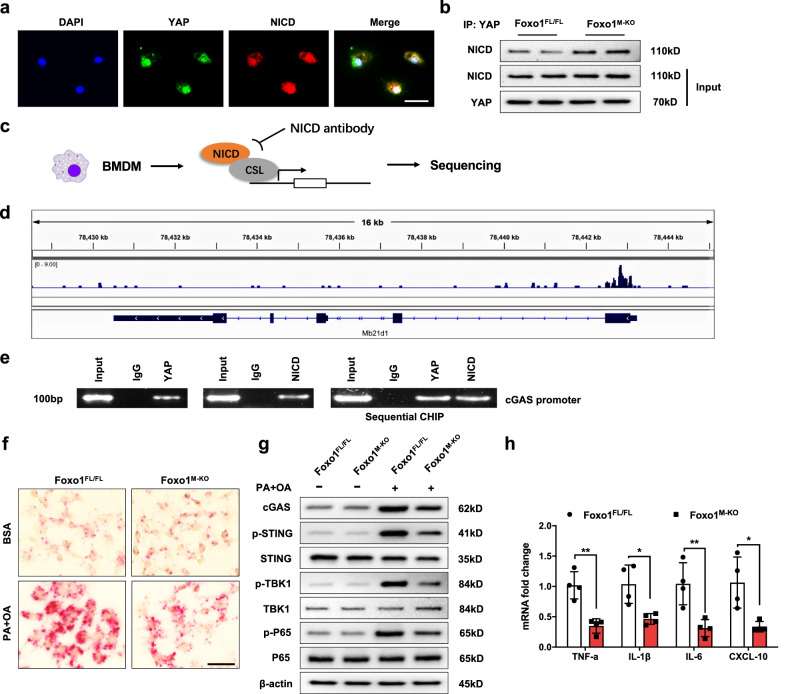
Innate immune activation is critical for initiating hepatic inflammation during nonalcoholic steatohepatitis (NASH) progression. However, the mechanisms by which immunoregulatory molecules recognize lipogenic, fibrotic, and inflammatory signals remain unclear. Here, we show that high-fat diet (HFD)-induced oxidative stress activates Foxo1, YAP, and Notch1 signaling in hepatic macrophages. Macrophage Foxo1 deficiency (Foxo1M-KO) ameliorated hepatic inflammation, steatosis, and fibrosis, with reduced STING, TBK1, and NF-κB activation in HFD-challenged livers. However, Foxo1 and YAP double knockout (Foxo1/YAPM-DKO) or Foxo1 and Notch1 double knockout (Foxo1/Notch1M-DKO) promoted STING function and exacerbated HFD-induced liver injury. Interestingly, Foxo1M-KO strongly reduced TGF-β1 release from palmitic acid (PA)- and oleic acid (OA)-stimulated Kupffer cells and decreased Col1α1, CCL2, and Timp1 expression but increased MMP1 expression in primary hepatic stellate cells (HSCs) after coculture with Kupffer cells. Notably, PA and OA challenge in Kupffer cells augmented LIMD1 and LATS1 colocalization and interaction, which induced YAP nuclear translocation. Foxo1M-KO activated PGC-1α and increased nuclear YAP activity, modulating mitochondrial biogenesis. Using chromatin immunoprecipitation (ChIP) coupled with massively parallel sequencing (ChIP-Seq) and in situ RNA hybridization, we found that NICD colocalizes with YAP and targets Mb21d1 (cGAS), while YAP functions as a novel coactivator of the NICD, which is crucial for reprogramming STING function in NASH progression. These findings highlight the importance of the macrophage Foxo1-YAP-Notch1 axis as a key molecular regulator that controls lipid metabolism, inflammation, and innate immunity in NASH.


Conflict of interest statement

The authors declare no competing interests.

Figures

Fig. 1
Fig. 1. Macrophage Foxo1 deficiency reduces hepatic steatosis and inflammation in HFD-induced NASH.
a The protein expression of nuclear Foxo1, p-JNK, JNK, p-P65, and P65 was upregulated in liver macrophages from wild-type (WT) mice after they were fed a high-fat diet (HFD) for 24 weeks, as determined by western blot analysis. The data are representative of three experiments. Notes: After 12 weeks of HFD feeding, nuclear Foxo1 expression was substantially increased in Kupffer cells. b Immunofluorescence staining showed that macrophage Foxo1 expression was increased in steatotic livers (n = 6 mice/group). Scale bars, 100 μm, and 30 μm. c The Foxo1M-KO mice displayed lower liver-to-body weight ratios (n = 6 samples/group). d The levels of liver TG and TC (mg/g) in the Foxo1M-KO livers were reduced (n = 6 samples/group). e Representative histological staining (H&E and Oil Red O) revealed that Foxo1M-KO alleviated hepatic steatosis and reduced hepatocyte ballooning and lipid accumulation in the liver (n = 6 mice/group). Scale bars, 100 μm. f The NAS (NAFLD activity score) based on histological images was measured and found to be significantly decreased in the Foxo1M-KO group (n = 6 mice/group). g Serum ALT and AST levels were decreased in the HFD-fed Foxo1M-KO mice (IU/L) (n = 6 samples/group). h Quantitative PCR revealed that the levels of Srebp1c, Slc27a1, Fabp1, CD36, Fas, and Accα were significantly reduced, and the expression of Cpt1α, Acox1, and Acadm was increased in steatotic livers (n = 6 samples/group). Notes: Foxo1M-KO significantly reduced the mRNA levels of genes responsible for fatty acid uptake and synthesis and increased fatty acid β-oxidation gene expression. All the data are presented as the mean±SD. Statistical analysis was performed using the permutation t test. *P < 0.05, **P < 0.01, ***P < 0.001, ****P < 0.0001.
Fig. 2
Fig. 2. Macrophage Foxo1 deficiency inhibits STING-mediated liver inflammation and fibrosis in HFD-induced NASH.
a The expression of p-STING, p-TBK1, and p-P65 was diminished in the livers of the Foxo1M-KO mice after 24 weeks of HFD feeding. Notes: Foxo1M-KO diminished p-STING, p-TBK1, and p-P65 expression in the HFD-challenged livers. b Immunofluorescence staining showed that CD11b+ macrophages were decreased in the livers of the Foxo1M-KO mice after 24 weeks of HFD feeding (n = 6 mice/group). Quantification of CD11b+ macrophages; scale bars, 100 μm. c Quantitative RT‒PCR revealed that TNF-α, IL-1β, IL-6, and CXCL-10 levels were reduced in the Foxo1M-KO mice after 24 weeks of HFD feeding, and IL-10 levels were increased in the Foxo1M-KO mice (n = 6 samples/group). d Representative histological and immunohistochemical staining (Sirius Red, Masson, and α-SMA) showing reduced liver fibrosis in the livers of the HFD-challenged Foxo1M-KO mice (n = 6 mice/group). Scale bars, 100 μm. e Quantitative RT‒PCR revealed that the expression of α-SMA, Col1a1, CCL2, and Timp1 was reduced in steatotic Foxo1M-KO livers (n = 6 samples/group). f Hepatic Kupffer cells were stimulated with a mixture of 0.2 mM palmitic acid (PA) and 0.4 mM oleic acid (OA) for 24 h and then cocultured with primary hepatic stellate cells (HSCs). Compared with Foxo1FL/FL, Foxo1M-KO markedly reduced TGF-β1 release from PA/OA-stimulated macrophages in the coculture supernatant (n = 4 samples/group). g Reduced HSC levels of the mRNAs encoding Col1α1, CCL2, Timp1, and augmented matrix metalloproteinase 1 (MMP1) after coculture were found (n = 4 samples/group). Notes: Foxo1M-KO inhibits the mRNA expression of fibrogenic genes and reduces liver fibrosis in HFD-induced NASH. All the data are presented as the mean±SD. Statistical analysis was performed using the permutation t test. *P < 0.05, **P < 0.01, ***P < 0.001.
Fig. 3
Fig. 3. Macrophage Foxo1 deficiency promotes the Notch1 and Hippo signaling pathways in response to HFD challenge.
Total RNA was extracted from liver macrophages from the Foxo1FL/FL and Foxo1M-KO mice after 24 weeks of HFD feeding. Deep RNA sequencing (RNA-seq) was subsequently performed. A modified Fisher’s exact test (enrichment score) was used for the functional enrichment analyses. Significantly upregulated or downregulated genes were determined by an adjusted P value of less than 0.05, which was set as the threshold to define DEGs, KEGG, and GO analysis. a The log2-fold changes in gene expression in the HFD-challenged liver macrophages from the Foxo1M-KO mice compared to the Foxo1FL/FL controls. Differentially expressed genes (DEGs) (n = 912, P < 0.05) in the HFD-challenged liver macrophages are indicated (red, upregulated, n = 415; green, downregulated, n = 497). b, c Gene Ontology (GO) enrichment analysis of cellular components and biological processes in the HFD-challenged liver macrophages from the Foxo1M-KO mice. d Kyoto Encyclopedia of Genes and Genomes (KEGG) pathway enrichment analysis of transcripts differentially expressed in the HFD-challenged liver macrophages from the Foxo1M-KO mice. e Heatmap showing the genes whose expression changed in the HFD-challenged liver macrophages from the Foxo1M-KO mice. Notes: RNA-seq analysis revealed that the Foxo1M-KO promotes the expression of genes related to the Notch1 and Hippo signaling pathways in the HFD-challenged macrophages.
Fig. 4
Fig. 4. Macrophage Foxo1 deficiency increases YAP/NICD activity and inhibits STING activation in HFD-induced oxidative stress.
a Nuclear YAP and NICD expression were substantially increased in macrophages after HFD feeding. The data are representative of three experiments. b Liver macrophages were isolated from WT mice and stimulated with a mixture of 0.2 mM palmitic acid (PA) and 0.4 mM oleic acid (OA) for 24 h. PA/OA stimulation activated JNK and increased nuclear Foxo1 and PGC-1α expression in macrophages. c PA and OA stimulation increased the expression of p-LATS1 and LIMD1, leading to reduced cytoplasmic YAP phosphorylation and increased nuclear YAP expression in macrophages. d Immunofluorescence staining showing macrophage LIMD1 (green) and LATS1 (red) colocalization in PA/OA-stimulated macrophages. DAPI was used to visualize nuclei (blue). Scale bars, 30 μm. e Immunoprecipitation analysis showed that PA/OA challenge augmented the colocalization and interaction of LIMD1 and LATS1 in macrophages. f Liver macrophages from WT mice were transfected with CRISPR/Cas9-mediated LIMD1 KO or control vector after PA/OA challenge. Moreover, LIMD1 KO increased cytoplasmic YAP phosphorylation and reduced nuclear YAP expression. g Disruption of macrophage Foxo1 markedly increased PGC-1α, YAP, and NICD levels and reduced p-STING expression in response to PA/OA challenge. h Liver macrophages from Foxo1M-KO mice were transfected with CRISPR/Cas9-mediated PGC-1α KO or control vector after PA/OA stimulation. Immunoprecipitation analysis revealed that CRISPR/Cas9-mediated PGC-1α KO in the Foxo1M-KO cells reduced the interaction of YAP with the NICD and augmented p-STING expression. Notes: Foxo1M-KO activates YAP/NICD and inhibits STING activation in PA/OA-stimulated macrophages. All Western blots represent three experiments, and the data are presented as the mean±SD.
Fig. 5
Fig. 5. The YAP–NICD interaction targets cGAS and modulates STING-mediated inflammation.
a Immunofluorescence staining showing the colocalization of YAP (green) and NICD (red) in the nuclei of macrophages after PA/OA stimulation. Scale bars, 30 μm. b Immunoprecipitation analysis showed that disruption of Foxo1 increased YAP binding to the NICD in Foxo1M-KO but not in Foxo1FL/FL macrophages. The data are representative of three experiments. c Experimental design of the NICD ChIP-seq analysis. BMMs were collected and fixed after culture with PA/OA for 24 h. Following chromatin shearing and NICD antibody selection, the precipitated DNA fragments bound by NICD-containing protein complexes were subjected to sequencing. d Localization of NICD-binding sites on the mouse Mb21d1 (cGAS) gene. Five exons, 4 introns, the 3’ untranslated region (UTR), the 5’ UTR, and the transcription start site (TSS) of the mouse cGAS gene on chromosome 9 are shown. e ChIP‒PCR analysis of NICD and YAP binding to the cGAS promoter. Protein-bound chromatin was prepared from BMMs and immunoprecipitated with NICD or YAP antibodies. For sequential ChIP, the protein-bound chromatin was first immunoprecipitated with the NICD antibody, followed by elution with a second immunoprecipitation using the YAP antibody. Then, the immunoprecipitated DNA was analyzed by PCR. Normal IgG was used as a negative control. f RNA in situ hybridization assays showed that the transcript expression of the target gene cGAS was increased in the Foxo1FL/FL macrophages after PA/OA stimulation (n = 4 samples/group). Scale bars, 50 μm. g Fatty acid stimulation increased cGAS, p-STING, p-TBK1, and p-P65 expression in the PA-stimulated Foxo1FL/FL macrophages from the Foxo1FL/FL or Foxo1M-KO mice, whereas Foxo1 deletion diminished cGAS, p-STING, p-TBK1, and p-P65 expression. h qRT‒PCR analysis showed that TNF‒α, IL‒1β, IL‒6, and CXCL‒10 levels were decreased in the macrophages exposed to PA/OA (n = 4 samples/group). Notes: ChIP-seq analysis revealed that the YAP–NICD axis targets cGAS and regulates the STING-mediated inflammatory response in response to PA/OA stimulation. All western blots represent three experiments, and the data are presented as the mean±SD. Statistical analysis was performed using the permutation t test. *P < 0.05, **P < 0.01.
Fig. 6
Fig. 6. Disruption of macrophage Notch1 signaling activates cGAS and increases STING-mediated liver inflammation and fibrosis in HFD-induced NASH.
a Foxo1/Notch1M-DKO increased cGAS, p-STING, p-TBK1, p-P65, and nuclear PGC-1α, LIMD1, and YAP expression in steatotic livers after 24 weeks of HFD feeding. b Immunofluorescence staining showed that Foxo1/Notch1M-DKO increased CD11b+ macrophage accumulation in steatotic livers (n = 6 mice/group). Quantification of CD11b+ macrophages; scale bars, 100 μm. c Foxo1/Notch1M-DKO increased TNF-α, IL-1β, IL-6, and CXCL-10 expression and decreased IL-10 levels in steatotic livers (n = 6 samples/group). d The liver/body weight ratio was significantly increased in the HFD-fed Foxo1/Notch1M-DKO mice (n = 6 samples/group). e TG and TC (mg/g) lipid levels were significantly increased in the HFD-fed Foxo1/Notch1M-DKO mice (n = 6 samples/group). f Representative histological staining (H&E and Oil Red O) showing that the livers from the HFD-fed Foxo1/Notch1M-DKO mice exhibited increased lipid accumulation (n = 6 mice/group). Scale bars, 100 μm. g NASs (NAFLD activity scores) were measured based on histological images and were significantly increased in the Foxo1/Notch1M-DKO group (n = 6 mice/group). h Serum ALT and AST levels were increased in the HFD-fed Foxo1/Notch1M-DKO mice (IU/L) (n = 6 samples/group). i Representative histological and immunohistochemical staining (Sirius Red and Masson) of steatotic liver tissues showing augmented liver fibrosis in the Foxo1/Notch1M-DKO mice (n = 6 mice/group). Scale bars, 100 μm. j Increased mRNA expression of profibrotic genes, including αSMA, Col1α1, TGF-β1, CCL2, and TIMP1, in the Foxo1/Notch1M-DKO livers after HFD feeding (n = 6 samples/group). Notes: Foxo1/Notch1M-DKO activates cGAS, increases the STING-mediated inflammatory response, and exacerbates liver fibrosis in HFD-induced NASH. All the data are presented as the mean ± SD. Statistical analysis was performed using the permutation t test. *P < 0.05, **P < 0.01, ***P < 0.001, ****P < 0.0001.
Fig. 7
Fig. 7. YAP is required for macrophage Foxo1-mediated immune regulation of STING function in lipotoxicity-induced mitochondrial oxidative stress.
BMMs were isolated from Foxo1M-KO mice, transfected with CRISPR/Cas9-mediated YAP knockout (p-CRISPR-YAP KO) or control vector, and then cocultured with primary hepatocytes after incubation with a 0.2 mM palmitic acid (PA) and 0.4 mM oleic acid (OA) mixture for 24 h. a CRISPR/Cas9-mediated YAP knockout augmented the expression of cGAS, p-STING, p-TBK1, and p-P65 in the PA-stimulated macrophages. The data are representative of three experiments. b The mRNA levels of IL-6, TNF-α, CCL2, and IL-β were elevated in the PA- and OA-stimulated Foxo1M-KO macrophages. c ELISA analysis revealed that HMGB1 release was markedly increased in the p-CRISPR-YAP-KO cells but not in the control cells (n = 4 samples/group). d Immunofluorescence staining for ROS production showed that p-CRISPR-YAP KO in Foxo1M-KO macrophages increased ROS production in hepatocytes after coculture and exposure to PA/OA (n = 4 samples/group). Quantification of ROS-producing macrophages (green). Scale bars, 100 μm. e The expression of TFAM, COX-1, and UCP3 was diminished in hepatocytes after coculture with p-CRISPR-YAP KO-transfected Foxo1M-KO macrophages. The data are representative of three experiments. f Quantitative RT‒PCR analysis revealed that p-CRISPR-YAP KO reduced mtDNA levels in hepatocytes after coculture with p-CRISPR-YAP KO or control vector-transfected macrophages (n = 4 samples/group). Notes: YAP deletion in Foxo1M-KO macrophages increased ROS production and reduced TFAM, Cox-1, UCP3, and mtDNA levels related to mitochondrial biogenesis in hepatocytes after coculture following PA/OA challenge. g Oil Red O staining revealed increased intracellular lipids in hepatocytes after coculture with the p-CRISPR-YAP-KO or control vector-transfected macrophages (n = 4 samples/group). Scale bars, 100 μm. All the data are presented as the mean±SD. Statistical analysis was performed using the permutation t test. *P < 0.05, **P< 0.01, ***P < 0.001.
Fig. 8
Fig. 8. The Foxo1–YAP axis modulates STING-mediated liver inflammation and steatosis in HFD-induced NASH.
a Representative histological staining (H&E and Oil Red O) showing that the Foxo1M-KO mice exhibited decreased lipid accumulation, whereas the Foxo1/YAPM-DKO mice exhibited increased hepatic steatosis after 24 weeks of HFD feeding (n = 6 mice/group). Scale bars, 100 μm. b The NAS (NAFLD activity score) based on histological images was measured and found to be significantly increased in the Foxo1/YAPM-DKO group (n = 6 mice/group). c The liver/body weight ratios were significantly greater in the Foxo1/YAPM-DKO mice (n = 6 samples/group). d The TG and TC levels (mg/g) were significantly increased in the Foxo1/YAPM-DKO mice (n = 6 samples/group). e The Foxo1/YAPM-DKO mice exhibited significantly increased serum ALT and AST levels (IU/L) (n = 6 samples/group). f Representative immunofluorescence and immunohistochemistry images (α-SMA and Masson) showing significantly increased liver fibrosis in the Foxo1/YAPM-DKO livers (n = 6 mice/group). Scale bars, 100 μm. g Quantitative RT‒PCR analysis showed that Foxo1/YAPM-DKO increased cGAS, p-STING, p-TBK1, and p-P65 expression in steatotic livers (n = 6 samples/group). h Western blot analysis revealed that Foxo1/YAPM-DKO increased cGAS, p-STING, p-TBK1, and p-P65 expression and increased nuclear PGC-1α, LIMD1, and NICD expression in steatotic livers. The data are representative of three experiments. i Immunofluorescence staining showed increased CD11b+ macrophage accumulation in ischemic livers (n = 6 mice/group). Quantification of CD11b+ macrophages; scale bars, 100 μm. j The mRNA levels of TNF-α, IL-1β, IL-6, and CXCL-10 were increased, and the IL-10 level was decreased in the steatotic Foxo1/YAPM-DKO livers (n = 6 samples/group). Notes: Foxo1/YAPM-DKO exacerbates STING-mediated liver inflammation, steatosis, and fibrosis in mice with HFD-induced NASH. All the data are presented as the mean±SD. Statistical analysis was performed using the permutation t test. *P < 0.05, **P < 0.01, ***P < 0.001.

References

    1. Fuchs, M. & Sanyal, A. J. Lipotoxicity in NASH. J. Hepatol.56, 291–293 (2012). 10.1016/j.jhep.2011.05.019 - DOI - PubMed
    1. Baffy, G. Kupffer cells in non-alcoholic fatty liver disease: the emerging view. J. Hepatol.51, 212–223 (2009). 10.1016/j.jhep.2009.03.008 - DOI - PMC - PubMed
    1. West, A. P. & Shadel, G. S. Mitochondrial DNA in innate immune responses and inflammatory pathology. Nat. Rev. Immunol.17, 363–375 (2017). 10.1038/nri.2017.21 - DOI - PMC - PubMed
    1. Alisi, A., Carsetti, R. & Nobili, V. Pathogen- or damage-associated molecular patterns during nonalcoholic fatty liver disease development. Hepatology54, 1500–1502 (2011). 10.1002/hep.24611 - DOI - PubMed
    1. Petrasek, J. et al. STING-IRF3 pathway links endoplasmic reticulum stress with hepatocyte apoptosis in early alcoholic liver disease. Proc. Natl. Acad. Sci. USA110, 16544–16549 (2013). 10.1073/pnas.1308331110 - DOI - PMC - PubMed

MeSH terms