. 2024 Aug 9;7(1):967.
doi: 10.1038/s42003-024-06671-y.

Mitochondrial permeability transition dictates mitochondrial maturation upon switch in cellular identity of hematopoietic precursors

Affiliations

Mitochondrial permeability transition dictates mitochondrial maturation upon switch in cellular identity of hematopoietic precursors

Sandeep P Dumbali et al. Commun Biol. .

Abstract

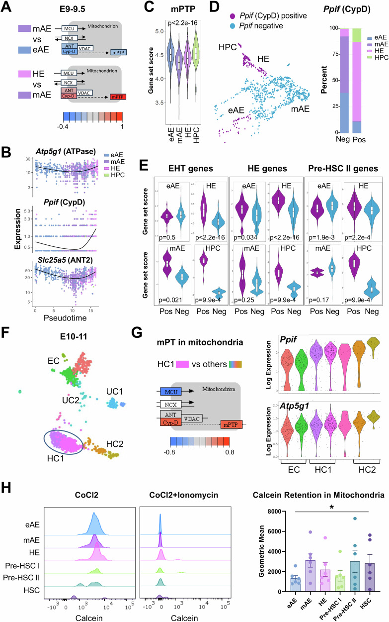
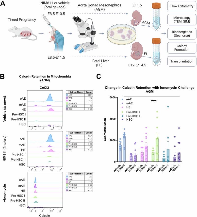
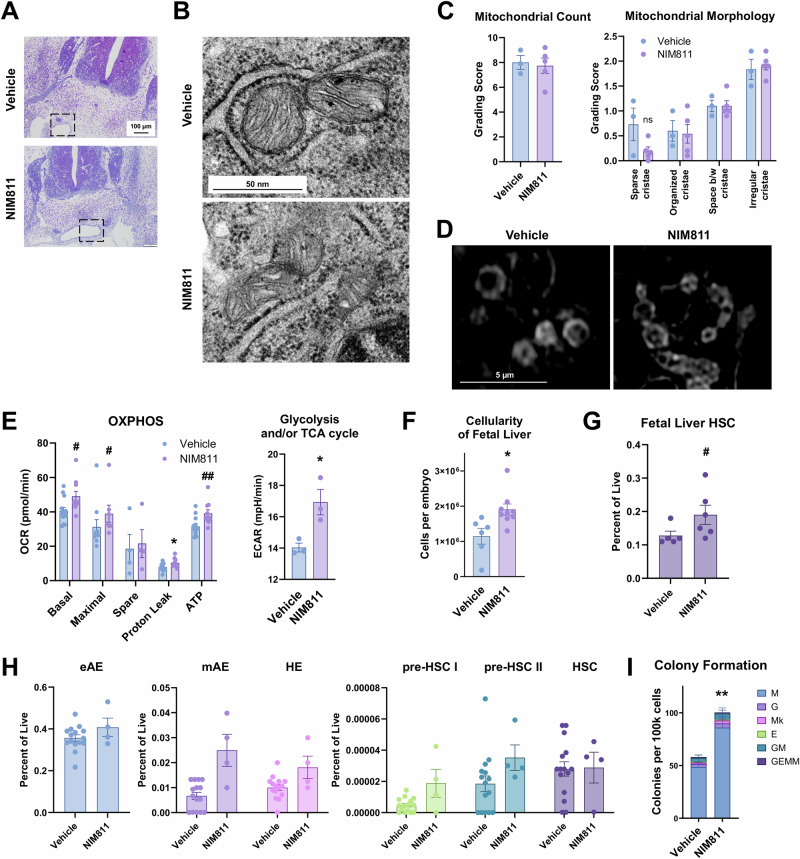
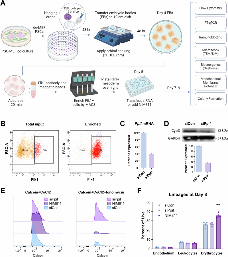
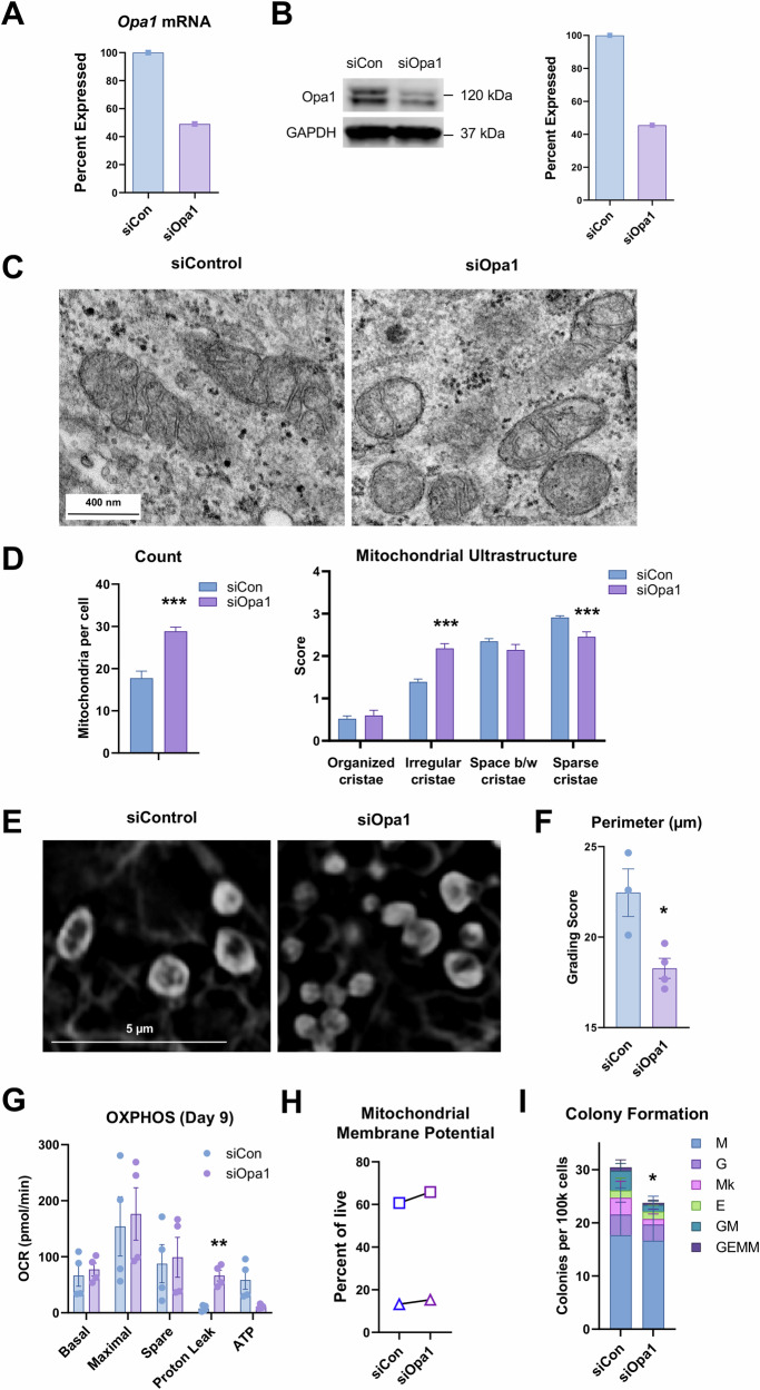
The mitochondrial permeability transition pore (mPTP) is a supramolecular channel that regulates exchange of solutes across cristae membranes, with executive roles in mitochondrial function and cell death. The contribution of the mPTP to normal physiology remains debated, although evidence implicates the mPTP in mitochondrial inner membrane remodeling in differentiating progenitor cells. Here, we demonstrate that strict control over mPTP conductance shapes metabolic machinery as cells transit toward hematopoietic identity. Cells undergoing the endothelial-to-hematopoietic transition (EHT) tightly control chief regulatory elements of the mPTP. During EHT, maturing arterial endothelium restricts mPTP activity just prior to hematopoietic commitment. After transition in cellular identity, mPTP conductance is restored. In utero treatment with NIM811, a molecule that blocks sensitization of the mPTP to opening by Cyclophilin D (CypD), amplifies oxidative phosphorylation (OXPHOS) in hematopoietic precursors and increases hematopoiesis in the embryo. Additionally, differentiating pluripotent stem cells (PSCs) acquire greater organization of mitochondrial cristae and hematopoietic activity following knockdown of the CypD gene, Ppif. Conversely, knockdown of Opa1, a GTPase critical for proper cristae architecture, induces cristae irregularity and impairs hematopoiesis. These data elucidate a mechanism that regulates mitochondrial maturation in hematopoietic precursors and underscore a role for the mPTP in the acquisition of hematopoietic fate.


Conflict of interest statement

The authors declare no competing interests.

Figures

Fig. 1
Fig. 1. Mitochondrial permeability transition pore (mPTP) factors are down-regulated as arterial endothelium matures and subsequently up-regulated with acquisition of hematopoietic fate in HE and pre-HSCs.
A mPTP and other channels involved in mitochondrial membrane permeability are depicted from a KEGG pathway analysis of differentially expressed genes from E9.0-E9.5 scRNA-seq data, where red-blue scale is based on log2 fold change and white denotes no change. Cells were classified as early arterial endothelium (eAE), mature arterial endothelium (mAE), hemogenic endothelium (HE), or hematopoietic progenitor cells (HPC) based upon a classifier established in a recent publication, and differential gene expression between cell populations was calculated. Cell numbers included eAE = 314, mAE = 417, HE = 178, and HPC = 24. B Biphasic regulation of transcripts required for the mPTP is seen as cells transit over a developmental trajectory from endothelial to hematopoietic identity. C A score for genes required for mPTP was calculated as reported previously. Kruskal-Wallis, *p < 2.2e-16. D Clusters were classified as positive if >50% of cells expressed Ppif (gene encoding CypD). Ppif is lowest in mAE just prior to differentiation into HE. E Ppif-positive clusters are enriched for genes that define EHT, HE, and pre-HSCs, using published gene sets,. Wilcoxon, p-values are depicted on each plot. F Independent scRNA-seq dataset of 968 E10-11 AGM cells shows a cluster containing all HSC activity (pink, defined as HC1 in the original report). EC endothelial cells, HC1 HSC-enriched, HC2 mature HSPCs, UC1,2 unknown cluster 1 and 2. G HC1 has a high expression of Ppif relative to all other stages of differentiation and appears elevated in HC2 (mature HSPCs). Red-blue scale in KEGG pathway is based on log2 fold change, where white denotes no change. H Dye efflux measurements at E11.5 show reduced mitochondrial inner membrane permeability in mAE and elevated permeability in eAE, HE, and pre-HSCs. Representative plots are shown of calcein intensity with and without ionomycin. Ionomycin triggers opening of the mPTP and quenching of calcein fluorescence by cobalt chloride (CoCl2). High mean intensity indicates more dye retention and less mPTP opening. Error bars are shown as mean ± SEM from N = 6; six independent experiments where at least 5 embryos were pooled for each sample per experiment. One-way ANOVA, *p = 0.04.
Fig. 2
Fig. 2. In utero administration of NIM811 promotes mPTP closure in EHT populations.
A Overview of NIM811-mediated mPTP inhibition in developing AGM and fetal liver. Created with BioRender. B Representative flow cytometry plots show that calcein retention is elevated by NIM811. C AGM cells treated with NIM811 in utero undergo greater change in calcein intensity following in vitro stimulation with ionomycin. Two-way ANOVA, Holm-Sidak method, ***p < 0.001 for NIM811 vs. vehicle, N = 8 vehicle, N = 10 NIM811.
Fig. 3
Fig. 3. Inhibition of mPTP induces metabolic reprogramming during EHT to elevate hematopoietic progenitor activity.
A Sections of E11.5 AGM were prepared to verify location of dorsal aorta. Dashed boxes represent areas where luminal cells were examined by TEM. B TEM images show mitochondrial ultrastructure and abundance in arterial endothelium lining the dorsal aorta. Cells depicted line the aorta lumen. C Blinded scoring of mitochondrial count and morphology by pathologist. Error bars are shown as mean ± SEM from N = 3 vehicle, N = 5 NIM811; each data point represents an individual embryo from different litters. D SIM images of mitochondria within AGM cells isolated from E11.5 embryos. E Seahorse assays reveal elevated oxygen consumption rate (OCR) and extracellular acidification rate (ECAR) indicative of an increased bioenergetic state after NIM811 in AGM cells. Error bars are shown as mean ± SEM from N ≥ 4 for OCR and N = 3 for ECAR; at least three independent experiments are shown representing >5 pooled embryos as data points. T-test, *p < 0.05; Mann–Whitney Rank Sum, #p < 0.05, ##p < 0.01. F Cellularity of the fetal liver at E14.5 is increased with NIM811. Error bars are shown as mean ± SEM from N = 6 vehicle, N = 9 NIM811; at least six independent experiments are shown. T-test, *p < 0.05. G HSC frequency in fetal liver is increased by NIM811. Error bars are shown as mean ± SEM from N = 5 vehicle, N = 6 NIM811. Mann–Whitney Rank Sum, #p < 0.05. H EHT population frequencies in the AGM are not significantly altered by NIM811. Error bars are shown as mean ± SEM from N = 15 vehicle, N = 4 NIM811. I CFU activity in E11.5 AGM is elevated by NIM811. Error bars are shown as mean ± SEM from N = 4; independent experiments with at least 5 pooled embryos in each experiment. T-test, **p = 0.003, N = 4.
Fig. 4
Fig. 4. NIM811 increases stem and progenitor cell reconstitution in adult transplant recipients.
A Peripheral blood donor chimerism from transplanted fetal liver is unaltered. Error bars are shown as mean ± SEM from N = 4 vehicle recipients, N = 3 NIM811 recipients. Each data point represents one recipient transplanted with a small portion (3%) of 3 pooled fetal livers from embryos different from those transplanted into the other recipients. T-test, n.s. B Six weeks after transplant, chimerism in bone marrow is not significantly altered by NIM811. C Progenitors are significantly increased in recipients of NIM811 treated fetal liver. Two-way ANOVA, Holm-Sidak method, *p = 0.02 for NIM811 vs. vehicle. D Long-term HSCs are expanded following engraftment of NIM811 fetal liver. T-test, *p = 0.04.
Fig. 5
Fig. 5. Transient knockdown of Ppif gene encoding the CypD mPTP regulatory factor in mesoderm decreases mitochondrial permeability.
A Process of directed differentiation and enrichment of Flk1+ mesoderm. Flk1+ cells from embryoid bodies (EBs) received siRNAs or NIM811 at day 5 of differentiation and were transferred to assays between day 7 and 9. Created with BioRender. B Purity check of Flk1+ cells before and after magnetic bead enrichment. C Efficiency of Ppif knockdown measured by RT-qPCR. Error bars are shown as mean ± SEM from N = 3 independent knockdown experiments. D Western blot for CypD (encoded by Ppif) shows approximately 75% knockdown at the protein level, normalized to GAPDH. Error bars are shown as mean ± SEM from N = 3 independent knockdown experiments. E Ppif knockdown decreases mitochondrial permeability, most notably in the context of ionomycin-induced calcium overload and is more protective than NIM811. Conversely, NIM811 reduces mPTP opening at baseline but is unable to fully block mPT induced by ionomycin challenge. F Erythroid cells are increased by NIM811 at day 8 of differentiation. Two-way ANOVA, Holm-Sidak method, **p = 0.004 for NIM811 vs. siCon and siPpif.
Fig. 6
Fig. 6. Ppif knockdown alters mitochondrial ultrastructure and enhances hematopoietic activity from PSCs.
A Cristae was visualized by TEM on day 9. B Pathology scoring shows a reduction in irregular cristae and spaces between cristae by siPpif and NIM811 treatments. Kruskal-Wallis One Way ANOVA on Ranks, Dunn’s posthoc method, *p < 0.05, **p < 0.01, ***p < 0.001. C Analysis by SIM corroborates TEM observations. D Ppif knockdown increased frequency of detectable cristae in mitochondria. Error bars are shown as mean ± SEM from N = 3 siCon, N = 6 siPpif. Three independent knockdown experiments. Fisher’s exact test, *p = 0.01. E OXPHOS was increased at day 9 by transient knockdown of Ppif. Error bars are shown as mean ± SEM from N = 3 independent experiments. T-test, *p < 0.05, #p < 0.10. F No difference was observed in mitochondrial membrane potential using TMRM dye in pairwise analyses of two different cell lines. G Hematopoietic activity in colony formation assays trended upward with Ppif knockdown. Error bars are shown as mean ± SEM from N = 3; Three independent knockdown experiments. Wilcoxon signed rank, #p = 0.09.
Fig. 7
Fig. 7. Transient knockdown of the Opa1 cristae remodeling factor attenuates mitochondrial capacity and hematopoietic activity.
Flk1+ mesoderm enriched from EBs received siRNAs at day 5 of differentiation and were transferred to assays at day 9. A RT-qPCR shows knockdown of Opa1 mRNA. Error bars are shown as mean ± SEM from N = 3 independent knockdown experiments. B Western blot for Opa1 indicates knockdown of protein to approximately 40% of control cells, normalized to GAPDH. Error bars are shown as mean ± SEM from N = 3 independent knockdown experiments. C TEM images of mitochondria from control and Opa1 knockdown mesoderm (D) TEM scoring indicates increase in numbers of mitochondria and irregular cristae by siOpa1. Mann–Whitney Rank Sum Test, ***p < 0.001. E SIM of mitochondria from independently differentiated mesoderm. F Perimeter of mitochondria was reduced by Opa1 knockdown, suggesting that transient loss of Opa1 results in smaller mitochondria. Error bars are shown as mean ± SEM from N = 3 siCon, N = 4 siOpa1. Three independent knockdown experiments. T-test, *p = 0.02. G Opa1 knockdown did not dramatically alter respiration but did elevate proton leak at day 9. Error bars are shown as mean ± SEM from N = 4 independent knockdown experiments. T-test, **p = 0.006. H Mitochondrial membrane potential as measured by TMRM was unchanged by Opa1 knockdown in pairwise analyses of two different cell lines. I Hematopoietic colony formation activity trended downward with transient knockdown of Opa1. Error bars are shown as mean ± SEM from N = 3 independent knockdown experiments. T-test, *p < 0.05.

References

    1. Orkin, S. H. & Zon, L. I. Hematopoiesis: an evolving paradigm for stem cell biology. Cell132, 631–644 (2008). 10.1016/j.cell.2008.01.025 - DOI - PMC - PubMed
    1. Dzierzak, E. & Bigas, A. Blood development: hematopoietic stem cell dependence and independence. Cell Stem Cell22, 639–651 (2018). 10.1016/j.stem.2018.04.015 - DOI - PubMed
    1. Nunnari, J. & Suomalainen, A. Mitochondria: in sickness and in health. Cell148, 1145 (2012). 10.1016/j.cell.2012.02.035 - DOI - PMC - PubMed
    1. Hinge, A. et al. Asymmetrically segregated mitochondria provide cellular memory of hematopoietic stem cell replicative history and drive HSC attrition. Cell Stem Cell26, 420–430.e6 (2020). 10.1016/j.stem.2020.01.016 - DOI - PMC - PubMed
    1. Simsek, T. et al. The distinct metabolic profile of hematopoietic stem cells reflects their location in a hypoxic niche. Cell Stem Cell7, 380–390 (2010). 10.1016/j.stem.2010.07.011 - DOI - PMC - PubMed

Publication types

MeSH terms

Substances

LinkOut - more resources