. 2024 Aug 9;73(10):210.
doi: 10.1007/s00262-024-03794-3.

Preclinical development of a novel CCR8/CTLA-4 bispecific antibody for cancer treatment by disrupting CTLA-4 signaling on CD8 T cells and specifically depleting tumor-resident Tregs

Affiliations

Preclinical development of a novel CCR8/CTLA-4 bispecific antibody for cancer treatment by disrupting CTLA-4 signaling on CD8 T cells and specifically depleting tumor-resident Tregs

Cuicui Guo et al. Cancer Immunol Immunother. .

Abstract

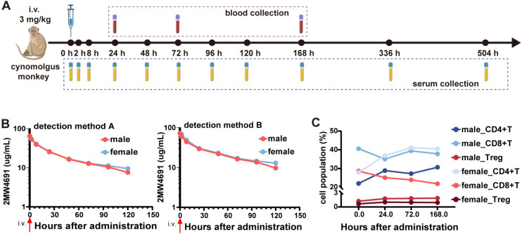
Anti-CTLA-4 antibodies faced challenges due to frequent adverse events and limited efficacy, which spurred the exploration of next-generation CTLA-4 therapeutics to balance regulatory T cells (Tregs) depletion and CD8 T cells activation. CCR8, identified primarily on tumor-infiltrating Tregs, has become a target of interest due to the anti-tumor effects demonstrated by CCR8 antibody-mediated Tregs depletion. Single-cell RNA sequencing analysis reveals that CCR8-positive Tregs constitute a small subset, with concurrent expression of CCR8 and CTLA-4. Consequently, we proposed a novel bispecific antibody targeting CCR8 and CTLA-4 that had the potential to enhance T cell activation while selectively depleting intratumor Tregs. The candidate molecule 2MW4691 was developed in a tetravalent symmetric format, maintaining a strong binding affinity for CCR8 while exhibiting relatively weaker CTLA-4 binding. This selective binding ability allowed 2MW4691 to target and deplete tumor-infiltrating Tregs with higher specificity. In vitro assays verified the antibody's capacity for antibody-dependent cellular cytotoxicity (ADCC) to Tregs with high level of CTLA-4 expression, but not CD8 T cells with relatively low level of CTLA-4 on cell surface. Also, 2MW4691 inhibited the CTLA-4 pathway and enhanced T cell activation. The in vivo therapeutic efficacy of 2MW4691 was further demonstrated using hCCR8 or hCTLA-4 humanized mouse models and hCCR8/hCTLA-4 double knock-in mouse models. In cynomolgus monkeys, 2MW4691 was well-tolerated, exhibited the anticipated pharmacokinetic profile, and had a minimal impact on the peripheral T cell population. The promising preclinical results supported the further evaluation of 2MW4691 as a next-generation Treg-based therapeutics in clinical trials.

Keywords: ADCC; Bispecific antibody; Immunotherapy; Treg depletion; Tumor inhibition.


Conflict of interest statement

Cuicui Guo, Xiaodong Dai, Xiumei Xiong, Yulei Du, and Xun Gui are employees of Mabwell (Shanghai) Bioscience Co., Ltd. Cuicui Guo and Xun Gui may hold shares in Mabwell (Shanghai) Bioscience Co., Ltd, suggesting a financial interest in the outcomes of the research. No competing interests were declared by the other authors, indicating that beyond their employment, they do not have any additional financial stakes that could be perceived as influencing the research outcomes.

Figures

Fig. 1
Fig. 1
CCR8 and CTLA-4 expression levels in NSCLC and CRC by scRNA-seq. (A) Schematic overview of GSE139555 study design. Six NSCLC patients were included in the single-cell sequencing analysis. (B) Uniform manifold approximation and projection (UMAP) plots of NSCLC patient cells showing seven clusters. (C-E) UMAP plots show FOXP3 (C), CCR8 (D), and CTLA-4 (E) expression in NSCLC. (F) Schematic overview of the CRC scRNA-seq design. The data sources are from GSE146771. (G) UMAP plots of 10 CRC patient cells showing nine clusters. (H-J) UMAP plots show FOXP3 (H), CCR8 (I), and CTLA-4 (J) expression in CRC
Fig. 2
Fig. 2
Bispecific antibody design, construction, and screening. (A) Binding ability characterization of CCR8 antibodies was performed using Jurkat-hCCR8 stable cells, which were incubated with serially diluted antibodies and detected by FACS using APC-labeled secondary antibody. (B) ADCC activity of B9B11 was evaluated by PBMC-mediated HEK293T-hCCR8 lysis. PBMCs were incubated with HEK293T-hCCR8 cells (E:T = 15: 1) for 5 h in the presence of CCR8 antibodies, and the reactions were detected by LDH detection reagents. (C) B9B11 binding to cynoCCR8 was evaluated using Jurkat-cynoCCR8 stable cells. Serial dilutions of CCR8 antibodies were incubated with Jurkat-cynoCCR8 and detected by FACS using APC-labeled goat anti-human IgG Fc antibody. (D) The format schematic diagram of CCR8/CTLA-4 bispecific antibodies. A total of eight distinguished bispecific antibody formats were designed and constructed. (E) Comparison of bispecific antibodies binding ability to CHOK1-hCCR8. Threefold dilutions of bispecific antibodies and parent antibody were incubated with CHOK1-hCCR8 cells and detected by FACS using APC-labeled secondary antibody. (F) The binding activity measurement of bispecific antibodies to CHOK1-hCTLA-4. Serially diluted antibodies starting from 100 nM were incubated with CHOK1-hCTLA-4 cells and detected by FACS using APC-labeled secondary antibody. (G) The blocking ability of bispecific antibodies to CHOK1-hCTLA-4 /CD80. 2 μg/mL biotin-labeled hCD80 was incubated with CHOK1-hCTLA-4 cells in the present of serial dilutions of antibodies and detected by FACS using APC streptavidin. (H) The blockade of association of CHOK1-hCTLA-4 with CD86 by bispecific antibodies. CHOK1-hCTLA-4 cells were incubated with 2 μg/mL biotin-labeled hCD86, followed by the addition of serially diluted antibodies and detection by FACS using APC streptavidin
Fig. 3
Fig. 3
In vitro efficacy evaluation of 2MW4691. (A) ADCC effect of 2MW4691 on CHOK1-hCCR8 was performed based on reporter-based bioassay. Jurkat-NFAT-luc/hFcγRIIIa cells were incubated with CHOK1-hCCR8 cells (E:T = 7.5:1) in the presence of serial dilutions of antibodies for 6 h before luminescence detection. (B) ADCC effects of 2MW4691 on CHOK1-hCTLA4. Jurkat-NFAT-luc/hFcγRIIIa cells were stimulated with serially diluted antibodies in the presence of CHOK1-hCTLA-4 cells for 6 h before measuring luminescence. (C) Analysis of CTLA-4 and CCR8 expression on Tregs and CD8 T cells. ScRNA-seq data from the TNBC dataset GSE110686 were collected to analyze the expression pattern of CTLA-4 and CCR8 in the two kinds of cells. (D) RNA sequencing data from the COAD dataset DSE139555 were gathered to analyze the expression of CTLA-4 and CCR8 in Tregs and CD8 T cells. (E) Tregs isolated from PBMCs were activated by CD3/CD28 beads for 72 h and the CTLA-4 and CCR8 expression on Tregs was analyzed by FACS using PE-labeled ipilimumab and B9B11. (F) CTLA-4 and CCR8 expression on CD3/CD28-activated CD8 T cells was analyzed by FACS using PE-labeled ipilimumab and B9B11. (G) Activated Tregs were applied to the PBMC-mediated ADCC assay to evaluate the function 2MW4691. Calcein AM-stained Tregs were cocultured with PBMCs at a ratio of 1:50 in the presence of antibodies for 5 h before measuring Calcein AM release by M5e Microplate Reader. (H) PBMCs were cocultured with activated CD8 T (E:T = 50:1) in the presence of antibodies for 5 h, and cell lysis was characterized by measuring Calcein AM release. (I) The measurements of secretion of IL-2 in SEB stimulation assay. PBMCs (1 × 105/well) from two different donors were stimulated by 400 ng/mL SEB in the presence of antibodies for 96 h. Released IL-2 in the supernatant was measured by HTRF® technology
Fig. 4
Fig. 4
In vivo efficacy determination of 2MW4691. (A) Two groups (n = 7/group) of C57BL/6-hCCR8 mice, bearing MC38 tumor cells, were treated with 6.66 mg/kg 2MW4691 or 5 mg/kg isotype at a frequency of once a week. Tumor size was measured three times a week and shown as mean ± SEM. Student’s t-test was applied and P value was labeled. (B) Mice’s body weight was measured during treatment. (C) The tumor tissue was isolated from mice of the isotype_AF group on day 17 after tumor inoculation and mice of the 2MW4691 group on day 24. (D) C57BL/6-hCTLA-4 mice (n = 6/group) were treated with 1.33 mg/kg 2MW4691 or 1 mg/kg isotype control on days 6, 13, and 20 after MC38 cells inoculation. The tumor volume change was recorded three times a week and shown as mean ± SEM. Student’s t-test was applied and P value was marked. (E) The average mice body weight change of each group was recorded during treatment. (F) Resected tumor tissues were photographed when isotype_AF treated mice were sacrificed on day 21 and 2MW4691 treated mice on day 28. (G) C57BL/6-hCCR8/hCTLA-4 mice (n = 7/group) bearing MC38 cells were intraperitoneally administrated with 2MW4691 (0.4 mg/kg), B9B11_AF (0.3 mg/kg), ipilimumab (0.3 mg/kg), B9B11_AF plus ipilimumab (0.15 + 0.15 mg/kg), and isotype_AF (0.3 mg/kg), respectively. The treatments occurred on days 9, 16, 19 post-tumor transplantation. Tumor volume was measured three times a week and shown as mean ± SEM. (H) Mice’s body weight was measured and recorded during treatment. (I) Statistical analysis of tumor volumes on day 21 after tumor inoculation was determined by one-way ANOVA with Dunnett’s multiple comparisons test. (J) Mice of isotype_AF and B9B11_AF groups were sacrificed on day 21 after tumor inoculation and mice of the other groups on day 24. The tumor tissue isolated from mice was photographed. (K) 0.3 mg/kg isotype_AF, 0.4 mg/kg 2MW4691, and 0.16 mg/kg HBM4033 was intraperitoneally administrated into C57BL/6-hCCR8/hCTLA-4 mice (n = 6/group) bearing MC38 tumors twice a week for 2 weeks. Tumor size was measured three times a week and displayed as mean ± SEM. Student’s t-test was applied and P value was marked. (L) The average body weight of mice of each was recorded during treatment. (M) Tumor tissues were removed from mice of 2MW4691 and HBM4003 groups when mice were sacrificed on day 26 post-tumor inoculation. The tumors were weighed and photographed. Statistical analysis of tumor weight was determined by Student’s t-test. *P < 0.05, ** P < 0.01, **P < 0.001
Fig. 5
Fig. 5
PK and toxicity evaluation of 2MW4691. (A) Schematic illustration of the workflow of a single-dose pharmacokinetics and toxicity of 2MW4691 in NHPs. One male and one female cynomolgus monkeys were intravenously administrated 3 mg/kg 2MW4691 via intravenous infusion. Serums were collected 0, 2, 8, 24, 48, 72, 96, 120, 168, 336, and 504-h post-administration. Blood samples with anticoagulation were collected before treatment and at 24, 72, and 168 h after infusion. (B) Pharmacokinetics of 2MW4691 in NPHs after single intravenous administration. The concentration of 2MW4691 in serum was quantitated by two ELISA methods, one of which was detected using a general secondary antibody and the other using the biotin-labeled antigen. (C) Population analysis of CD4 T cells, CD8 T cells, and Tregs in peripheral blood of cynomolgus monkeys by FACS using CD45, CD3, CD4, CD8, CD25, and FOXP3 markers. The proportion change of such three kinds of cells was measured

References

    1. Robert C, Thomas L, Bondarenko I, O’Day S, Weber J, Garbe C, Lebbe C, Baurain JF, Testori A, Grob JJ, Davidson N, Richards J, Maio M, Hauschild A, Miller WH Jr, Gascon P, Lotem M, Harmankaya K, Ibrahim R, Francis S, Chen TT, Humphrey R, Hoos A, Wolchok JD (2011) Ipilimumab plus dacarbazine for previously untreated metastatic melanoma. N Engl J Med 364(26):2517–2526. 10.1056/NEJMoa1104621 10.1056/NEJMoa1104621 - DOI - PubMed
    1. Hodi FS, O’Day SJ, McDermott DF, Weber RW, Sosman JA, Haanen JB, Gonzalez R, Robert C, Schadendorf D, Hassel JC, Akerley W, van den Eertwegh AJ, Lutzky J, Lorigan P, Vaubel JM, Linette GP, Hogg D, Ottensmeier CH, Lebbé C, Peschel C, Quirt I, Clark JI, Wolchok JD, Weber JS, Tian J, Yellin MJ, Nichol GM, Hoos A, Urba WJ (2010) Improved survival with ipilimumab in patients with metastatic melanoma. N Engl J Med 363(8):711–723. 10.1056/NEJMoa1003466 10.1056/NEJMoa1003466 - DOI - PMC - PubMed
    1. Larkin J, Chiarion-Sileni V, Gonzalez R, Grob JJ, Cowey CL, Lao CD, Schadendorf D, Dummer R, Smylie M, Rutkowski P, Ferrucci PF, Hill A, Wagstaff J, Carlino MS, Haanen JB, Maio M, Marquez-Rodas I, McArthur GA, Ascierto PA, Long GV, Callahan MK, Postow MA, Grossmann K, Sznol M, Dreno B, Bastholt L, Yang A, Rollin LM, Horak C, Hodi FS, Wolchok JD (2015) Combined nivolumab and ipilimumab or monotherapy in untreated melanoma. N Engl J Med 373(1):23–34. 10.1056/NEJMoa1504030 10.1056/NEJMoa1504030 - DOI - PMC - PubMed
    1. Postow MA, Chesney J, Pavlick AC, Robert C, Grossmann K, McDermott D, Linette GP, Meyer N, Giguere JK, Agarwala SS, Shaheen M, Ernstoff MS, Minor D, Salama AK, Taylor M, Ott PA, Rollin LM, Horak C, Gagnier P, Wolchok JD, Hodi FS (2015) Nivolumab and ipilimumab versus ipilimumab in untreated melanoma. N Engl J Med 372(21):2006–2017. 10.1056/NEJMoa1414428 10.1056/NEJMoa1414428 - DOI - PMC - PubMed
    1. Weber J, Mandala M, Del Vecchio M, Gogas HJ, Arance AM, Cowey CL, Dalle S, Schenker M, Chiarion-Sileni V, Marquez-Rodas I, Grob J-J, Butler MO, Middleton MR, Maio M, Atkinson V, Queirolo P, Gonzalez R, Kudchadkar RR, Smylie M, Meyer N, Mortier L, Atkins MB, Long GV, Bhatia S, Lebbé C, Rutkowski P, Yokota K, Yamazaki N, Kim TM, de Pril V, Sabater J, Qureshi A, Larkin J, Ascierto PA (2017) Adjuvant nivolumab versus ipilimumab in resected stage III or IV melanoma. N Engl J Med 377(19):1824–1835. 10.1056/NEJMoa1709030 10.1056/NEJMoa1709030 - DOI - PubMed

LinkOut - more resources