

Effects of exposure length, cortical and trabecular bone contact areas on primary stability of infrazygomatic crest mini-screws at different insertion angles
- PMID: 39123162
- PMCID: PMC11316306
- DOI: 10.1186/s12903-024-04626-7
Effects of exposure length, cortical and trabecular bone contact areas on primary stability of infrazygomatic crest mini-screws at different insertion angles
Abstract
Background: The infrazygomatic crest mini-screw has been widely used, but the biomechanical performance of mini-screws at different insertion angles is still uncertain. The aim of this study was to analyse the primary stability of infrazygomatic crest mini-screws at different angles and to explore the effects of the exposure length (EL), screw-cortical bone contact area (SCA), and screw-trabecular bone contact area (STA) on this primary stability.
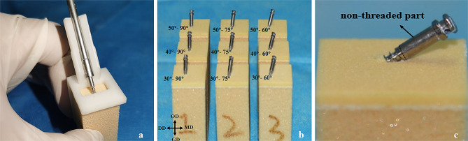
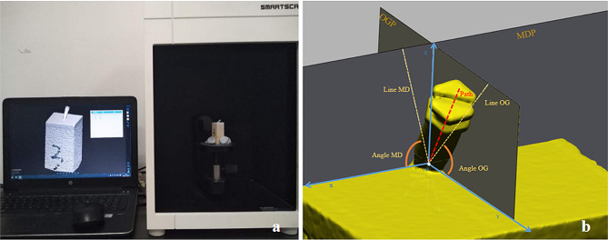
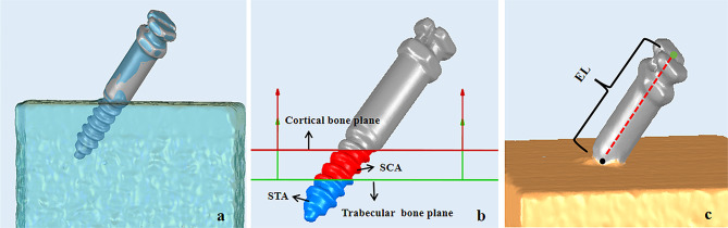
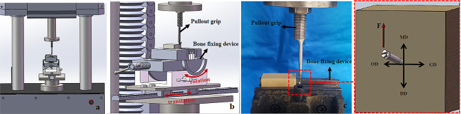
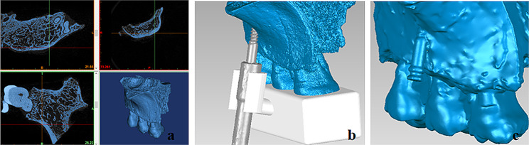
Methods: Ninety synthetic bones were assigned to nine groups to insert mini-screws at the cross-combined angles in the occlusogingival and mesiodistal directions. SCA, STA, EL, and lateral pull-out strength (LPS) were measured, and their relationships were analysed. Twelve mini-screws were then inserted at the optimal and poor angulations into the maxillae from six fresh cadaver heads, and the same biomechanical metrics were measured for validation.
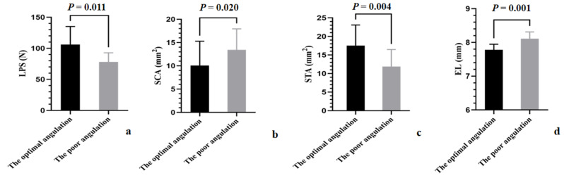
Results: In the synthetic-bone test, the LPS, SCA, STA, and EL had significant correlations with the angle in the occlusogingival direction (rLPS = 0.886, rSCA = -0.946, rSTA = 0.911, and rEL= -0.731; all P < 0.001). In the cadaver-validation test, significant differences were noted in the LPS (P = 0.011), SCA (P = 0.020), STA (P = 0.004), and EL (P = 0.001) between the poor and optimal angulations in the occlusogingival direction. The STA had positive correlations with LPS (rs = 0.245 [synthetic-bone test] and r = 0.720 [cadaver-validation test]; both P < 0.05).
Conclusions: The primary stability of the infrazygomatic crest mini-screw was correlated with occlusogingival angulations. The STA significantly affected the primary stability of the infrazygomatic crest mini-screw, but the SCA and EL did not.
Keywords: Bone; Insertion angles; Mini-screws; Stability.
© 2024. The Author(s).
Conflict of interest statement
The authors declare no competing interests.
Figures

Similar articles
-
Comparison of bone thickness in infrazygomatic crest area at various miniscrew insertion angles in Dravidian population - A cone beam computed tomography study.Int Orthod. 2020 Mar;18(1):105-114. doi: 10.1016/j.ortho.2019.12.001. Epub 2020 Jan 8. Int Orthod. 2020. PMID: 31926867
-
An in vitro study of a combined patient-specific device for safe and accurate insertion of infrazygomatic crest miniscrews.Angle Orthod. 2025 Jan 1;95(1):43-50. doi: 10.2319/022624-147.1. Angle Orthod. 2025. PMID: 39322232 Free PMC article.
-
Clinical analysis of successful insertion of orthodontic mini-implants in infrazygomatic crest.BMC Oral Health. 2023 Jun 1;23(1):348. doi: 10.1186/s12903-023-03081-0. BMC Oral Health. 2023. PMID: 37264370 Free PMC article.
-
Assessment of infrazygomatic bone depth for mini-screw insertion.Clin Oral Implants Res. 2009 Jun;20(6):638-42. doi: 10.1111/j.1600-0501.2008.01691.x. Epub 2009 Mar 4. Clin Oral Implants Res. 2009. PMID: 19281501
-
Optimizing Infrazygomatic Miniscrew Insertion Parameters: Systematic Review and Meta-Regression Analysis of Bone Thickness by Insertion Height, Angulation, and Anatomical Position.J Clin Med. 2025 Jun 5;14(11):4005. doi: 10.3390/jcm14114005. J Clin Med. 2025. PMID: 40507767 Free PMC article. Review.
References
-
- Chen Y, Kao C, Huang T. Evaluation of ten Extra-alveolar Temporary Anchorage device insertion sites by Cone Beam Volumetric Computer Tomography: a pilot study. J Dent Sci. 2010;5:21–9. 10.1016/S1991-7902(10)60004-9.10.1016/S1991-7902(10)60004-9 - DOI
-
- Miyawaki S, Koyama I, Inoue M, Mishima K, Sugahara T, Takano-Yamamoto T. Factors Associated with the Stability of Titanium screws placed in the posterior region for Orthodontic Anchorage. Am J Orthod Dentofac Orthop. 2003;124:373–8. 10.1016/s0889-5406(03)00565-1.10.1016/s0889-5406(03)00565-1 - DOI - PubMed
MeSH terms
Grants and funding
LinkOut - more resources
Full Text Sources
