Review
. 2024 Jul 24;16(15):2634.
doi: 10.3390/cancers16152634.

Decoding Clonal Hematopoiesis: Emerging Themes and Novel Mechanistic Insights

Affiliations
Review

Decoding Clonal Hematopoiesis: Emerging Themes and Novel Mechanistic Insights

Shalmali Pendse et al. Cancers (Basel). .

Abstract

Clonal hematopoiesis (CH), the relative expansion of mutant clones, is derived from hematopoietic stem cells (HSCs) with acquired somatic or cytogenetic alterations that improve cellular fitness. Individuals with CH have a higher risk for hematological and non-hematological diseases, such as cardiovascular disease, and have an overall higher mortality rate. Originally thought to be restricted to a small fraction of elderly people, recent advances in single-cell sequencing and bioinformatics have revealed that CH with multiple expanded mutant clones is universal in the elderly population. Just a few years ago, phylogenetic reconstruction across the human lifespan and novel sensitive sequencing techniques showed that CH can start earlier in life, decades before it was thought possible. These studies also suggest that environmental factors acting through aberrant inflammation might be a common theme promoting clonal expansion and disease progression. However, numerous aspects of this phenomenon remain to be elucidated and the precise mechanisms, context-specific drivers, and pathways of clonal expansion remain to be established. Here, we review our current understanding of the cellular mechanisms driving CH and specifically focus on how pro-inflammatory factors affect normal and mutant HSC fates to promote clonal selection.

Keywords: Asxl1; CHIP; Dnmt3a; Tet2; clonal evolution; clonal hematopoiesis; clonal selection; hematopoietic stem cells; inflammation; self-renewal.


Conflict of interest statement

The authors declare no conflicts of interest, financial or otherwise.

Figures

Figure 1
Figure 1
Diseases associated with clonal hematopoiesis. Individuals with clonal hematopoiesis have a higher risk of hematologic and non-hematologic diseases and an overall higher rate of mortality. Hematologic diseases associated with CH include myelodysplastic syndrome and myeloid and lymphoid malignancies. Non-hematologic diseases associated with CH include, among others, cardiovascular disease, acute kidney disease, chronic obstructive pulmonary disease, chronic liver disease, osteoporosis, type 2 diabetes, and COVID-19. Created with https://biorender.com.
Figure 2
Figure 2
Frequencies of the most common somatic variants associated with myeloid and lymphoid clonal hematopoiesis. (A) The top 25 driver genes mutated in myeloid CH (M-CH) and (B) lymphoid CH (L-CH), respectively (adapted from [28]). Created with https://biorender.com.
Figure 3
Figure 3
The number of mutant clones throughout an individual’s lifetime can change dynamically and grow, shrink, or remain stable for extended periods. Clonal hematopoiesis is initiated when HSCs acquire mutations that confer a competitive advantage over wild-type HSCs. (A) Over time, this mutation allows the mutant clone (blue) to expand relative to normal HSCs (red). (B) Alternatively, mutant clones can remain stable for extended periods and/or the entire lifespan. (C) Mutant clones that started to expand can also shrink again. This can happen if the environmental conditions become less favorable and/or if another clone with increased evolutionary fitness outcompetes the original clone. The red question mark indicates changes in clonal growth rates. The reasons for these changes are currently unclear. Created with https://biorender.com.
Figure 4
Figure 4
Inflammation drives clonal expansion of mutant HSCs. Normal HSCs (round red cells) self-renew to maintain the stem cell pool and differentiate to produce mature blood cells (left). Eventually, HSCs acquire mutations. While most mutations remain inconsequential, others provide a fitness advantage (round blue cells) under certain circumstances. In the absence of stress, these mutant HSCs self-renew and differentiate similarly to normal HSCs (middle). When exposed to inflammatory factors, mutant HSCs can outcompete normal HSCs through different cytokine-mediated and context-dependent mechanisms. The following clonal outgrowth of mature mutant blood cells leads to aberrant production of pro-inflammatory cytokines, reinforcing inflammation. As mutant HSCs are more stress-resilient in this pro-inflammatory environment, this selective advantage further promotes clonal expansion and perpetuates the cycle. Created with https://biorender.com.
Figure 5
Figure 5
Differentially regulated pathways, genes, and cell fates in Asxl1-mutant HSPCs. Differentially regulated signaling pathways, genes, and their effects on cell fates, including cell death, self-renewal, proliferation, and differentiation of Asxl1 mutant relative to normal HSPCs. Cell-extrinsic factors driving the clonal expansion of Asxl1 mutants are currently unknown. Transparent boxes and annotations indicate unknown signaling pathways, genes, and cell fates. (A) In a zebrafish model, asxl1-mutant HSPCs exhibit increased proliferation and self-renewal. These cells express higher levels of anti-inflammatory nr4a1 and socs3a and the pro-differentiation genes junba and fosab. Deletion of nr4a1 showed that the fitness advantage of asxl1-mutant clones depends on nr4a1 [71]. (B) Using a mutant Asxl1 knock-in mouse model, clonal outgrowth and increased proliferation of Asxl1-mutant HSPCs were mediated by Akt/mTOR activation. However, transplants impaired Asxl1-mutant HSPC function [96]. Created with https://biorender.com.
Figure 6
Figure 6
Pro-inflammatory cytokine-induced changes in signaling, gene expression, and cell fates that occur in Dnmt3a-mutant, but not in wild-type HSPCs. Dnmt3a-mutant and normal HSPCs respond differently to inflammatory cytokines. Depicted are the currently known differentially regulated signaling pathways, genes, and their effects on survival, proliferation, self-renewal, and differentiation of Dnmt3a-mutant relative to normal HSPCs in response to inflammatory cytokines. Several genes, including Socs3, Nr4a1, Fos, Jun, and JunB, are deregulated in Dnmt3a-mutant HSPCs; these genes are sometimes up- or downregulated, suggesting context-dependent effects of pro-inflammatory cytokines. Transparent boxes and labels indicate unknown pathways, genes, and/or HSPC fates not investigated. (A) In fatty bone marrow, elevated levels of IL-6 promote Dnmt3afl-R882H/+ HSPC self-renewal [97]. (B) Knock-out of TNF-α receptor 1 (TNF-αR1), but not TNF-αR2 increases HSPC self-renewal, without changing myeloid vs. lymphoid differentiation. TNF-αR1 signaling induces Jun, Nfkb2, and CD69 more strongly in Dnmt3aR878H/+ than in normal HSPCs [98]. (C) In young Dnmt3aR878H/+ HSPCs, OSM activates STAT3, induces the expression of pro- and anti-inflammatory genes, and inhibits JunB expression. Despite these changes, OSM did not change HSPC fates, including cell death, proliferation, self-renewal, and differentiation [90]. (D,E) In response to IFN-γ, Dnmt3a−/− HSPCs die less and proliferate slower than wild-type HSPCs. In addition, Dnmt3a−/− HSPC self-renewal increased and differentiation decreased compared to wild-type HSPCs exposed to IFN-γ. (D) M. avium infection-mediated IFN-γ induces pro-inflammatory genes and inhibits pro-differentiation genes at the same time [92]. (E) IFN-γ induces TP53 signaling in Dnmt3a−/− HSPCs and inhibits cell cycle progression via Txnip-p21 [99]. Created with https://biorender.com.
Figure 7
Figure 7
Pro-inflammatory cytokine-induced changes in signaling, gene expression, and cell fates that occur in Tet2-mutant, but not in wild-type HSPCs. The effects of inflammatory cytokines on Tet2-mutant HSPCs across multiple studies show that changes in signaling, gene expression, and HSPC fates vary between cytokines and relative to normal HSPCs. Shown are known differentially regulated signaling pathways, changes in gene expression, and their effects on Tet2-mutant relative to wild-type HSPC fates including cell death, self-renewal, proliferation, and differentiation after exposure to inflammatory cytokines. Tet2-mutant HSPCs have increased self-renewal and proliferate more compared to wild-type HSPCs in response to inflammatory signals. Transparent boxes and labels indicate unknown effects on signaling pathways, genes, and HSPC fates. (A) IL-6 mediated downregulation of pro- and upregulation of anti-apoptotic genes reduces cell death of Tet+/− and Tet2−/− HSPCs [100]. (B) TNF-α-induced NF-κB signaling reduces cell death in Tet2−/− relative to wild-type HSPCs and correlates with the up-and downregulation of anti- and pro-apoptotic genes, respectively [101]. (C) IL-1β induces the expression of pro- as well as anti-inflammatory genes in Vav-Cre Tet2fl/fl HSPCs [91]. (D) The increase in self-renewal, proliferation, DNA replication, and DNA repair gene expression programs of Tet2+/− HSPCs depends on IL-1α receptor 1-mediated signaling [102]. (E) Studies using Tet2fl/fl;Sting−/− mice show that the cGAS-STING pathway is required for the competitive advantage of Tet2−/− HSPCs. Genes encoding pro-inflammatory cytokines, receptors, and signaling factors depend on STING activation in Tet2fl/fl HSPCs [103]. Created with https://biorender.com.
Figure 8
Figure 8
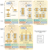
Summary of known differentially regulated cell fates of Dnmt3a-, Tet2-, and Asxl1-mutant mouse HSPCs in response to pro-inflammatory cytokines [71,90,91,92,96,97,98,99,100,101,102,103,111]. IL-6: Interleukin-6; TNF-α: Tumor Necrosis Factor-α; OSM: Oncostatin M; IFN-γ: Interferon-γ; IL-1β: Interleukin-1β; dsDNA: Double stranded DNA.

References

    1. Jagannathan-Bogdan M., Zon L.I. Hematopoiesis. Development. 2013;140:2463–2467. doi: 10.1242/dev.083147. - DOI - PMC - PubMed
    1. de Haan G., Lazare S.S. Aging of hematopoietic stem cells. Blood. 2018;131:479–487. doi: 10.1182/blood-2017-06-746412. - DOI - PubMed
    1. Bonnet D., Dick J.E. Human acute myeloid leukemia is organized as a hierarchy that originates from a primitive hematopoietic cell. Nat. Med. 1997;3:730–737. doi: 10.1038/nm0797-730. - DOI - PubMed
    1. Mitchell E., Chapman M.S., Williams N., Dawson K.J., Mende N., Calderbank E.F., Jung H., Mitchell T., Coorens T.H.H., Spencer D.H., et al. Clonal dynamics of haematopoiesis across the human lifespan. Nature. 2022;606:343–350. - PMC - PubMed
    1. Steensma D.P., Bejar R., Jaiswal S., Lindsley R.C., Sekeres M.A., Hasserjian R.P., Ebert B.L. Clonal hematopoiesis of indeterminate potential and its distinction from myelodysplastic syndromes. Blood. 2015;126:9–16. - PMC - PubMed

LinkOut - more resources