Review
. 2024 Jul 25;13(15):4346.
doi: 10.3390/jcm13154346.

Ultrasound and Intrapleural Enzymatic Therapy for Complicated Pleural Effusion: A Case Series with a Literature Review

Affiliations
Review

Ultrasound and Intrapleural Enzymatic Therapy for Complicated Pleural Effusion: A Case Series with a Literature Review

Riccardo Inchingolo et al. J Clin Med. .

Abstract

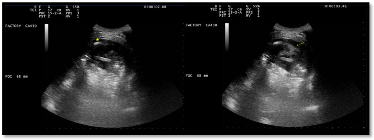
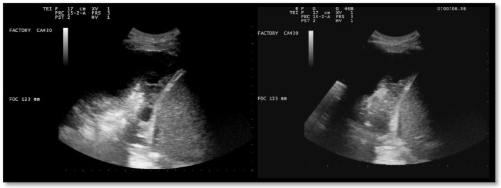
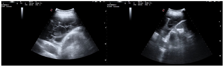
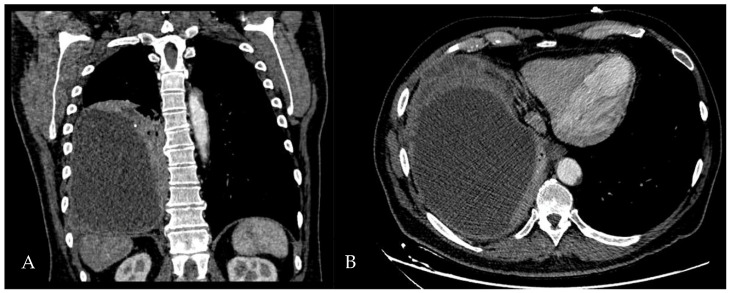
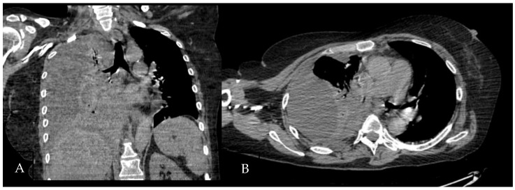
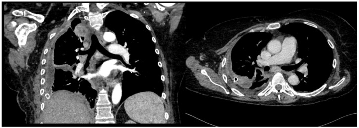
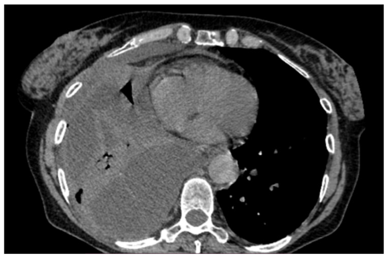
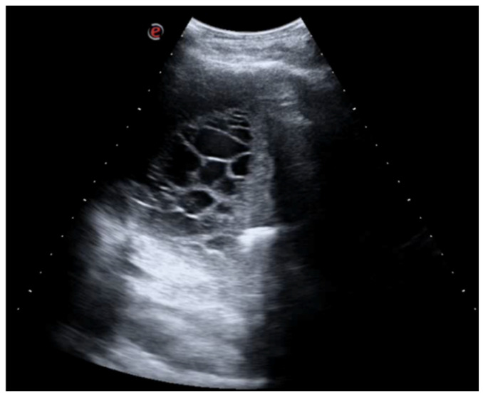
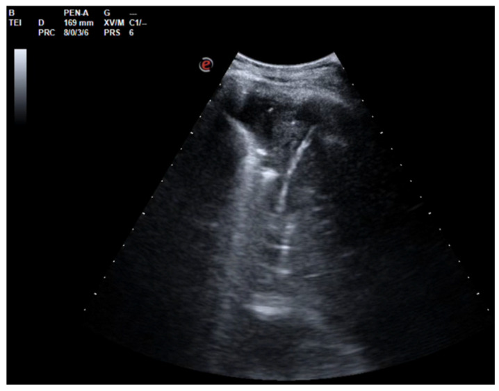
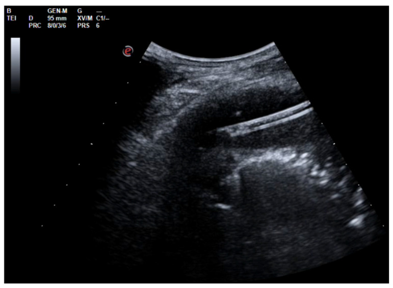
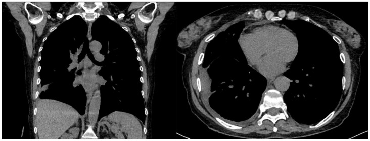
Pleural effusion is the most common manifestation of pleural disease, and chest ultrasound is crucial for diagnostic workup and post-treatment monitoring. Ultrasound helps distinguish the various types of pleural effusion and enables the detection of typical manifestations of empyema, which presents as a complicated, septated effusion. This may benefit from drainage and the use of intrapleural enzyme therapy or may require more invasive approaches, such as medical or surgical thoracoscopy. The mechanism of action of intrapleural enzymatic therapy (IPET) is the activation of plasminogen to plasmin, which breaks down fibrin clots that form septa or the loculation of effusions and promotes their removal. In addition, IPET has anti-inflammatory properties and can modulate the immune response in the pleural space, resulting in reduced pleural inflammation and improved fluid reabsorption. In this article, we briefly review the literature on the efficacy of IPET and describe a case series in which most practical applications of IPET are demonstrated, i.e., as a curative treatment but also as an alternative, propaedeutic, or subsequent treatment to surgery.

Keywords: DNase; alteplase; complex pleural effusion; empyema; intrapleural enzymatic therapy; lung ultrasound; t-PA; urokinase.


Conflict of interest statement

The authors declare no conflicts of interest.

Figures

Figure 1
Figure 1
Chest US at admission. Multiloculated pleural effusion.
Figure 2
Figure 2
Chest US during pleural lavage with urokinase. Star: distal end of chest drain; Arrow: spread of fluid.
Figure 3
Figure 3
Chest US 48 h (left) and 72 h (right) after intrapleural instillation of urokinase.
Figure 4
Figure 4
Complex and septated pleural effusion after VATS treated with IPFT.
Figure 5
Figure 5
Chest CT scan of the mediastinal window in the coronal (A) and axial (B) plane, showing fluid collection in the medium–lower parts of the right hemithorax (maximum axial dimeters of 19 × 13 cm; craniocaudal extension of 16 cm) with thickened walls and contrast enhancement. There is also a complete atelectasis of middle and lower lobes and partial atelectasis of the upper lobe.
Figure 6
Figure 6
Chest CT scan of the mediastinal window in the coronal (A) and axial (B) plane, showing a large right pleural effusion, occupying almost the entire hemithorax with organized appearance. There is also a complete right lower lobe atelectasis and almost-complete right middle and upper lobe atelectasis with sparing of anterior sectors.
Figure 7
Figure 7
Lung re-expansion after IPFT and VATS.
Figure 8
Figure 8
Large right pleural effusion on chest CT scan.
Figure 9
Figure 9
Chest ultrasound at admission showing multiloculated pleural effusion.
Figure 10
Figure 10
Chest US 48 h after urokinase. Minimum pleural effusion with residual fibrinous material and signs of lung re-expansion.
Figure 11
Figure 11
Chest US 48 h after urokinase. Chest tube in pleural cavity with residual pleural effusion.
Figure 12
Figure 12
Pre-discharge chest CT.

Similar articles

References

    1. Roberts M.E., Rahman N.M., Maskell N.A., Bibby A.C., Blyth K.G., Corcoran J.P., Edey A., Evison M., de Fonseka D., Hallifax R., et al. British Thoracic Society Guideline for Pleural Disease. Thorax. 2023;78:1143–1156. doi: 10.1136/thorax-2023-220304. - DOI - PubMed
    1. Demi L., Wolfram F., Klersy C., De Silvestri A., Ferretti V.V., Muller M., Miller D., Feletti F., Wełnicki M., Buda N., et al. New International Guidelines and Consensus on the Use of Lung Ultrasound. J. Ultrasound Med. 2023;42:309–344. doi: 10.1002/jum.16088. - DOI - PMC - PubMed
    1. Sorino C., Mondoni M., Lococo F., Marchetti G., Feller-Kopman D. Optimizing the Management of Complicated Pleural Effusion: From Intrapleural Agents to Surgery. Respir. Med. 2022;191:106706. doi: 10.1016/j.rmed.2021.106706. - DOI - PubMed
    1. Fitzgerald D.B., Polverino E., Waterer G.W. Expert Review on Nonsurgical Management of Parapneumonic Effusion: Advances, Controversies, and New Directions. Semin. Respir. Crit. Care Med. 2023;44:468–476. doi: 10.1055/s-0043-1769095. - DOI - PubMed
    1. Smargiassi A., Inchingolo R., Zanforlin A., Valente S., Soldati G., Corbo G.M. Description of Free-Flowing Pleural Effusions in Medical Reports after Echographic Assessment. Respiration. 2013;85:439–441. doi: 10.1159/000346991. - DOI - PubMed

LinkOut - more resources