. 2024 Jul 26;13(15):4382.
doi: 10.3390/jcm13154382.

Spontaneous Non-Aneurysmal Convexity Subarachnoid Hemorrhage: A Scoping Review of Different Etiologies beyond Cerebral Amyloid Angiopathy

Affiliations

Spontaneous Non-Aneurysmal Convexity Subarachnoid Hemorrhage: A Scoping Review of Different Etiologies beyond Cerebral Amyloid Angiopathy

Marialuisa Zedde et al. J Clin Med. .

Abstract

Spontaneous convexity subarachnoid hemorrhage (cSAH) is a vascular disease different from aneurysmal SAH in neuroimaging pattern, causes, and prognosis. Several causes might be considered in individual patients, with a limited value of the patient's age for discriminating among these causes. Cerebral amyloid angiopathy (CAA) is the most prevalent cause in people > 60 years, but reversible cerebral vasoconstriction syndrome (RCVS) has to be considered in young people. CAA gained attention in the last years, but the most known manifestation of cSAH in this context is constituted by transient focal neurological episodes (TFNEs). CAA might have an inflammatory side (CAA-related inflammation), whose diagnosis is relevant due to the efficacy of immunosuppression in resolving essudation. Other causes are hemodynamic stenosis or occlusion in extracranial and intracranial arteries, infective endocarditis (with or without intracranial infectious aneurysms), primary central nervous system angiitis, cerebral venous thrombosis, and rarer diseases. The diagnostic work-up is fundamental for an etiological diagnosis and includes neuroimaging techniques, nuclear medicine techniques, and lumbar puncture. The correct diagnosis is the first step for choosing the most effective and appropriate treatment.

Keywords: CAA-related inflammation; MRI; PRES; RCVS; cerebral amyloid angiopathy; cerebral venous thrombosis; convexity subarachnoid hemorrhage; endocarditis; intracranial stenosis.


Conflict of interest statement

The authors declare no conflicts of interest.

Figures
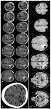
Figure 1
Figure 1
cSAH in a patient with probable CAA according with Boston criteria 2.0 [12]. Panel (a) shows a non-contrast computed tomography (NCCT) axial slice with the hyperdense content in one left anterior frontal sulcus, suggesting cSAH. Panels (b,c) show sequential axial FLAIR slices before (b) and after GBCA. Spontaneous contrast enhancement in more than one left anterior frontal sulcus is accompanied by cortical effacement and subcortical white matter hyperintensity in the same site. Panel (d) shows an SWI sequence reconstructed in axial plane with minimum, finding several lobar microbleeds, at least two macrobleeding foci and multifocal cortical superficial siderosis.
Figure 2
Figure 2
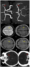
cSAH in a patient with left distal M1 MCA acute steno-occlusion. Panels (a,b) show CT angiography reconstructed with the maximum intensity projection/MPR tool in the axial and coronal planes, respectively, showing the focal lack of left MCA opacification corresponding to vessel occlusion (red arrows). Panels (c,d) show axial FLAIR-MRI slices with spontaneous hyperintensity in the precentral sulcus (yellow arrows), suggestive of cSAH. The same sulcus is relatively hyperintense in T2* (panel (e)) and not clearly discernible in the T1 sequence (panel (f)). Panels (g,h) illustrate MR angiography on the axial and coronal plane, reconstructed with the MIP/MPR tool (the red arrows focus on the occlusion).
Figure 3
Figure 3
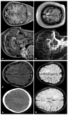
cSAH in a patient with infective endocarditis. Panel (a) shows NCCT with a linear hyperdense content in the right central sulcus. Panel (b) shows the corresponding axial reconstructed CT angiography with normal finding. Panel (c) underlines multiple splenic infarctions. Panel (d) shows multiple views of transesophageal echocardiography with a huge vegetation on the posterior leaf in the mitral valve.
Figure 4
Figure 4
cSAH in a patient with CVT. Panels (a,b) show, respectively, NCCT and FLAIR MRI with, respectively, hyperdense and hyperintense left superior frontal sulcus, suggestive of SAH. Panel (c) shows sagittal and coronal view of post GBCA with an extensive superior sagittal sinus thrombosis. Panel (d) highlights in SWI the cortical venous congestion in the right hemisphere. Panel (e) focuses on MR angiography reconstructed in sagittal plane, confirming the SSS thrombosis and the lack of intravascular contrast in the posterior third of the SSS on T1-MRI.
Figure 5
Figure 5
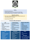
Diagnostic clues for cSAH.

References

    1. Khurram A., Kleinig T., Leyden J. Clinical associations and causes of convexity subarachnoid hemorrhage. Stroke. 2014;45:1151–1153. doi: 10.1161/STROKEAHA.113.004298. - DOI - PubMed
    1. Beitzke M., Gattringer T., Enzinger C., Wagner G., Niederkorn K., Fazekas F. Clinical presentation, etiology, and long-term prognosis in patients with nontraumatic convexal subarachnoid hemorrhage. Stroke. 2011;42:3055–3060. doi: 10.1161/STROKEAHA.111.621847. - DOI - PubMed
    1. Kumar S., Goddeau R.P., Selim M.H., Jr., Thomas A., Schlaug G., Alhazzani A., Searls D.E. Atraumatic convexal subarachnoid hemorrhage: Clinical presentation, imaging patterns, and etiologies. Neurology. 2010;74:893–899. doi: 10.1212/WNL.0b013e3181d55efa. - DOI - PMC - PubMed
    1. Greenberg S.M., Vonsattel J.P., Stakes J.W., Gruber M., Finklestein S.P. The clinical spectrum of cerebral amyloid angiopathy: Presentations without lobar hemorrhage. Neurology. 1993;43:2073–2079. doi: 10.1212/WNL.43.10.2073. - DOI - PubMed
    1. Hostettler I.C., Wilson D., Fiebelkorn C.A., Aum D., Ameriso S.F., Eberbach F., Beitzkeet M., Kleinig T., Phan T., Marchina S., et al. Risk of intracranial haemorrhage and ischaemic stroke after convexity subarachnoid haemorrhage in cerebral amyloid angiopathy: International individual patient data pooled analysis. J. Neurol. 2022;269:1427–1438. doi: 10.1007/s00415-021-10706-3. - DOI - PMC - PubMed

Publication types

LinkOut - more resources