. 2024 Jul 25;25(15):8108.
doi: 10.3390/ijms25158108.

Anti-Neuroinflammatory Effects of Ginkgo biloba Extract EGb 761 in LPS-Activated BV2 Microglial Cells

Affiliations

Anti-Neuroinflammatory Effects of Ginkgo biloba Extract EGb 761 in LPS-Activated BV2 Microglial Cells

Lu Sun et al. Int J Mol Sci. .

Abstract

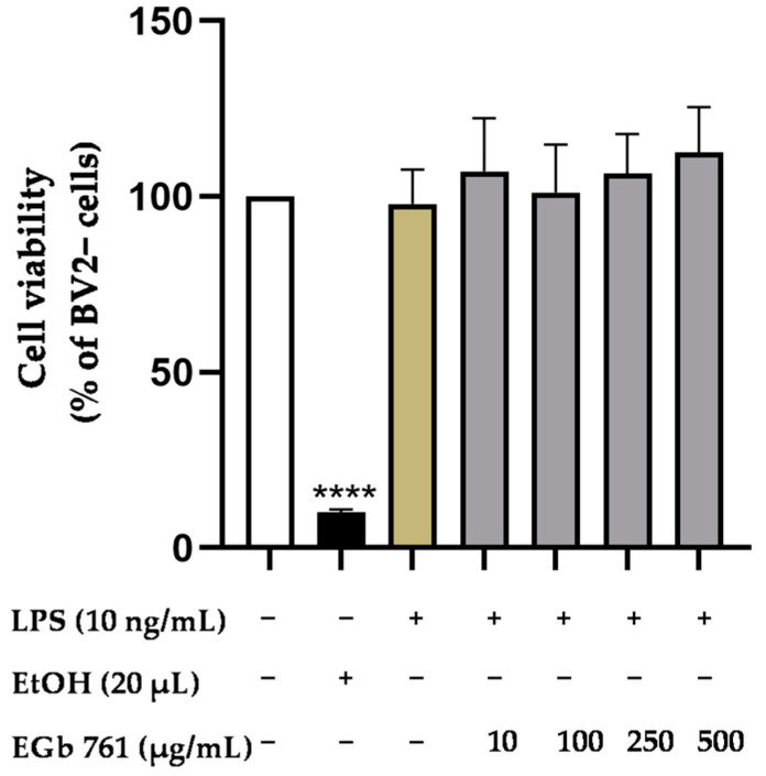
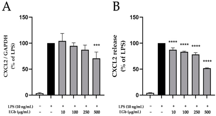
Inflammatory processes in the brain can exert important neuroprotective functions. However, in neurological and psychiatric disorders, it is often detrimental due to chronic microglial over-activation and the dysregulation of cytokines and chemokines. Growing evidence indicates the emerging yet prominent pathophysiological role of neuroinflammation in the development and progression of these disorders. Despite recent advances, there is still a pressing need for effective therapies, and targeting neuroinflammation is a promising approach. Therefore, in this study, we investigated the anti-neuroinflammatory potential of a marketed and quantified proprietary herbal extract of Ginkgo biloba leaves called EGb 761 (10-500 µg/mL) in BV2 microglial cells stimulated by LPS (10 ng/mL). Our results demonstrate significant inhibition of LPS-induced expression and release of cytokines tumor necrosis factor-α (TNF-α) and Interleukin 6 (IL-6) and chemokines C-X-C motif chemokine ligand 2 (CXCL2), CXCL10, c-c motif chemokine ligand 2 (CCL2) and CCL3 in BV2 microglial cells. The observed effects are possibly mediated by the mitogen-activated protein kinases (MAPK), p38 MAPK and ERK1/2, as well as the protein kinase C (PKC) and the nuclear factor (NF)-κB signaling cascades. The findings of this in vitro study highlight the anti-inflammatory properties of EGb 761 and its therapeutic potential, making it an emerging candidate for the treatment of neuroinflammatory diseases and warranting further research in pre-clinical and clinical settings.

Keywords: EGb 761; MAPK pathway; NF-κB pathway; chemokines; cytokines; microglia; neuroinflammation.


Conflict of interest statement

MDL and GPHD are employed by Dr. Willmar Schwabe GmbH & Co KG, the producer of EGb 761®. All other authors declare no conflicts of interest.

Figures

Figure 1
Figure 1
Effects of EGb 761 on cell viability in LPS-stimulated BV2 microglial cells. Cell viability was measured after 24 h of treatment by color change due to MTT reduction. Values are presented as mean ± SD of four independent experiments. Statistical analysis was performed using one-way ANOVA with Dunnett’s post hoc test with **** p < 0.0001 compared to untreated cells.
Figure 2
Figure 2
Effects of EGb 761 on TNFα expression (A) and release (B) in LPS-stimulated BV2 cells. Cells were stimulated as described in Materials and Methods. (A) After 4 h of stimulation, RNA was isolated and expression of TNFα was determined using qPCR. (B) After 24 h of stimulation, supernatants were collected and the release of TNFα was measured by ELISA. Values are presented as the mean ± SD of nine and three independent experiments for qPCR and ELISA, respectively. Statistical analysis was performed using one-way ANOVA with Dunnett’s post hoc tests with * p < 0.05 and **** p < 0.0001 compared to LPS.
Figure 3
Figure 3
Effects of EGb 761 on IL-6 expression (A) and synthesis (B) in LPS-stimulated BV2 cells. Cells were stimulated as described in Materials and Methods. (A) After 4 h of stimulation, RNA was isolated and expression of IL-6 was determined using qPCR. (B) After 24 h of stimulation, supernatants were collected and the release of IL-6 was measured by ELISA. Values are presented as the mean ± SD of five and three independent experiments for qPCR and ELISA, respectively. Statistical analysis was performed using one-way ANOVA with Dunnett’s post hoc tests with ** p < 0.01, and **** p < 0.0001 compared to LPS.
Figure 4
Figure 4
Effects of EGb 761 on CCL2 expression (A) and synthesis (B) in LPS-stimulated BV2 cells. Cells were stimulated as described in Materials and Methods. (A) After 4 h of stimulation, RNA was isolated and expression of CCL2 was determined using qPCR. (B) After 24 h of stimulation, supernatants were collected and the release of CCL2 was measured by ELISA. Values are presented as the mean ± SD of six independent experiments. Statistical analysis was performed using one-way ANOVA with Dunnett’s post hoc tests with * p < 0.05, ** p < 0.01, *** p < 0.001 and **** p < 0.0001 compared to LPS.
Figure 5
Figure 5
Effects of EGb 761 on CCL3 expression (A) and synthesis (B) in LPS-stimulated BV2 cells. Cells were stimulated as described in Materials and Methods. (A) After 4 h of stimulation, RNA was isolated and expression of CCL3 was determined using qPCR. (B) After 24 h of stimulation, supernatants were collected and the release of CCL3 was measured by ELISA. Values are presented as the mean ± SD of nine and three independent experiments for qPCR and ELISA, respectively. Statistical analysis was performed using one-way ANOVA with Dunnett’s post hoc tests with *** p < 0.001 and **** p < 0.0001 compared to LPS.
Figure 6
Figure 6
Effects of EGb 761 on CXCL2 expression (A) and protein release (B) in LPS-stimulated BV2 cells. Cells were stimulated as described in Materials and Methods. (A) After 4 h of stimulation, RNA was isolated and expression of CXCL2 was determined using qPCR. (B) After 24 h of stimulation, supernatants were collected and the release of CXCL2 was measured by ELISA. Values are presented as the mean ± SD of six independent experiments. Statistical analysis was performed using one-way ANOVA with Dunnett’s post hoc tests with *** p < 0.001 and **** p < 0.0001 compared to LPS.
Figure 7
Figure 7
Effects of EGb 761 on CXCL10 expression (A) and release (B) of CXCL10 in LPS-stimulated BV2 cells. Cells were stimulated as described in Materials and Methods. (A) After 4 h of stimulation, RNA was isolated and expression of CXCL10 was determined using qPCR. (B) After 24 h of stimulation, supernatants were collected and the release of CXCL10 was measured by ELISA. Values are presented as the mean ± SD of nine and three independent experiments for qPCR and ELISA. Statistical analysis was performed using one-way ANOVA with Dunnett’s post hoc tests with ** p < 0.01 and **** p < 0.0001 compared to LPS.
Figure 8
Figure 8
Effects of EGb 761 on the phosphorylation of ERK 1/2 (A) and p38 MAPK (B) in LPS-stimulated BV2 cells. Cells were stimulated and Western blot was performed as described in Materials and Methods. Values are presented as the mean ± SD of three independent experiments, and protein levels were referenced to Vinculin. Statistical analysis was performed using one-way ANOVA with Dunnett’s post hoc tests with * p < 0.05 compared to LPS.
Figure 9
Figure 9
Effects of EGb 761 on phosphorylation of PKC (pan) (βII Ser660) (A) and NF-κBp65 (B) in LPS-stimulated BV2 cells. Cells were stimulated and Western blot was performed as described in Materials and Methods. Values are presented as the mean ± SD of three independent experiments, and protein levels were referenced to Vinculin. Statistical analysis was performed using one-way ANOVA with Dunnett’s post hoc tests with * p < 0.05 and ** p < 0.01 compared to LPS.

References

    1. Ozben T., Ozben S. Neuro-Inflammation and Anti-Inflammatory Treatment Options for Alzheimer’s Disease. Clin. Biochem. 2019;72:87–89. doi: 10.1016/j.clinbiochem.2019.04.001. - DOI - PubMed
    1. Jia X., Gao Z. Microglia in Depression: Current Perspectives. Life Sci. 2021;64:911–925. doi: 10.1007/s11427-020-1815-6. - DOI - PubMed
    1. Hartmann A., Hirsch E.C. Neuroinflammatory Processes in Parkinson’s Disease. Park. Relat. Disord. 2005;11:9–15. doi: 10.1016/j.parkreldis.2004.10.013. - DOI - PubMed
    1. Cannon J.R., Greenamyre J.T. The Role of Environmental Exposures in Neurodegeneration and Neurodegenerative Diseases. Park. Relat. Disord. 2011;124:225–250. doi: 10.1093/toxsci/kfr239. - DOI - PMC - PubMed
    1. Celorrio M., Rojo-Bustamante E., Fernández-Suárez D., Sáez E., Estella-Hermoso de Mendoza A., Müller C.E., Ramírez M.J., Oyarzábal J., Franco R., Aymerich M.S. GPR55: A Therapeutic Target for Parkinson’s Disease? Neuropharmacology. 2017;125:319–332. doi: 10.1016/j.neuropharm.2017.08.017. - DOI - PubMed

LinkOut - more resources