. 2024 Jul 27;25(15):8197.
doi: 10.3390/ijms25158197.

Insulin-Activated Signaling Pathway and GLUT4 Membrane Translocation in hiPSC-Derived Cardiomyocytes

Affiliations

Insulin-Activated Signaling Pathway and GLUT4 Membrane Translocation in hiPSC-Derived Cardiomyocytes

Giulia Querio et al. Int J Mol Sci. .

Abstract

Human induced pluripotent stem cell-derived cardiomyocytes (hiPSC-CM) are a cell model now widely used to investigate pathophysiological features of cardiac tissue. Given the invaluable contribution hiPSC-CM could make for studies on cardio-metabolic disorders by defining a postnatal metabolic phenotype, our work herein focused on monitoring the insulin response in CM derived from the hiPSC line UKBi015-B. Western blot analysis on total cell lysates obtained from hiPSC-CM showed increased phosphorylation of both AKT and AS160 following insulin treatment, but failed to highlight any changes in the expression dynamics of the glucose transporter GLUT4. By contrast, the Western blot analysis of membrane fractions, rather than total lysates, revealed insulin-induced plasma membrane translocation of GLUT4, which is known to also occur in postnatal CM. Thus, these findings suggest that hiPSC-derived CMs exhibit an insulin response reminiscent to that of adult CMs regarding intracellular signaling and GLUT4 translocation to the plasma membrane, representing a suitable cellular model in the cardio-metabolic research field. Moreover, our studies also demonstrate the relevance of analyzing membrane fractions rather than total lysates in order to monitor GLUT4 dynamics in response to metabolic regulators in hiPSC-CMs.

Keywords: GLUT4; cardiac maturation; cardiomyocyte; hiPSC-CM; insulin; lactate medium; metabolism; plasma membrane translocation; postnatal cardiac cell.


Conflict of interest statement

The authors declare no conflicts of interest.

Figures

Figure 1
Figure 1
Differentiated hiPSC-CMs show core elements of the insulin signaling pathway. (a) Schematic representation of the differentiation protocol. Cells were differentiated with standard protocol, and then, from days 11 to 14, incubated with lactate medium before the experiments. Bright field acquisitions of undifferentiated (left) and differentiated (right) cells (20×, scale bar 5 µm). Bar graph and representative blots of lactate-purified hiPSC-CMs treated or not with insulin (INS, 100 nM, 30 min) showing (b,c) pAKT/AKT (CTRL: 100.00; INS: 136.86 ± 9.80) and (d,e) pAS160/AS160 (CTRL: 100.00; INS: 137.93 ± 8.12). n = 3 independent experiments, * p < 0.05; ** p < 0.01.
Figure 2
Figure 2
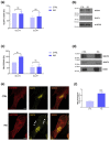
Lactate-purified hiPSC-CMs show significant GLUT4 membrane translocation after insulin stimulation. Bar graph and representative blots of (a,b) total lysates and (c,d) membrane fractions showing higher GLUT4 levels in membrane fractions of insulin-treated cells. GLUT1 shows no variations after insulin stimulation. (Total lysates: CTRL-GLUT4: 100.00; INS-GLUT4: 97.33 ± 14.33, CTRL-GLUT1: 72.18 ± 12.04; INS-GLUT1: 79.51 ± 22.60; Membrane fractions: CTRL-GLUT4: 100.00; INS-GLUT4: 150.34 ± 5.49, CTRL-GLUT1: 57.26 ± 14.42; INS-GLUT1: 61.04 ± 17.60). (e) Representative immunostaining showing GLUT4 membrane translocation in insulin-treated cells (white arrows) (40×, scale bar 10 µm), and (f) the bar graph summarizing the immunofluorescence experiments analysis in 34 α-actinin-positive cells for both conditions (CTRL: 1.00 ± 0.13; INS: 2.31 ± 0.28). n = 3 independent experiments, * p < 0.05; *** p < 0.001.

References

    1. Mitcheson J.S., Hancox J.C., Levi A.J. Cultured Adult Cardiac Myocytes: Future Applications, Culture Methods, Morphological and Electrophysiological Properties. Cardiovasc. Res. 1998;39:280–300. doi: 10.1016/S0008-6363(98)00128-X. - DOI - PubMed
    1. Louch W.E., Sheehan K.A., Wolska B.M. Methods in Cardiomyocyte Isolation, Culture, and Gene Transfer. J. Mol. Cell. Cardiol. 2011;51:288–298. doi: 10.1016/j.yjmcc.2011.06.012. - DOI - PMC - PubMed
    1. Bistola V., Nikolopoulou M., Derventzi A., Kataki A., Sfyras N., Nikou N., Toutouza M., Toutouzas P., Stefanadis C., Konstadoulakis M.M. Long-Term Primary Cultures of Human Adult Atrial Cardiac Myocytes: Cell Viability, Structural Properties and BNP Secretion in Vitro. Int. J. Cardiol. 2008;131:113–122. doi: 10.1016/j.ijcard.2007.10.058. - DOI - PubMed
    1. Adegunsoye A., Gonzales N.M., Gilad Y. Induced Pluripotent Stem Cells in Disease Biology and the Evidence for Their In Vitro Utility. Annu. Rev. Genet. 2023;57:341–360. doi: 10.1146/annurev-genet-022123-090319. - DOI - PubMed
    1. Zhang J., Wilson G.F., Soerens A.G., Koonce C.H., Yu J., Palecek S.P., Thomson J.A., Kamp T.J. Functional Cardiomyocytes Derived From Human Induced Pluripotent Stem Cells. Circ. Res. 2009;104:e30–e41. doi: 10.1161/CIRCRESAHA.108.192237. - DOI - PMC - PubMed

MeSH terms

LinkOut - more resources