. 2024 Jul 27;25(15):8201.
doi: 10.3390/ijms25158201.

A Barth Syndrome Patient-Derived D75H Point Mutation in TAFAZZIN Drives Progressive Cardiomyopathy in Mice

Affiliations

A Barth Syndrome Patient-Derived D75H Point Mutation in TAFAZZIN Drives Progressive Cardiomyopathy in Mice

Paige L Snider et al. Int J Mol Sci. .

Abstract

Cardiomyopathy is the predominant defect in Barth syndrome (BTHS) and is caused by a mutation of the X-linked Tafazzin (TAZ) gene, which encodes an enzyme responsible for remodeling mitochondrial cardiolipin. Despite the known importance of mitochondrial dysfunction in BTHS, how specific TAZ mutations cause diverse BTHS heart phenotypes remains poorly understood. We generated a patient-tailored CRISPR/Cas9 knock-in mouse allele (TazPM) that phenocopies BTHS clinical traits. As TazPM males express a stable mutant protein, we assessed cardiac metabolic dysfunction and mitochondrial changes and identified temporally altered cardioprotective signaling effectors. Specifically, juvenile TazPM males exhibit mild left ventricular dilation in systole but have unaltered fatty acid/amino acid metabolism and normal adenosine triphosphate (ATP). This occurs in concert with a hyperactive p53 pathway, elevation of cardioprotective antioxidant pathways, and induced autophagy-mediated early senescence in juvenile TazPM hearts. However, adult TazPM males exhibit chronic heart failure with reduced growth and ejection fraction, cardiac fibrosis, reduced ATP, and suppressed fatty acid/amino acid metabolism. This biphasic changeover from a mild-to-severe heart phenotype coincides with p53 suppression, downregulation of cardioprotective antioxidant pathways, and the onset of terminal senescence in adult TazPM hearts. Herein, we report a BTHS genotype/phenotype correlation and reveal that absent Taz acyltransferase function is sufficient to drive progressive cardiomyopathy.

Keywords: Barth syndrome; mitochondria; p53 pathway; patient-tailored Tafazzin mutant allele; progressive cardiomyopathy.


Conflict of interest statement

Chin is the Founder and CEO of TransCellular Therapeutics Inc., which had no participation or input in this project. Payne is a Consultant for Larimar Therapeutics, Inc., which had no participation or input in this project. Conway, Rubart and Brault all declare no conflicts of interest.

Figures

Figure 1
Figure 1
Phenotyping TazPM♂ point mutant knock-in mice. (A) TazPM♂ next to larger wt♂ (left) littermate at 1 month of age. (B) Between 1 and 3 three months of age, TazPM♂ on average are 4–6 g < wt♂ littermates (n = 10 TazPM♂ and 11 wt; p < 0.0001). (C) Kaplan–Meier survival curves of age-matched wt♂ (n = 82) and TazPM♂ (n = 29) reveal curtailed TazPM♂ lifespans (p < 0.0001). (D) Mass spectrometry quantification of MLCL:CL ratio in blood from 4-month TazPM♂ vs. wt♂ (n = 6/genotype) confirms deficient CL biosynthesis. (E) Relative 3-methylglutaconic acid levels/gram of creatinine in TazPM♂ and wt♂ urine (n = 13/genotype). (F) Oil red-O stain reveals diminished microvesicular lipid deposition in TazPM♂ (lower panel) but not in wt♂ (upper panel) littermate adult livers (n = 17 litters). (G) Giemsa stain of juvenile TazPM♂ peripheral blood revealed a <50% reduction in neutrophils compared to wt♂. (H) Western analysis of steady-state Taz levels in surviving adult TazPM♂ (n = 6) and wt♂ (n = 7) littermate ventricles (atria and valvular tissues removed), gastrocnemius skeletal muscles and livers, normalized to housekeeping GAPDH levels. Quantification and statistical analysis are shown in Supplemental Figure S1. Statistical significance set at ** p < 0.01, *** p < 0.005 and **** p < 0.001. Scale bars: (F) = 500 μm, (G) = 20 μm.
Figure 2
Figure 2
Adult TazPM♂ cardiac phenotype. (A) Transthoracic echocardiographic measurements (n = 8/genotype) reveal a dilated “pumpkin-shaped” LV in 10-month TazPM♂ as compared to wt♂ littermates. Prominent TazPM♂ LV trabeculations indicated via *. (B,C) Quantification demonstrates both decreased fractional shortening ((B), p = 0.007) and ejection fraction ((C), p = 0.009) in TazPM♂ (red) compared to wt♂ (black) littermates (n = 8/genotype). (D) 10-month heart/body weight measurements show a reduction from ~8 mg/g in wt♂ to ~7.1 mg/g in TazPM♂ (n = 6/genotype, p = 0.02). (E) Histology confirmed all 10-month TazPM♂ hearts (n = 5/5) exhibit significant ventricular chamber dilatation with prominent LV trabeculations with deep intertrabecular recesses (arrow) compared to wt♂ littermates. (F) Masson’s trichrome (n = 6/genotype) revealed increased fibrosis (blue) in TazPM♂ but not wt♂ LV. (G) Morphometric measurement of fibrosis (n = 6/genotype) as % of the entire LV myocardium. (H,I) Cardiac fibrosis was confirmed via immuno-histochemical detection of ectopic deposition of profibrotic Periostin (brown) in 10-month TazPM♂ ((H), n = 6/6) compared to wt♂ ((I)) littermate hearts. Sirus Red/methyl green inset (I), wherein red indicates fibrosis overlapping Periostin expression. (J,K) Oil Red-O (n = 6/genotype) reveals significant steatosis (red) in only adult TazPM♂ CMs ((K), arrows). (L,M) Immunohistochemistry revealed Nppa protein (brown) is ectopically upregulated within mainly the adult TazPM♂ trabecular LV zone (M, indicated by arrow) but absent in wt♂ littermates (L). (N,O) Non-radioactive in situ hybridization revealed ectopic Bmp10 mRNA is induced in adult TazPM♂ ventricle cardiomyocytes ((O), arrow). Statistical significance set at * p < 0.05 and ** p < 0.01. Abbreviations: LV, left ventricle. Scale bars: (E) = 200 μm, (F,H,I) = 100 μm, (J,K) = 20 μm, (L,M) = 500 μm.
Figure 3
Figure 3
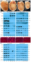
Progressive metabolism dysfunction and heart failure in TazPM males. (A,B) Representative images of whole adult (A) and juvenile (B) littermate wt♂ and TazPM♂ (red *) hearts. (C) Western analysis using antibodies against key profibrotic and cardiac output prognostic biomarkers in adult (6–8 months) and juvenile (4 weeks) wt♂ and TazPM♂ ventricles. (DG) Fluorescent immunohistochemistry revealed Gal3 and CathD (red) are both upregulated in TazPM♂ cardiomyocytes (E,G) but are absent in wt♂ adult littermates (D,F). Nuclei counterstained with DAPI (blue). (DG) were magnified ×200. (H) Western analysis using antibodies against fatty acid and amino acid metabolism effectors. GAPDH was used as a loading control and triplicate age-matched adult and juvenile wt♂ (n = 7) and TazPM♂ (n = 6) littermate ventricles were examined. Quantification and statistical analysis are shown in Supplemental Figure S4.
Figure 4
Figure 4
Dysmorphic TazPM♂ mitochondrial structure and function is offset via upregulation of cardioprotective pathways. (AC), Electron microscopy showing adult wt♂ (A) and TazPM♂ swollen (B), and honeycomb (C) mitochondrial morphology. (D,E) Quantification of “honeycomb” mitochondria (D) and measurement of the mitochondrial area as % of LV in wt♂ (n = 6) vs. TazPM♂ (n = 7) LVs (E). (F) Western blot image of the individual mitochondrial complexs’ expression in wt♂ and TazPM♂ mitochondrial fractions probed using an OxPhos rodent antibody cocktail kit, and mitochondrial VDAC as the loading control. Note that C1 (*) levels are severely reduced. (G) Adenine nucleotide contents (μmol.g−1 tissue) in adult wt♂ and TazPM♂ ventricles (n = 5/genotype). Note, TazPM♂ ATP levels were significantly reduced (p = 0.0017) while ADP and AMP levels remained unaltered (p = ns). (H,I) Western analysis showing key mitochondrial effector (H) and cardioprotective (I) expression levels within triplicate wt♂ and TazPM♂ adult and juvenile ventricles (n = 6 per genotype/age). GAPDH was used as a loading control. Quantification and statistical analysis are shown in Supplemental Figure S6. Statistical significance set at ** p < 0.01, *** p < 0.005 and **** p < 0.001. Abbreviations: M, mitochondria; sM, swollen mitochondria; hD, honeycomb mitochondria; mF, myofibrils. Scale bars, (AC) = 500 nm.
Figure 5
Figure 5
Induction of autophagic and senescent adaptive stress responses in surviving TazPM♂ hearts. (A) Western analysis showing key autophagy and senescence biomarker expression levels within triplicate wt♂ and TazPM♂ adult and juvenile ventricles (n = 6 per genotype/age). GAPDH was used as a loading control. Quantification and statistical analysis are shown in Supplemental Figure S7. (B,C) Immunofluorescence to detect autophagic protein LC3B (arrows, red fluorescence) upregulation in juvenile TazPM♂ (C) but not wt♂ (B) ventricles. Nuclei are marked by DAPI (blue fluorescence). (B,C) were magnified ×630. (D,E) Representative image of senescence-associated βgal (SA-βgal) staining in adult wt♂ (D) and TazPM♂ (E) ventricles. Note, only TazPM♂ LVs are positive for SA-βgal (blue). (D,E) were magnified ×400.
Figure 6
Figure 6
Transient p53 pathway activation in surviving TazPM♂ hearts. (A) Western analysis showing p53 and key p53 effector expression levels within triplicate wt♂ and TazPM♂ adult and juvenile ventricles (n = 6/genotype/age). GAPDH was used as a loading control. (B) Representative western analysis of whole cell (juvenile ventricle) and isolated mitochondrial (juvenile mitos) fractions from juvenile wt♂ and TazPM♂ ventricles (n = 3/genotype/isolation method). Total p53 and Bcl2 are upregulated in TazPM♂ whole cell fraction but are suppressed in isolated TazPM♂ mitochondrial fractions. VDAC was used as a loading control and to verify mitochondrial enrichment. LamininB1 was used as a loading and purity control since nuclear LamininB1 is only present in whole cell fractions. (C) Representative western of nuclear and cytosolic fractions from juvenile wt♂ and TazPM♂ ventricles (n = 3/genotype/isolation method). Total p53 is slightly down in TazPM♂ nuclear but is robustly upregulated in TazPM♂ cytosolic fraction. Nuclear LamininB1 was used as a loading and nuclear fraction purity control, and cytosolic Tubulin was used as a loading and cytosolic fraction purity control. Quantification and statistical analysis are shown in Supplemental Figure S7.

References

    1. Brown D.A., Perry J.B., Allen M.E., Sabbah H.N., Stauffer B.L., Shaikh S.R., Cleland J.G., Colucci W.S., Butler J., Voors A.A., et al. Expert consensus document: Mitochondrial function as a therapeutic target in heart failure. Nat. Rev. Cardiol. 2017;14:238–250. doi: 10.1038/nrcardio.2016.203. - DOI - PMC - PubMed
    1. Ren M., Phoon C.K., Schlame M. Metabolism and function of mitochondrial cardiolipin. Prog. Lipid. Res. 2014;55:1–16. doi: 10.1016/j.plipres.2014.04.001. - DOI - PubMed
    1. Murphy E., Ardehali H., Balaban R.S., DiLisa F., Dorn G.W., Kitsis R.N., Otsu K., Ping P., Rizzuto R., Sack M.N., et al. American Heart Association Council on Basic Cardiovascular Sciences, Council on Clinical Cardiology, and Council on Functional Genomics and Translational Biology. Mitochondrial Function, Biology, and Role in Disease: A Scientific Statement From the American Heart Association. Circ. Res. 2016;118:1960–1991. doi: 10.1161/RES.0000000000000104. - DOI - PMC - PubMed
    1. Towbin J.A., Jefferies J.L. Cardiomyopathies Due to Left Ventricular Noncompaction, Mitochondrial and Storage Diseases, and Inborn Errors of Metabolism. Circ. Res. 2017;121:838–854. doi: 10.1161/CIRCRESAHA.117.310987. - DOI - PubMed
    1. Barth P.G., Scholte H.R., Berden J.A., Van der Klei-Van Moorsel J.M., Luyt-Houwen I.E.M., Van’t Veer-Korthof E.T., Van der Harten J.J., Sobotka-Plojhar M.A. An X-linked mitochondrial disease affecting cardiac muscle, skeletal muscle and neutrophil leucocytes. J. Neurol. Sci. 1983;62:327–355. doi: 10.1016/0022-510X(83)90209-5. - DOI - PubMed