. 2024 Jul 27;25(15):8206.
doi: 10.3390/ijms25158206.

A Novel Luciferase-Based Reporter Gene Technology for Simultaneous Optical and Radionuclide Imaging of Cells

Affiliations

A Novel Luciferase-Based Reporter Gene Technology for Simultaneous Optical and Radionuclide Imaging of Cells

Natasa Gaspar et al. Int J Mol Sci. .

Abstract

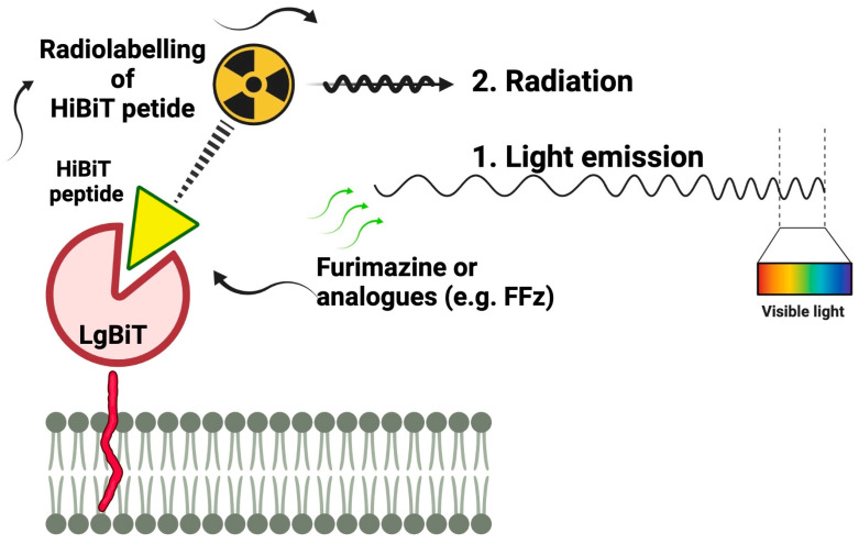
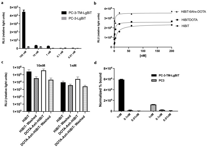
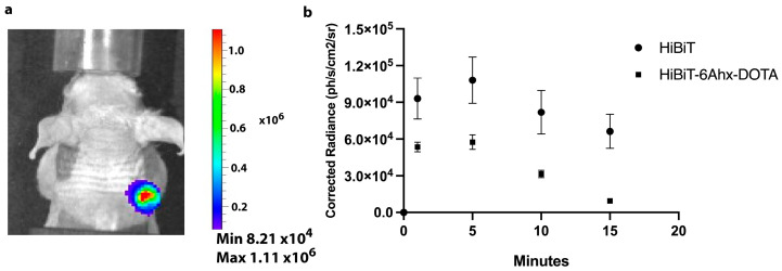
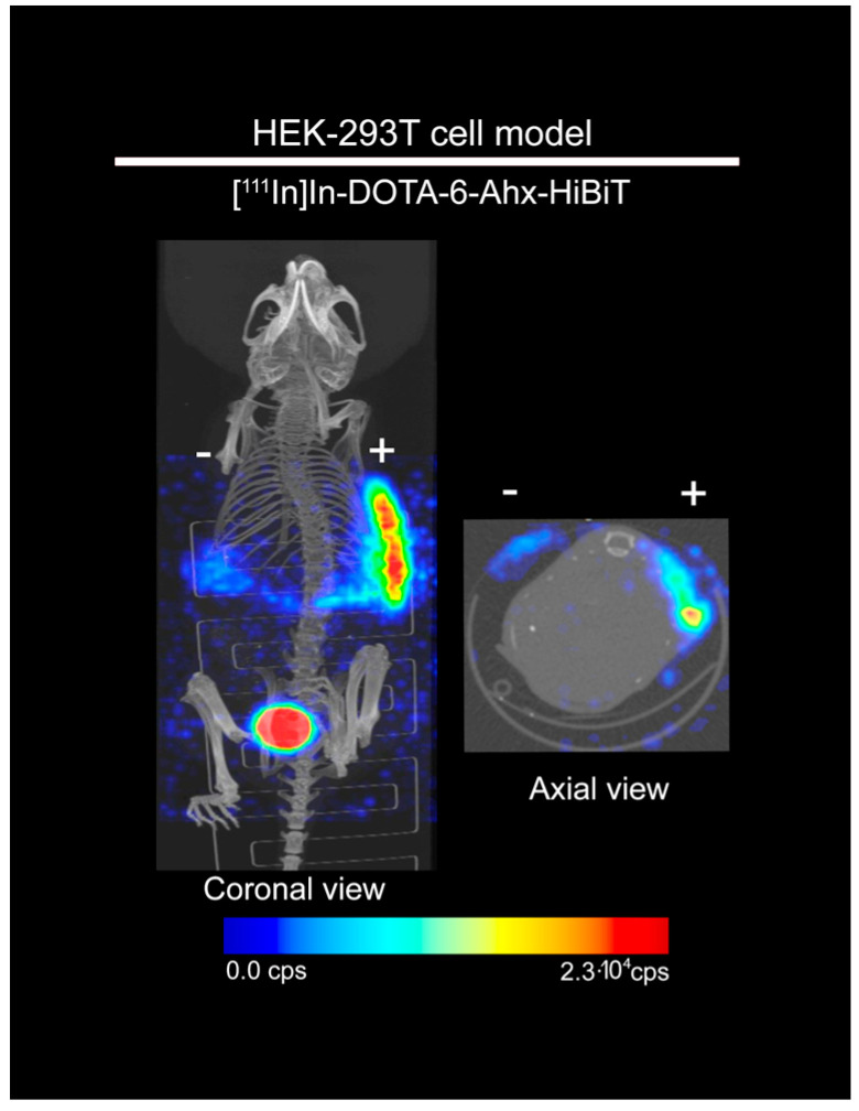
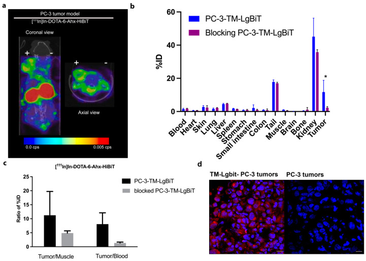
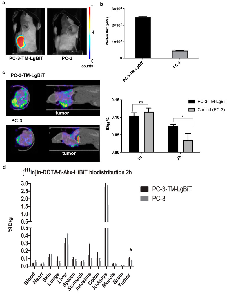
Multimodality reporter gene imaging combines the sensitivity, resolution and translational potential of two or more signals. The approach has not been widely adopted by the animal imaging community, mainly because its utility in this area is unproven. We developed a new complementation-based reporter gene system where the large component of split NanoLuc luciferase (LgBiT) presented on the surface of cells (TM-LgBiT) interacts with a radiotracer consisting of the high-affinity complementary HiBiT peptide labeled with a radionuclide. Radiotracer uptake could be imaged in mice using SPECT/CT and bioluminescence within two hours of implanting reporter-gene-expressing cells. Imaging data were validated by ex vivo biodistribution studies. Following the demonstration of complementation between the TM-LgBiT protein and HiBiT radiotracer, we validated the use of the technology in the highly specific in vivo multimodal imaging of cells. These findings highlight the potential of this new approach to facilitate the advancement of cell and gene therapies from bench to clinic.

Keywords: bioluminescence imaging; luciferase complementation; peptide tracers; radionuclide imaging; reporter gene.


Conflict of interest statement

Authors Thomas A. Kirkland, Mary P. Hall and Lance P. Encell were employed by the company Promega Corporation. The remaining authors declare that the research was conducted in the absence of any commercial or financial relationships that could be construed as a potential conflict of interest.

Figures

Figure 1
Figure 1
Schematic representation of the novel reporter gene technology. LgBiT protein is anchored to the membrane with PDGFR transmembrane domain and exposed to the extracellular space.
Figure 2
Figure 2
Characterization of TM-LgBiT reporter gene and HiBiT tracers. Luminescence signals were detected using the IVIS, validating the expression of reporter (LgBiT) when expressed on the membrane or within the cytosol of PC-3 cells upon administration of different concentrations of HiBiT peptide (a). To assess how the conjugation affects the HiBiT probe characteristics, functional and affinity data were obtained using one site specific binding function using purified LgBiT. Calculated KD values were as follows: HiBiT = 6.8 nM; DOTA-VSGWRLFKKIS = 1.3 nM; and DOTA-6-Ahx-VSGWRLFKKIS = 0.7 nM. (b). DOTA-6-Ahx-VSGWRLFKKIS and HiBiT probes’ binding specificity was evaluated on tracer-positive (TM-LgBiT) and -negative PC-3 cells based on obtained luminescence signals. DOTA-6-Ahx-VSGWRLFKKIS and HiBit bioluminescence signal in TM-Lgbit PC-3 cells was not significantly different (c). Specific binding after 1 h of incubation with 1 nM/150 MBq of [111In]In-DOTA-6-Ahx-VSGWRLFKKIS in cells expressing TM-LgBiT in comparison to control cells. At 1 nM concentration, the radioactive signal of PC-3-TM-LgBiT cells is significantly higher than in PC-3 controls (p-value < 0.05). The results shown were performed in triplicate, and the values are indicated as means ± SD (d).
Figure 3
Figure 3
Longitudinal bioluminescence imaging (BLI) of HiBiT probe distribution in mice implanted with target (TM-LgBiT)-positive tumors (a). A strong and specific bioluminescent signal from NanoLuc luciferase was detected at the tumor site 30 min after peptide (HiBiT and DOTA-6-Ahx-VSGWRLFKKIS) injection upon administration of fluorofurimazine (minutes after fluorofurimazine injection indicated in the x axis) (b).
Figure 4
Figure 4
[111In]In-DOTA-6-Ahx-VSGWRLFKKIS SPECT/CT imaging of mice implanted with target-positive and -negative HEK-293T cells. [111In]In-DOTA-6-Ahx-VSGWRLFKKIS showed higher uptake (3.6-fold) in HEK-293T target-positive cells (right flank, +) compared with the target-negative HEK-293T cells (left flank, −) at 1 h after subcutaneous administration of [111In]In-DOTA-6-Ahx-VSGWRLFKKIS probe.
Figure 5
Figure 5
[111In]In-DOTA-6-Ahx-VSGWRLFKKIS (Hibit) SPECT/CT imaging and ex vivo biodistribution in PC-3 tumor xenografts. [111In]In-DOTA-6-Ahx-VSGWRLFKKIS showed higher uptake in the TM-LgBiT-positive tumor (left flank, +) compared with the TM-LgBiT-negative tumor (right flank, −) at 1 h after i.v. administration of [111In]In-DOTA-6-Ahx-VSGWRLFKKIS tracer (a). Ex vivo biodistribution confirmed the [111In]In-DOTA-6-Ahx-VSGWRLFKKIS specificity since a significant difference in tumor uptake (* p-value < 0.05) was detected when performing blocking studies (b). Prostate cancer (PC-3) was employed to establish a xenograft model with both target-positive (PC-3-TM-LgBiT) and -negative (PC-3) tumors. Tumor-to-muscle and tumor-to-blood ratios demonstrate specific uptake of [111In]In-DOTA-6-Ahx-VSGWRLFKKIS in PC-3 TM-LgBiT tumor, with a reduced ratio in mice that were pre-administered with 100-fold excess of cold In-DOTA-6-Ahx-VSGWRLFKKIS (c). Tissue IF of PC-3-TM-LgBiT tumor cells and PC-3 tumor cells using an anti-HA antibody confirm the expression of TM-LgBiT in mouse tumors ( microscope magnification 20X) (d).
Figure 6
Figure 6
Multimodality imaging of LgBiT-TM-expressing cells. Prostate cancer (PC-3) was employed to establish a xenograft model with both target-positive (PC-3-TM-LgBiT) and -negative (PC-3) tumors. Mice receiving 1 nMol/20 mBq of [111In]In-DOTA-6-Ahx-VSGWRLFKKIS were imaged for bioluminescence 15 min after addition of fluorofurimazine substrate (a). Tumor-to-background ratio of bioluminescence signal in target-positive and -negative PC3-tumor-bearing mice after administration of fluorofurimazine substrate (b). SPECT images of target-positive (PC-3-TM-LgBiT) (left) and -negative (PC-3) (right) tumors at 2 h after injection of [111In]In-DOTA-6-Ahx-VSGWRLFKKIS. Graph reporting the tumor %ID/g calculated from images of tumors at 1 h and 2 h after injection (c). Ex vivo distribution study at 2 h after administration of [111In]In-DOTA-6-Ahx-VSGWRLFKKIS revealed significant differences between target-positive (PC-3-TM-LgBiT) and -negative (PC-3) tumor uptake (* p-value < 0.05; ns = not significantly different) (d).

References

    1. Gambhir S.S. Molecular imaging of cancer with positron emission tomography. Nat. Rev. Cancer. 2002;2:683–693. doi: 10.1038/nrc882. - DOI - PubMed
    1. Kang J., Chung J.-K. Molecular-Genetic Imaging Based on Reporter Gene Expression. J. Nucl. Med. Off. Publ. Soc. Nucl. Med. 2008;49((Suppl. 2)):164S–179S. doi: 10.2967/jnumed.107.045955. - DOI - PubMed
    1. Shaikh F.A., Kurtys E., Kubassova O., Roettger D. Reporter gene imaging and its role in imaging-based drug development. Drug Discov. Today. 2020;25:582–592. doi: 10.1016/j.drudis.2019.12.010. - DOI - PubMed
    1. Ashmore-Harris C., Iafrate M., Saleem A., Fruhwirth G.O. Non-invasive Reporter Gene Imaging of Cell Therapies, including T Cells and Stem Cells. Mol. Ther. 2020;28:1392–1416. doi: 10.1016/j.ymthe.2020.03.016. - DOI - PMC - PubMed
    1. De A., Lewis X.Z., Gambhir S.S. Noninvasive imaging of lentiviral-mediated reporter gene expression in living mice. Mol. Ther. 2003;7:681–691. doi: 10.1016/S1525-0016(03)00070-4. - DOI - PMC - PubMed

Grants and funding

LinkOut - more resources