Review
. 2024 Jul 28;25(15):8238.
doi: 10.3390/ijms25158238.

Skin Aging and the Upcoming Role of Ferroptosis in Geroscience

Affiliations
Review

Skin Aging and the Upcoming Role of Ferroptosis in Geroscience

Rita Rezzani et al. Int J Mol Sci. .

Abstract

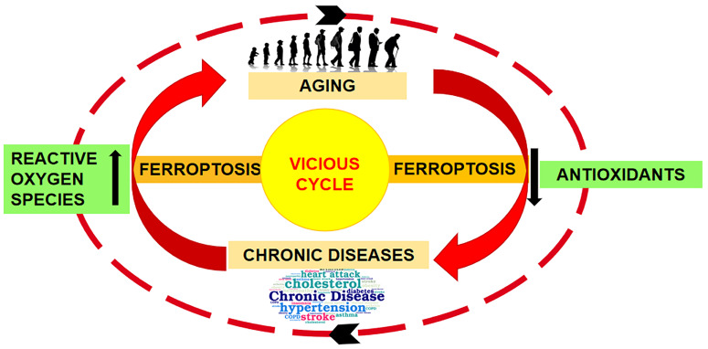
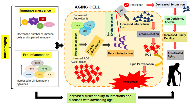
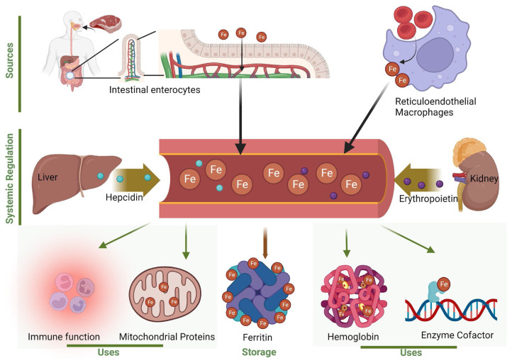
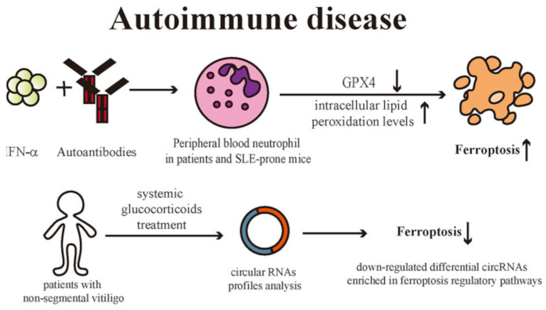
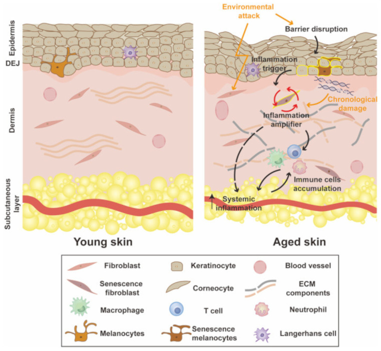
The skin is considered the most important organ system in mammals, and as the population ages, it is important to consider skin aging and anti-aging therapeutic strategies. Exposure of the skin to various insults induces significant changes throughout our lives, differentiating the skin of a young adult from that of an older adult. These changes are caused by a combination of intrinsic and extrinsic aging. We report the interactions between skin aging and its metabolism, showing that the network is due to several factors. For example, iron is an important nutrient for humans, but its level increases with aging, inducing deleterious effects on cellular functions. Recently, it was discovered that ferroptosis, or iron-dependent cell death, is linked to aging and skin diseases. The pursuit of new molecular targets for ferroptosis has recently attracted attention. Prevention of ferroptosis is an effective therapeutic strategy for the treatment of diseases, especially in old age. However, the pathological and biological mechanisms underlying ferroptosis are still not fully understood, especially in skin diseases such as melanoma and autoimmune diseases. Only a few basic studies on regulated cell death exist, and the challenge is to turn the studies into clinical applications.

Keywords: aging; autoimmune diseases; cutaneous diseases; ferroptosis; gut microbiota; melanoma; skin.


Conflict of interest statement

The authors declare no conflicts of interest.

Figures

Figure 1
Figure 1
Aging and ferroptosis. The illustration represents the aging induction of ferroptosis, which, in turn, disrupts the imbalance between oxidative stress and antioxidant defense, thereby implementing, in a vicious cycle, the aging-related damage. Illustration from Mazhar et al., 2021 [7]. (This is an open access article distributed under the terms of the Creative Commons Attribution 4.0 International license).
Figure 2
Figure 2
Aging, iron dyshomeostasis, ferroptosis, and hepidicin. The illustration represents a possible interaction among aging, iron dyshomeostasis, ferroptosis, and hepidicin, a hepatic iron-regulatory hormone. Aging increases iron stores in tissues, and the intracellular iron induces redox imbalances and cellular injury, leading to ferroptosis, which, in turn, promotes aging and associated morbidity. The aging-related increment in intracellular iron levels may be linked to the increased production of hepcidin due to underlying chronic inflammation. (GPX4): glutathione peroxidase-4; (GSH): glutathione; (NK): natural killer; (B): B lymphocytes; (CD4 T): CD4 T lymphocyte; (M): microfold cells; (MQ): macrophages; (COX2): cyclooxygenase-2; (TNF-α): tumor necrosis factor alpha; (NF-kB): nuclear factor kappa-light-chain-enhancer of activated B cells; (iNOS): inducible nitric oxide synthase; (IL-1): interleukin-1; (IL-6): interleukin-6; (ROS): reactive oxygen species; (FPN): ferroportin. Illustration from Mazhar et al., 2021 [7]. (This is an open access article distributed under the terms of the Creative Commons Attribution 4.0 International license).
Figure 3
Figure 3
Adult human skin. Representative photomicrograph of face skin from adult donors (under 65 years old). Haematoxylin-eosin staining. The skin biopsies were obtained from head cadaveric specimens (MedCure, Amsterdam, The Netherlands). Specimens were stored at −20 °C, defrosted before the anatomical dissecting session, and analyzed at the Anatomical Facility “Luigi Fabrizio Rodella” of the University of Brescia (Italy). The human cadaveric studies have been performed in accordance with the ethical standards laid down in the 1964 Declaration of Helsinki and its later amendments. Bar: 20 µm. (E): epidermis; (D): dermis; (white stars): epidermal network ridges. Illustration adapted from Favero et al., 2024 [18].
Figure 4
Figure 4
Human skin functions. The illustration summarizes the main human skin functions. Illustration adapted from Ahmed and Mikail et al,. 2024 [3].
Figure 5
Figure 5
Human skin microbiota. The skin microbiota affects the cutaneous physical barrier function. The microbiota enhances the skin’s chemical barrier by producing lipases that digest sebum triglycerides into free fatty acids, which in turn amplify skin acidity and limit colonization by transient and pathogenic species. Moreover, the skin microbiota stimulates innate and adaptive immune defenses. Illustration adapted from Harris-Tryon and Grice, 2022 [28].
Figure 6
Figure 6
Human skin aging physiopathological process. Human skin aging is related to biomechanical, structural, and physical change at nano-, micro-, and macro-scales. Illustration adapted from Park, 2022 [32].
Figure 7
Figure 7
Elderly human skin. Representative photomicrograph of face skin from elderly donors (over 65 years old). Haematoxylin-eosin staining. The skin biopsies were obtained from head cadaveric specimens (MedCure, Amsterdam, The Netherlands). Specimens were stored at −20 °C, defrosted before the anatomical dissecting session, and analyzed at the Anatomical Facility “Luigi Fabrizio Rodella” of the University of Brescia (Italy). The human cadaveric studies have been performed in accordance with the ethical standards laid down in the 1964 Declaration of Helsinki and its later amendments. Bar: 20 µm. (E): epidermis; (D): dermis; (white stars): epidermal network ridges; (arrows): melanocytes; (**): fibroblasts; (*): mast cells. Illustration adapted from Favero et al., 2024 [18].
Figure 8
Figure 8
Human epidermal thickness evaluation in adult and elderly specimens. The plot shows the epidermal thickness distribution performed on adult (under 65 years old) and elderly (over 65 years old) donor specimens. The face skin sections were stained with hematoxylin-eosin following standard procedures. The thickness of the epidermal layer of each specimen was calculated in micrometers (μm) by a blind examiner using an image analyzer (Image Pro Premier 9.1; Media Cybernetics, Rockville, MD, USA). The epidermal layer was measured from the free margin of skin to the dermal papillae and epidermal network ridge. The analysis was performed on five alternately stained sections for each skin specimen. Illustration adapted from Favero et al., 2024 [18].
Figure 9
Figure 9
Human dermal mast cell quantification in adult and elderly specimens. The plot summarizes the total number of mast cells in the dermal layer performed on adult (under 65 years old) and elderly (over 65 years old) donor specimens. The face skin sections were stained with toluidine blue. The numbers of mast cells were evaluated by a blind examiner using an optical BX50 microscope (Olympus, Hamburg, Germany) as the number of cells per field. At least five representative visual fields of five alternate sections for each skin specimen were analyzed. Illustration adapted from Favero et al., 2024 [18].
Figure 10
Figure 10
Geroscience and age-related diseases. The illustration represents the link between geroscience and aging. Genetics and environmental factors affect various cellular and physiological pathways fundamental to aging and inflammation. These factors, together with disease-specific risk factors, can increase the risk of aging-related chronic disease development. Illustration adapted from Campisi et al., 2019 [37].
Figure 11
Figure 11
Human skin dermal collagen. Representative photomicrographs of type I (a) and type III (b) collagen immunohistochemistry at the dermal layer were performed on adult (a) and old (b) skin specimens. Type I collagen immunostaining was widely distributed, whereas type III collagen immunostaining was light and sparse with an uneven distribution. Bars: 200 µm. Illustration from Cheng et al., 2011 [51]. (This is an open access article distributed under the terms of the Creative Commons CC-BY license).
Figure 12
Figure 12
Young and adult sun-protected skin. Representative photomicrographs of skin melanocytes and collagen 17 and collagen 17a immunofluorescence were performed on young (22 years old) and adult (years old) sun-protected skin specimens. Aged skin showed a flattened dermal-epidermal junction, a decreased number of melanocytes, and reduced immunostaining for collagen 17 and 17a. Bars: 50 µm. Illustration from Chin et al., 2023 [31]. (This is an open access article distributed under the terms of the Creative Commons CC-BY license).
Figure 13
Figure 13
Collagen 17 modulates interfollicular epidermis homeostasis. The illustration represents the possible mechanism of action of collagen 17 in regulating paw interfollicular epidermis homeostasis. Illustration from Watanabe et al., 2017 [54]. (This is an open access article distributed under the terms of the Creative Commons CC-BY license).
Figure 14
Figure 14
Schematic representation of human skin barrier immunity. The epidermal layer consists of T-resident memory cells (Trm), Langerhans cells, and keratinocytes; the latter form a stratified corneum with interspersed melanocytes. The dermal layer is populated by dermal dendritic cells (DCs), macrophages, Foxp3+ T regulatory cells (Tregs), CD4+ and CD8+ Trm, fibroblasts, and mast cells. The subcutaneous layer is composed of adipocytes. Illustration from Chambers et al., 2020 [23]. (License number 5835261420374).
Figure 15
Figure 15
Skin immune functions. The graph summarizes the implications of immune cells for skin immune functions. Illustration adapted from Agrawal et al., 2023 [105].
Figure 16
Figure 16
Iron homeostasis. The illustration represents an overview of the iron sources, the systemic processes that balance the iron level, and its cellular uses. The regulation of iron homeostasis involves the absorption, transport, storage, recycling, and utilization of iron. Illustration from Zeidan et al., 2024 [125]. (This is an open access article distributed under the terms of the Creative Commons CC-BY license).
Figure 17
Figure 17
Ultraviolet irradiation and ferritin. The illustration represents the ultraviolet irradiation injury at skin level. The skin’s ultraviolet irradiation increases cell-free iron, promoting ferritin degradation, which, in a vicious cycle, implements the free iron release. (NF-kB): nuclear factor kappa-light-chain-enhancer of activated B cells. Illustration adapted from Pouillot et al., 2014 [127].
Figure 18
Figure 18
Programmed or non-programmed cell death. The illustration compares the main morphologic features of programmed and non-programmed cell death: ferroptosis, pyroptosis, apoptosis, and necrosis. Illustration from Khorsandi et al., 2023 [130]. (This is an open access article distributed under the terms of the Creative Commons Attribution 4.0 International license).
Figure 19
Figure 19
The development of the ferroptosis concept. Timeline diagram of last decades’ history observations on regulated cell deaths and the advances that contributed to the emergence of the concept of ferroptosis. Ferroptosis was observed in various contexts before coining of the term in 2012. Illustration adapted from Hirschhorn and Stockwell, 2019 [135].
Figure 20
Figure 20
Ferroptosis hallmarks. The illustration summarizes the three main hallmarks that promote ferroptotic death: oxidizable phospholipids acylated with polyunsaturated fatty acids, redox-active iron, and defective lipid peroxide repair. Illustration adapted from Dixon and Stockwell, 2019 [143].
Figure 21
Figure 21
Ferroptosis cell pathways. The illustrations summarize the main conditions that can trigger cell ferroptosis. (TFR1): transferrin receptor 1; (FPN): ferroportin; (DMT1): divalent metal transporter 1; (STEAP3): six-transmembrane epithelial antigens of the prostate 3; (NCOA4): Nuclear receptor coactivator 4; (LIP): labile iron pool; (ROS): reactive oxygen species; (SLC3A2): Solute Carrier Family 3 Member 2; (SLC7A11): Solute Carrier Family 7 Member 11; (PUFAs): polyunsaturated fatty acids; (AA/AdA): arachidonic acid/adrenic acid (AdA); (AA/AdA-Pes): AA/AdAphosphatidylethanolamine (PE); (AA/AdA-PEs-OH): AA/AdA-PEs-alcohols; (AA/AdA-PEs-OOH): AA/AdA-PEs-hydroperoxides; (GPX4): glutathione peroxidase-4; (GSH): glutathione; (GSSG): glutathione disulfide; (LOXs): lipoxygenases; (ACSL4/LPCAT3): acyl-CoA synthetase long-chain family member 4/lysophosphatidylcholine acyltransferase 3. Illustration (A) is from Liu et al., 2023 [140]. (This is an open access article distributed under the term of the Creative Commons CC-BY license). Illustration (B) is from Khorsandi et al., 2023 [130]. (This is an open access article distributed under the terms of the Creative Commons Attribution 4.0 International license).
Figure 22
Figure 22
Ferroptosis and gut microbiota. The illustration represents the influence of gut microbiota on ferroptosis in various tissues, organs, and diseases. Illustration from Mao et al., 2024 [152]. (This is an open access article distributed under the terms of the Creative Commons Attribution 4.0 International license).
Figure 23
Figure 23
Dermatological manifestation of vitiligo. The illustration showed the main dermatological features of vitiligo. Cutaneous chronic depigmentation and white lesions of a vitiligo patient (A). Comparison of skin morphology between a healthy subject and a vitiligo patient. Haematoxylin-eosin staining. Black arrows indicate melanocytes (×400) (B). Comparison of skin melanocytes between a healthy subject and a vitiligo patient underling. Melan-A (red staining) immunofluorescence. Nuclei have been counterstained with 4′,6′-diamidino-2-phenylindole (blue staining). Bar = 100 µm. The white arrows indicated melanocytes (C). (B,C) underline the absence of melanocytes in vitiligo skin. Illustration from Chen et al., 2020 [158]. (This is an open access article distributed under the terms of the Creative Commons CC-BY license).
Figure 24
Figure 24
Melanocytes in vitiligo. The illustration represents schematically the main hallmarks that promote melanocyte destruction, resulting in the depletion of melanocytes in vitiligo lesional skin. (ROS): reactive oxygen species; (RCD): regulated cell death. Illustration adapted from Chen et al., 2020 [158].
Figure 25
Figure 25
Autoimmune diseases and ferroptosis. The illustration summarizes the ferroptosis pathway in vitiligo-affected patients. (IFN-α): interferon-α; (GPX4): glutathione peroxidase-4; (SLE): systemic lupus erythematosus; (circRNA): circular RNAs. Illustration from Liu et al., 2023 [140]. (This is an open access article distributed under the terms of the Creative Commons CC-BY license).
Figure 26
Figure 26
Young vs. old human skin. The illustration compares human young skin to human aged skin. Aged skin is characterized by an altered epithelial barrier, a thin and impaired dermal layer, inflammation, and excessive senescent fibroblasts and immune cells. Illustration from Zhang et al., 2024 [219]. (This is an open access article distributed under the terms of the Creative Commons CC-BY license).

Similar articles

Cited by

References

    1. Ahmed I.A., Mikail M.A., Zamakshshari N., Abdullah A.S.H. Natural Anti-Aging Skincare: Role and Potential. Biogerontology. 2020;21:293–310. doi: 10.1007/s10522-020-09865-z. - DOI - PubMed
    1. Ahmed I.A., Mikail M.A. Anti-Aging Skincare: The Natural and Organic Way. In: Koltover V.K.B.T.-A.-A.P., editor. Anti-Aging Pharmacology. Academic Press; Cambridge, MA, USA: 2023. pp. 269–284.
    1. Ahmed I.A., Mikail M.A. Diet and Skin Health: The Good and the Bad. Nutrition. 2024;119:112350. doi: 10.1016/j.nut.2023.112350. - DOI - PubMed
    1. Franco A.C., Aveleira C., Cavadas C. Skin Senescence: Mechanisms and Impact on Whole-Body Aging. Trends Mol. Med. 2022;28:97–109. doi: 10.1016/j.molmed.2021.12.003. - DOI - PubMed
    1. He X., Gao X., Xie W. Research Progress in Skin Aging, Metabolism, and Related Products. Int. J. Mol. Sci. 2023;24:15930. doi: 10.3390/ijms242115930. - DOI - PMC - PubMed

LinkOut - more resources