. 2024 Jul 30;25(15):8340.
doi: 10.3390/ijms25158340.

Establishment and Characterization of a Chicken Myoblast Cell Line

Affiliations

Establishment and Characterization of a Chicken Myoblast Cell Line

Dongxue Guo et al. Int J Mol Sci. .

Abstract

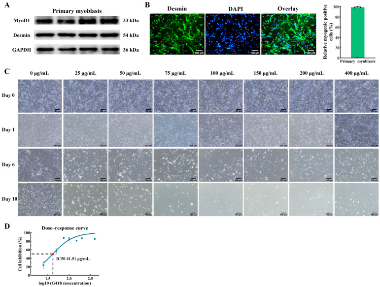
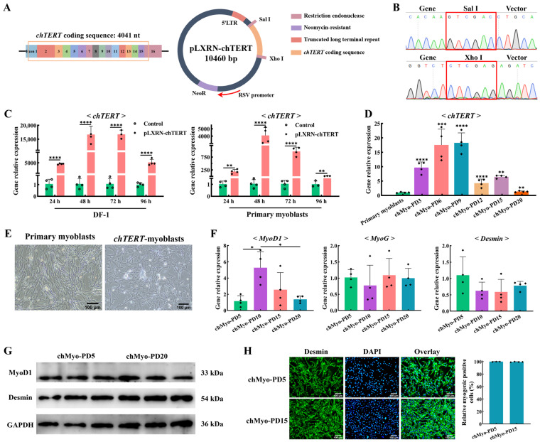
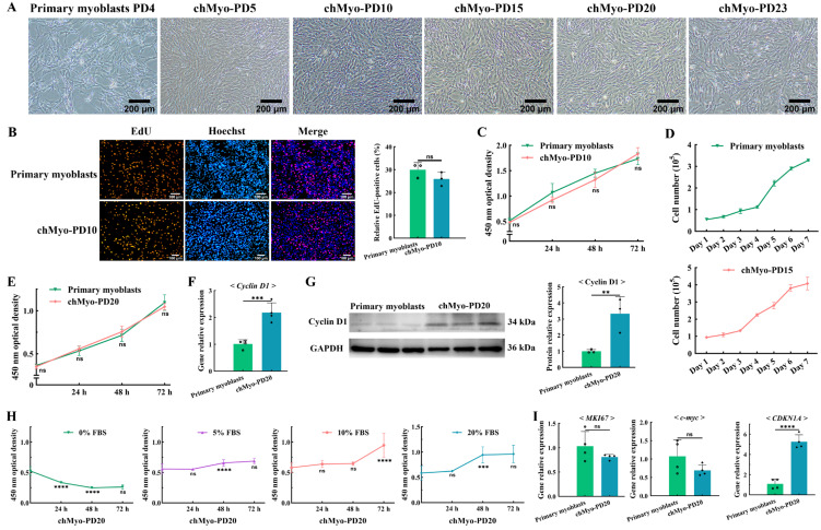
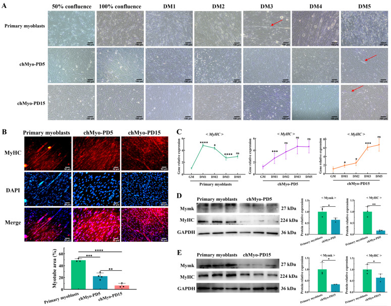
Skeletal muscle, which is predominantly constituted by multinucleated muscle fibers, plays a pivotal role in sustaining bodily movements and energy metabolism. Myoblasts, which serve as precursor cells for differentiation and fusion into muscle fibers, are of critical importance in the exploration of the functional genes associated with embryonic muscle development. However, the in vitro proliferation of primary myoblasts is inherently constrained. In this study, we achieved a significant breakthrough by successfully establishing a chicken myoblast cell line through the introduction of the exogenous chicken telomerase reverse transcriptase (chTERT) gene, followed by rigorous G418-mediated pressure screening. This newly developed cell line, which was designated as chTERT-myoblasts, closely resembled primary myoblasts in terms of morphology and exhibited remarkable stability in culture for at least 20 generations of population doublings without undergoing malignant transformation. In addition, we conducted an exhaustive analysis that encompassed cellular proliferation, differentiation, and transfection characteristics. Our findings revealed that the chTERT-myoblasts had the ability to proliferate, differentiate, and transfect after multiple rounds of population doublings. This achievement not only furnished a valuable source of homogeneous avian cell material for investigating embryonic muscle development, but also provided valuable insights and methodologies for establishing primary cell lines.

Keywords: G418 screening; chTERT; chicken; myoblast cell line; proliferation and differentiation.


Conflict of interest statement

The authors declare no conflicts of interest.

Figures

Figure 1
Figure 1
Myogenic identification of chicken primary myoblasts and cell screening by G418 with different concentrations. (A) Relative protein expression levels of MyoD1 and Desmin in the primary myoblasts after inducing differentiation for 48 h. (B) Primary myoblasts were stained with the myogenic cell marker Desmin (green) and DAPI (blue). The scale bar represents 100 μm. (C) The chicken primary myoblasts were screened by different concentrations of G418. “Day 0” represents the cells after 24 h of proliferation, and “Day n (n ≠ 0)” represents the cells after n days of G418 screening. Magnification 100×, scale bar 100 μm. (D) The determination of the minimum lethal dose of G418 on the chicken primary myoblasts.
Figure 2
Figure 2
Immortalization of chicken primary myoblasts and cell line identification. (A) Structure diagram of the pLXRN-chTERT plasmid. (B) Sequencing results of the pLXRN-chTERT plasmid. The red box indicates different restriction site. (C) The relative expression levels of chTERT in different chicken-derived cells transfected with the pLXRN-chTERT for 24, 48, 72, and 96 h (mean ± SD; ** p < 0.01; **** p < 0.0001; n = 4). (D) The relative expression levels of chTERT in chicken primary myoblasts and chTERT-myoblasts at different population doublings (chMyo-PDs) (mean ± SD; ** p < 0.01; *** p < 0.001; **** p < 0.0001; n = 4). (E) Light microscopy of chicken primary myoblasts (left) and chTERT-myoblasts (right). (F) The relative expression levels of MyoD, MyoG, and Desmin in chTERT-myoblasts at different PDs after inducing differentiation for 48 h (mean ± SD; * p < 0.05; n = 4). (G) Relative protein expression level of MyoD1 and Desmin in chTERT-myoblasts at PDs 5 and 20 after inducing differentiation for 48 h. (H) chTERT-myoblasts at PDs 5 and 20 were stained with the myogenic cell marker Desmin (green) and DAPI (blue).
Figure 3
Figure 3
Proliferation characteristics of chTERT-myoblasts. (A) Light microscopy of chicken primary myoblasts and chTERT-myoblasts at different population doublings (chMyo-PDs). (B) Proliferation of chicken primary myoblasts and chTERT-myoblasts at PD 10 were assessed by EdU (mean ± SD; ns represents non-significant; n = 3). (C) CCK-8 assay on chicken primary myoblasts and chTERT-myoblasts at PD 10 (mean ± SD; ns represents non-significant; n = 6). (D) Cell growth curve of chicken primary myoblasts and chTERT-myoblasts at PD 15 (mean ± SD; n = 3). (E) CCK-8 assay on chicken primary myoblasts and chTERT-myoblasts at PD 20 (mean ± SD; ns represents non-significant; n = 6). (F) Relative expression level of proliferation marker gene in chTERT-myoblasts at PD 20 by qPCR (mean ± SD; *** p < 0.001; n = 4). (G) Relative protein expression level of proliferation marker gene in chTERT-myoblasts at PD 20 (mean ± SD; ** p < 0.01; n = 3). (H) Serum-dependent analysis on chTERT-myoblasts at different population doublings (mean ± SD; ns represents non-significant; *** p < 0.001; **** p < 0.0001; n = 6). (I) Relative expression level of oncogenes and tumor suppressor genes in chTERT-myoblasts at PD 20 by qPCR (mean ± SD; ns represents non-significant; **** p < 0.0001; n = 4).
Figure 4
Figure 4
Differentiation characteristics of chTERT-myoblasts. (A) Light microscopy of chicken primary myoblasts and chTERT-myoblasts at PDs 5 (chMyo-PD5) and 15 (chMyo-PD15) in differentiation medium (DM) for different days. The red arrows indicate the formed myotubes. (B) After inducing differentiation for 48 h, myotubes derived from chicken primary myoblasts and chTERT-myoblasts at PDs 5 and 15 by performing immunofluorescence staining against MyHC (red), and nuclei were counterstained with DAPI (mean ± SD; ** p < 0.01; *** p < 0.001; **** p < 0.0001; n = 3). (C) Relative expression levels of differentiation marker gene on chicken primary myoblasts and chTERT-myoblasts at PDs 5 and 15 in differentiation medium (DM) for different days (mean ± SD; * p < 0.05; *** p < 0.001; **** p < 0.0001; ns represents non-significant; n = 4). (D) Relative protein expression levels of differentiation marker genes in chicken primary myoblasts and chTERT-myoblasts at PD 5 after inducing differentiation for 48 h (mean ± SD; * p < 0.05; ** p < 0.01; n = 3). (E) Relative protein expression levels of differentiation marker genes in chicken primary myoblasts and chTERT-myoblasts at PD 15 after inducing differentiation for 48 h (mean ± SD; * p < 0.05; n = 3).
Figure 5
Figure 5
Analysis of transfection efficiency of chTERT-myoblasts. (A) Schematic diagram of pcDNA3.1-SOX9 structure. The orange box represents the coding sequence of SOX9. (B) Schematic diagram of pCD2.1-circIGF2BP3 structure. The orange box represents the cyclization sequence of circIGF2BP3. (C) Relative expression levels of SOX9 in chTERT-myoblasts at PD 15 transfected with pcDNA3.1-SOX9 (mean ± SD; *** p < 0.001; **** p < 0.0001; n = 4). (D) Relative expression levels of circIGF2BP3 in chTERT-myoblasts at PD 15 transfected with pCD2.1-circIGF2BP3 (mean ± SD; *** p < 0.001; **** p < 0.0001; n = 4). (E) Bright-field image before cell transfection and the fluorescence images of chTERT-myoblasts at PD 15 transfected with pCD2.1-circIGF2BP3.
Figure 6
Figure 6
The schematic diagram of TERT promoting telomere elongation.

Similar articles

Cited by

References

    1. Oluwagbenga E.M., Fraley G.S. Heat stress and poultry production: A comprehensive review. Poult. Sci. 2023;102:103141. doi: 10.1016/j.psj.2023.103141. - DOI - PMC - PubMed
    1. Comai G., Tajbakhsh S. Molecular and Cellular Regulation of Skeletal Myogenesis. In: Taneja R., editor. Current Topics in Developmental Biology. Volume 110. Academic Press; Cambridge, MA, USA: 2014. pp. 1–73. - DOI - PubMed
    1. Chal J., Pourquié O. Making muscle: Skeletal myogenesis in vivo and in vitro. Development. 2017;144:2104–2122. doi: 10.1242/dev.151035. - DOI - PubMed
    1. Lehka L., Rędowicz M.J. Mechanisms regulating myoblast fusion: A multilevel interplay. Semin. Cell Dev. Biol. 2020;104:81–92. doi: 10.1016/j.semcdb.2020.02.004. - DOI - PubMed
    1. Richler C., Yaffe D. The in vitro cultivation and differentiation capacities of myogenic cell lines. Dev. Biol. 1970;23:1–22. doi: 10.1016/S0012-1606(70)80004-5. - DOI - PubMed

LinkOut - more resources