. 2024 Aug 1;25(15):8395.
doi: 10.3390/ijms25158395.

Copper(II) and Zinc(II) Complexes with Bacterial Prodigiosin Are Targeting Site III of Bovine Serum Albumin and Acting as DNA Minor Groove Binders

Affiliations

Copper(II) and Zinc(II) Complexes with Bacterial Prodigiosin Are Targeting Site III of Bovine Serum Albumin and Acting as DNA Minor Groove Binders

Lena Pantelic et al. Int J Mol Sci. .

Abstract

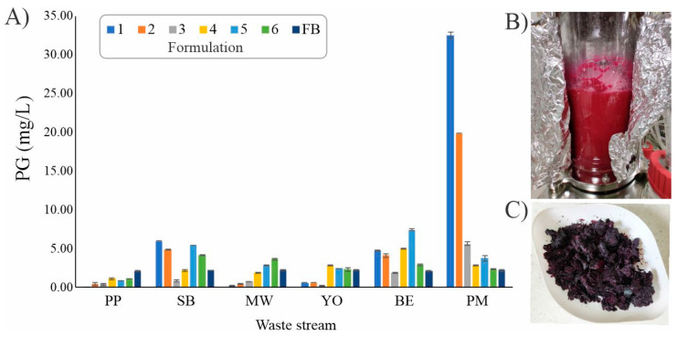
The negative environmental and social impacts of food waste accumulation can be mitigated by utilizing bio-refineries' approach where food waste is revalorized into high-value products, such as prodigiosin (PG), using microbial bioprocesses. The diverse biological activities of PG position it as a promising compound, but its high production cost and promiscuous bioactivity hinder its wide application. Metal ions can modulate the electronic properties of organic molecules, leading to novel mechanisms of action and increased target potency, while metal complex formation can improve the stability, solubility and bioavailability of the parent compound. The objectives of this study were optimizing PG production through bacterial fermentation using food waste, allowing good quantities of the pure natural product for further synthesizing and evaluating copper(II) and zinc(II) complexes with it. Their antimicrobial and anticancer activities were assessed, and their binding affinity toward biologically important molecules, bovine serum albumin (BSA) and DNA was investigated by fluorescence emission spectroscopy and molecular docking. The yield of 83.1 mg/L of pure PG was obtained when processed meat waste at 18 g/L was utilized as the sole fermentation substrate. The obtained complexes CuPG and ZnPG showed high binding affinity towards target site III of BSA, and molecular docking simulations highlighted the affinity of the compounds for DNA minor grooves.

Keywords: DNA interactions; Serratia marcescens; bio-refinery; metal complexation; prodigiosin; protein interactions; waste.


Conflict of interest statement

The authors declare no conflict of interest.

Figures

Figure 1
Figure 1
(A) The influence of various waste streams and their formulations on PG production in S. marcescens ATCC 27117: potato peel (PP), stale bread (SB), mixed waste (MW), yogurt (YO), peeled boiled eggs (BE), processed meat (PM); (B) the bioreactor at the end of the PM-1 fermentation; (C) lyophillized S. marcescens ATCC 27117 cells.
Figure 2
Figure 2
Structural formulae of copper(II) and zinc(II) complexes with PG [49].
Figure 3
Figure 3
BSA fluorescence emission spectra in the presence of an increasing concentration of CuPG (A) and in the presence of the site markers (BD). The red arrow shows the changes in the intensity after the addition of the complex. The inserted graph presents the F0/F dependence of complex concentration.
Figure 4
Figure 4
A three-dimensional representation of the most stable conformations of the CuPG complex bound to three different active sites (I (subdomain IIA), II (subdomain IIIA), III (subdomain IB)) of BSA (PDB: 4F5S).
Figure 5
Figure 5
The most stable docking positions of CuPG (A) and ZnPG (B) in subdomain IB (active site III) of BSA (PDB code: 4F5S) are represented in three dimensions. The complexes are shown as gray sticks for carbon atoms, with other atoms colored differently: nitrogen (N) in blue, oxygen (O) in red, chlorine (Cl) in green, copper (Cu) in pink and zinc (Zn) in violet. To enhance clarity, the remaining protein structure is not presented. The two-dimensional representations illustrate the interactions between CuPG (C) and ZnPG (D) and BSA, including interatomic distances (Å) from the molecular docking study. Different colors in the ZnPG interaction diagram represent various types of interactions, as indicated in the legend.
Figure 6
Figure 6
Fluorescence emission spectra of the ct-DNA–EthBr system in the presence of increasing concentrations of PG (A), CuPG (B) and ZnPG (C). The red arrow shows the changes in the intensity upon addition of the studied compound. The inserted graph represents the Stern–Volmer plot of the F0/F vs. [compound].
Figure 7
Figure 7
Fluorescence emission spectra for the ct-DNA–Hoe system in the presence of increasing concentrations of PG (A), CuPG (B) and ZnPG (C). The red arrow shows the changes in the intensity upon addition of the studied compound. The inserted graph represents the Stern–Volmer plot of the F0/F vs. [compound].
Figure 8
Figure 8
A representation of the three-dimensional structures of CuPG (A) and ZnPG (B) in their most stable conformations as intercalators within the hexanucleotide d(CGATCG)2 (PDB code: 1Z3F). The sugar–phosphate backbones of the two complementary strands are shown as helically twisted white bands, with nucleobases in blue. Two-dimensional diagrams illustrate the interactions between CuPG (C) and ZnPG (D) with the nucleotides, including interatomic distances (Å) obtained from molecular docking simulations. The nucleotides are designated as DA (deoxyadenosine), DG (deoxyguanosine), DC (deoxycytidine) and DT (deoxythymidine). Different colors represent various types of interactions, as indicated in the legend. The investigated compounds are represented as gray sticks (carbon atoms), with spheres of different colors indicating specific atoms: N (blue), O (red), Cl (green), Cu (pink) and Zn (violet).
Figure 9
Figure 9
A representation of the three-dimensional structures of CuPG (A) and ZnPG (B) in their most stable conformations as minor groove binders within the dodecamer d(CGCGAATTCGCG)2 (PDB code: 1BNA). The sugar–phosphate backbones of the two complementary strands are shown as helically twisted white bands, with nucleobases in blue. Two-dimensional diagrams illustrate the interactions between CuPG (C) and ZnPG (D) with the nucleotides, including interatomic distances (Å) obtained from molecular docking simulations. The nucleotides are designated as DA (deoxyadenosine), DG (deoxyguanosine), DC (deoxycytidine) and DT (deoxythymidine). Different colors represent various types of interactions, as indicated in the legend. The investigated compounds are represented as gray sticks (carbon atoms), with spheres of different colors indicating specific atoms: N (blue), O (red), Cl (green), Cu (pink) and Zn (violet).

Similar articles

Cited by

References

    1. Gustavsson J., Cederberg C., Sonesson U., van Otterdijk R., Meybeck A. Global Food Losses and Food Waste: Extent, Causes and Prevention. Food and Agriculture Organization of the United Nations; Rome, Italy: 2011.
    1. Said Z., Sharma P., Thi Bich Nhuong Q., Bora B.J., Lichtfouse E., Khalid H.M., Luque R., Nguyen X.P., Hoang A.T. Intelligent approaches for sustainable management and valorisation of food waste. Bioresour. Technol. 2023;377:128952. doi: 10.1016/j.biortech.2023.128952. - DOI - PubMed
    1. Vyas S., Matsakas L., Rova U., Christakopoulos P., Patel A. Insights into hydrophobic waste valorization for the production of value-added oleochemicals. Microb. Biotechnol. 2023;16:177. doi: 10.1111/1751-7915.14122. - DOI - PMC - PubMed
    1. Leong H.Y., Chang C.K., Khoo K.S., Chew K.W., Chia S.R., Lim J.W., Chang J.-S., Show P.L. Waste biorefinery towards a sustainable circular bioeconomy: A solution to global issues. Biotechnol. Biofuels. 2021;14:87. doi: 10.1186/s13068-021-01939-5. - DOI - PMC - PubMed
    1. Venil C.K., Zakaria Z.A., Ahmad W.A. Bacterial pigments and their applications. Process Biochem. 2013;48:1065–1079. doi: 10.1016/j.procbio.2013.06.006. - DOI

MeSH terms

LinkOut - more resources