. 2024 Aug 2;25(15):8470.
doi: 10.3390/ijms25158470.

Neutrophil-like Monocytes Increase in Patients with Colon Cancer and Induce Dysfunctional TIGIT+ NK Cells

Affiliations

Neutrophil-like Monocytes Increase in Patients with Colon Cancer and Induce Dysfunctional TIGIT+ NK Cells

Alessia Calabrò et al. Int J Mol Sci. .

Abstract

Myeloid-derived suppressor cells (MDSCs) are a heterogeneous family of immune cells including granulocytic (CD14neg/CD15+/HLA-DRneg) and monocytic subtypes (CD14+/CD15neg/HLA-DRneg). In the present study, we found a population of monocytes expressing the granulocyte marker CD15 that significantly increased in both peripheral blood (PB) and tumoral tissues of patients with colorectal cancer (CRC). Further phenotypical analysis confirmed the granulocytic-like features of this monocyte subpopulation that is associated with an increase in granulocyte-monocyte precursors (GMPs) in the PB of these patients (pts). Mechanistically, this granulocyte-like monocyte population suppressed NK cell activity by inducing TIGIT and engaging NKp30. Accordingly, an increased frequency of TIGIT+ NK cells with impaired functions was found in both the PB and tumoral tissue of CRC pts. Collectively, we provided new mechanistic explanations for tumor immune escape occurring in CRC by showing the increase in this new kind of MDSC, in both PB and CRC tissue, which is able to significantly impair the effector functions of NK cells, thereby representing a potential therapeutic target for cancer immunotherapy.

Keywords: CRC; MDSCs; NK cells; TIGIT; human; monocytes; neutrophil-like cells.


Conflict of interest statement

The authors declare no conflicts of interest.

Figures

Figure 1
Figure 1
M−MDSCs expressing the granulocyte marker CD15 increase in the blood and tumor tissue of CRC pts. (A) Gating strategy used for identification of M-MDSCs (left) and representative dot plot showing CD15+ monocytes in PB of CRC pts, HD and tissues of CRC pts (right). Bars represent the frequency ±  SEM of CD15+ monocytes or the MFI of CD15 antigen (n = 30) * p < 0.05; ** p < 0.01; *** p < 0.001. (B) CD15 expression was assessed by imaging flow cytometry of FACS-sorted purified neutrophils and CD15+ and CD15neg monocytes from CRC pts. Comparative brightfield (BF), nuclear dye DAPI, CD14, CD15 and channel merge profile are shown. (C) Hematoxylin and eosin staining on CD15+, CD15neg monocytes and neutrophils from CRC pts. (D) Flow cytometer analysis of morphological parameters (left) and scatter width (right) of the indicated populations from CRC pts.
Figure 2
Figure 2
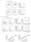
CD15+ monocytes display a granulocyte-like profile and are associated with an increase in circulating GMPs in CRC pts. (A,B) Histogram and relative statistical analysis showing the expression of the indicated markers associated with monocytic (A) and granulocytic (B) profiles, assessed on CD15+, CD15neg monocytes and neutrophils from CRC pts. Fluorescence minus one (FMO) staining was used as control. * p < 0.05; ** p < 0.01; *** p < 0.001. (C) Correlation between the frequency of CD15+ monocytes and neutrophils and monocytes and lymphocytes. (D) Bars represent the ratio between neutrophil-to-lymphocyte (NLR) or between CD15+ monocytes and lymphocytes (CD15+ monoLR) assessed on HD and CRC pts. * p < 0.05; ** p < 0.01. (E) Gating strategy used for identification of myeloid precursors and representative dot plot showing circulating granulocyte–monocyte progenitors (GMPs) in CRC pts (LIN neg: CD3, CD19, CD56). Bars represent the percentage ± SEM of GMPs (n = 5). ** p < 0.01. n.s.= not significant.
Figure 3
Figure 3
Frequency of dysfunctional TIGIT+ NK cells increases in CRC pts and correlates with that of CD15+ monocytes. (A) Representative dot plots and relative statistical analysis showing the expression of TIGIT on NK cells from PB of CRC pts and HD (upper panel) and tissues of CRC pts (lower panel). Bars represent MFI ± SEM of TIGIT+ NK cells (n = 12) * p < 0.05; (B) representative dot plots and relative statistical analysis showing the expression of CD107a and the production of IFN-γ by PMA/Iono-stimulated NK cells from CRC pts and HD with respect to TIGIT expression. Bars represent percentage ± SEM of CD107a+ and IFN-γ+ NK cells (n = 14) ** p < 0.01; *** p < 0.001; correlation between TIGIT+ NK cells and IFN-γ+ or CD107a+ NK cells from HD (blue) and CRC pts (red). (C) Correlation between the frequency of CD15+ monocytes and TIGIT+ NK cells in both PB and tissues.
Figure 4
Figure 4
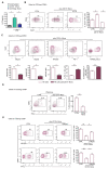
CD15+ monocytes induce dysfunctional NK cells via IL-10 and NKp30 engagement. (A) IL-10 concentration was measured in supernatants of CD15+ monocytes and CD15neg monocytes isolated from CRC pts (n = 4). Unstimulated monocytes were used as control. * p < 0.05. (B) Expression of TIGIT assessed on FACS-sorted NK cells from HD following 3 days of coculture with CD15+ monocytes from CRC pts in the presence of IL-10 blocking mAb. Bars indicate percentage ± SEM of TIGIT+ NK cells (n = 5) * p < 0.05. (C) Expression of activating receptors (NKp30, NKp46 and NKG2D) and inhibitory receptors (PD-1 and KIR2DL2/DL3) was assessed on HD-NK cells based on TIGIT expression following coculture with CD15+ monocytes from CRC. Bars indicate MFI ± SEM of the indicated makers. Grey contours represent negative controls for the indicated markers. (D) IFN-γ production was assessed on PMA/Iono-stimulated NK cells upon coculture with CD15+ monocytes. Bars represent frequency ± SEM of IFN-γ+ NK cells (n = 5), ** p < 0.01. (E) CD107a expression and IFN-γ production by NK cells following coculture with CD15+ monocytes in the presence or absence of NKp30 blocking mAb. Bars indicate percentage ± SEM of CD107a+ and IFN-γ + NK cells. PMA/Iono-stimulated NK cells from HD were used as control (n = 5) * p < 0.05; ** p < 0.01.
Figure 5
Figure 5
TIGIT limits NK cell-mediated tumor recognition in CRC pts. (A) Representative dot plots showing expression of TIGIT and DNAM-1 on NK cells from HD, CRC pts, or following coculture with CD15+ monocytes. Bars represent MFI ± SEM of DNAM-1 * p < 0.05. (B) Representative histograms showing the expression of PVR and Nectin-2 on Caco-2 and K562 cell line. FMO was used as control. (C) Representative dot plots and relative statistical analysis showing the expression of CD107a on NK cells from CRC pts following 6h of degranulation assay against K562 cells and Caco-2 cells in the presence or absence of TIGIT blocking mAb. Human IgG1 kappa isotype was used as control. Bars indicate percentage ± SEM of CD107a +NK cells (n = 3) * p < 0.05. n.s.= not significant.

References

    1. Ghorani E., Swanton C., Quezada S.A. Cancer cell-intrinsic mechanisms driving acquired immune tolerance. Immunity. 2023;56:2270–2295. doi: 10.1016/j.immuni.2023.09.004. - DOI - PubMed
    1. Zou W. Immunosuppressive networks in the tumour environment and their therapeutic relevance. Nat. Rev. Cancer. 2005;5:263–274. doi: 10.1038/nrc1586. - DOI - PubMed
    1. Zou W. Immune regulation in the tumor microenvironment and its relevance in cancer therapy. Cell. Mol. Immunol. 2022;19:1–2. doi: 10.1038/s41423-021-00738-0. - DOI - PMC - PubMed
    1. Lasser S.A., Ozbay Kurt F.G., Arkhypov I., Utikal J., Umansky V. Myeloid-derived suppressor cells in cancer and cancer therapy. Nat. Rev. Clin. Oncol. 2024;21:147–164. doi: 10.1038/s41571-023-00846-y. - DOI - PubMed
    1. Corzo C.A., Cotter M.J., Cheng P., Cheng F., Kusmartsev S., Sotomayor E., Padhya T., McCaffrey T.V., McCaffrey J.C., Gabrilovich D.I. Mechanism regulating reactive oxygen species in tumor-induced myeloid-derived suppressor cells. J. Immunol. 2009;182:5693–5701. doi: 10.4049/jimmunol.0900092. - DOI - PMC - PubMed

Substances

LinkOut - more resources