. 2024 Aug 5;25(15):8547.
doi: 10.3390/ijms25158547.

Connexin 43 Modulation in Human Chondrocytes, Osteoblasts and Cartilage Explants: Implications for Inflammatory Joint Disorders

Affiliations

Connexin 43 Modulation in Human Chondrocytes, Osteoblasts and Cartilage Explants: Implications for Inflammatory Joint Disorders

Elena Della Morte et al. Int J Mol Sci. .

Abstract

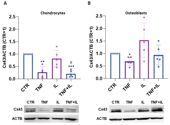
Connexin 43 (Cx43) is crucial for the development and homeostasis of the musculoskeletal system, where it plays multifaceted roles, including intercellular communication, transcriptional regulation and influencing osteogenesis and chondrogenesis. Here, we investigated Cx43 modulation mediated by inflammatory stimuli involved in osteoarthritis, i.e., 10 ng/mL Tumor Necrosis Factor alpha (TNFα) and/or 1 ng/mL Interleukin-1 beta (IL-1β), in primary chondrocytes (CH) and osteoblasts (OB). Additionally, we explored the impact of synovial fluids from osteoarthritis patients in CH and cartilage explants, providing a more physio-pathological context. The effect of TNFα on Cx43 expression in cartilage explants was also assessed. TNFα downregulated Cx43 levels both in CH and OB (-73% and -32%, respectively), while IL-1β showed inconclusive effects. The reduction in Cx43 levels was associated with a significant downregulation of the coding gene GJA1 expression in OB only (-65%). The engagement of proteasome in TNFα-induced effects, already known in CH, was also observed in OB. TNFα treatment significantly decreased Cx43 expression also in cartilage explants. Of note, Cx43 expression was halved by synovial fluid in both CH and cartilage explants. This study unveils the regulation of Cx43 in diverse musculoskeletal cell types under various stimuli and in different contexts, providing insights into its modulation in inflammatory joint disorders.

Keywords: Connexin 43; IL-1β; TNFα; cartilage explants; chondrocytes; osteoarthritis; osteoblasts; synovial fluid.


Conflict of interest statement

The authors declare that there are no conflicts of interest.

Figures

Figure 1
Figure 1
(A,B). Expression of Cx43 in TNFα and/or IL-1β-stimulated human articular chondrocytes (CHs) (A) and human osteoblasts (OBs) (B) at day 3, analyzed using Western blot. Specific bands were quantified through Image Lab Software v 6.1 (Bio-Rad, Milan, Italy) and data (n = 5 independent experiments/donors, each indicated by distinct dots) were normalized on ACTB and expressed as relative values (CTR = 1). The panels below show representative immunoblots. Statistical analysis was performed via one-way analysis of variance (ANOVA) using Tukey’s post hoc test. Data are shown as mean ± SD. Significances vs. CTR are shown as * p ≤ 0.05, ** p < 0.01 and *** p < 0.001; vs. IL # p ≤ 0.05.
Figure 2
Figure 2
(AC). Gene expression of GJA1 (A), MMP3 (B) and MMP13 (C) in TNFα-stimulated OB at day 1 analyzed using real-time PCR. Data (n = 6 independent experiments/donors, each indicated by distinct dots) are expressed as 2−ΔΔCt (TBP was used as a housekeeping gene). (D). Expression of Cx43 in OB pre-treated for 1 h with MG-132 and then stimulated with TNFα for 3 days, analyzed using Western blot. Specific bands were quantified through Image Lab Software v 6.1 (Bio-Rad, Milan, Italy) and data (n = 4 independent experiments/donors) were normalized on ACTB and expressed as relative values (CTR = 1). Statistical analyses were performed using paired t-test (AC) or one-way analysis of variance (ANOVA) using Tukey’s post hoc test (D). Data are shown as mean ± SD. Significance vs. CTR is shown as * p ≤ 0.05 and **** p < 0.0001.
Figure 3
Figure 3
Laser scanning confocal microscopy of OB; Cx43 and β-Tubulin were revealed with an Alexa Fluor® 488 (Cx43) and 568 (β-tubulin) conjugated antibody (green and red respectively), while nuclei were stained with DAPI (blue) (magnification 63×). The scale bar indicates 10 µm and the orthogonal views (yellow dashed lines) were obtained using Fiji software (ImageJ 1.51).
Figure 4
Figure 4
(A). Gene expression of GJA1 in CH treated with synovial fluid (SF50%) for 1 day analyzed using real-time PCR. Data (n = 4 independent experiments/donors, each indicated by distinct dots) are expressed as 2−ΔΔCt (TBP was used as a housekeeping gene). (B). Expression of Cx43 in CH treated with SF50% for 3 days analyzed using Western blot. A representative immunoblot is shown. Specific bands were quantified through Image Lab Software v 6.1 (Bio-Rad, Milan, Italy) and data (n = 4 independent experiments) were normalized on ACTB and expressed as relative values (CTR = 1). Data are shown as mean ± SD. Statistical analysis was performed using paired t-test. Significance vs. CTR is shown as * p ≤ 0.05 and *** p < 0.001.
Figure 5
Figure 5
(A,B). Expression of Cx43 in cartilage explants treated with TNFα (A) or SF50% (B) for 3 days analyzed using Western blot. Representative immunoblots are shown. Specific bands were quantified through Image Lab Software v 6.1 (Bio-Rad, Milan, Italy) and data (n = 8 and n = 5 independent experiments/donors, each indicated by distinct dots) were normalized on ACTB and expressed as relative values (CTR = 1). Data are shown as mean ± SD. Statistical analysis was performed using paired t-test. Significance vs. CTR is shown as * p ≤ 0.05.

References

    1. Fanani M.L., Wilke N. Regulation of phase boundaries and phase-segregated patterns in model membranes. Biochim. Biophys. Acta Biomembr. 2018;1860:1972–1984. doi: 10.1016/j.bbamem.2018.02.023. - DOI - PubMed
    1. Harris A.L. Emerging issues of connexin channels: Biophysics fills the gap. Q. Rev. Biophys. 2001;34:325–472. doi: 10.1017/S0033583501003705. - DOI - PubMed
    1. Zappalà A., Romano I.R., D’Angeli F., Musumeci G., Lo Furno D., Giuffrida R., Mannino G. Functional Roles of Connexins and Gap Junctions in Osteo-Chondral Cellular Components. Int. J. Mol. Sci. 2023;24:4156. doi: 10.3390/ijms24044156. - DOI - PMC - PubMed
    1. Sedovy M.W., Leng X., Leaf M.R., Iqbal F., Payne L.B., Chappell J.C., Johnstone S.R. Connexin 43 across the Vasculature: Gap Junctions and Beyond. J. Vasc. Res. 2023;60:101–113. doi: 10.1159/000527469. - DOI - PMC - PubMed
    1. Batra N., Kar R., Jiang J.X. Gap junctions and hemichannels in signal transmission, function and development of bone. Biochim. Biophys. Acta. 2012;1818:1909–1918. doi: 10.1016/j.bbamem.2011.09.018. - DOI - PMC - PubMed

MeSH terms

LinkOut - more resources