. 2024 Oct 2;32(10):3318-3330.
doi: 10.1016/j.ymthe.2024.08.009. Epub 2024 Aug 10.

Effective knockdown-replace gene therapy in a novel mouse model of DNM1 developmental and epileptic encephalopathy

Affiliations

Effective knockdown-replace gene therapy in a novel mouse model of DNM1 developmental and epileptic encephalopathy

Devin J Jones et al. Mol Ther. .

Abstract

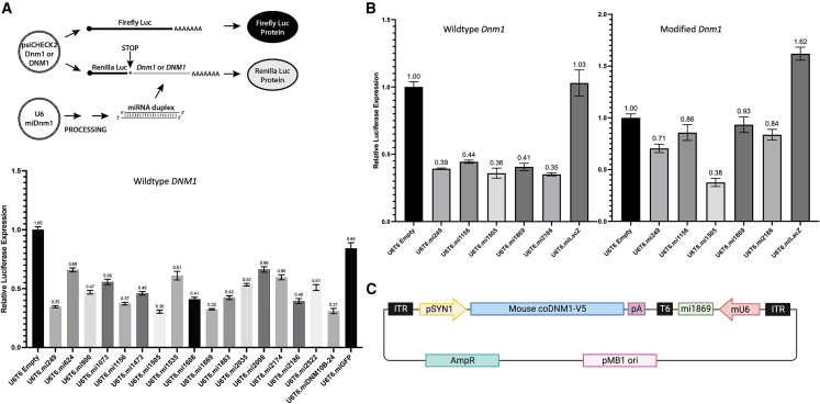
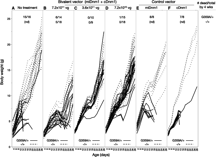
Effective gene therapy for gain-of-function or dominant-negative disease mutations may require eliminating expression of the mutant copy together with wild-type replacement. We evaluated such a knockdown-replace strategy in a mouse model of DNM1 disease, a debilitating and intractable neurodevelopmental epilepsy. To challenge the approach robustly, we expressed a patient-based variant in GABAergic neurons-which resulted in growth delay and lethal seizures evident by postnatal week three-and delivered to newborn pups an AAV9-based vector encoding a ubiquitously expressed, Dnm1-specific interfering RNA (RNAi) bivalently in tail-to-tail configuration with a neuron-specific, RNAi-resistant, codon-optimized Dnm1 cDNA. Pups receiving RNAi or cDNA alone fared no better than untreated pups, whereas the vast majority of mutants receiving modest doses survived with almost full growth recovery. Synaptic recordings of cortical neurons derived from treated pups revealed that significant alterations in transmission from inhibitory to excitatory neurons were rectified by bivalent vector application. To examine the mutant transcriptome and impact of treatment, we used RNA sequencing and functional annotation clustering. Mutants displayed abnormal expression of more than 1,000 genes in highly significant and relevant functional clusters, clusters that were abrogated by treatment. Together these results suggest knockdown-replace as a potentially effective strategy for treating DNM1 and related genetic neurodevelopmental disease.

Keywords: deno-associated virus; developmental and epileptic encephalopathy; dynamin-1; epilepsy; neurodevelopment.


Conflict of interest statement

Declaration of interests A patent application was submitted (Appl. No. 63/639,576: Products and methods to inhibit expression of dynamin-1 variants and replace dynamin-1). Inventors: S.Q.H., N.T., and W.N.F.

Figures

None
Graphical abstract
Figure 1
Figure 1
Visualization of 4-week growth curves for mutant and control littermates by Cre driver strain Dnm1 genotype indicated by solid line (heterozygous: G359A/+) or dotted line (hemizygous, −/+). Cre driver strain indicated by color (Gad-Cre, black; Nestin-Cre, blue; Nkx2.1-Cre, green; Pvalb-Cre, purple; Sox2-Cre, red). The shading indicates standard error, and Xs are shown to indicate (for Sox2-Cre and Gad2-Cre) when mice were found dead. Weighing was terminated for Gad2-Cre mice after 18 days as most of the G359A/+ mice had succumbed to lethal seizure.
Figure 2
Figure 2
Bivalent AAV vector development design (A) Renilla-Firefly dual-luciferase assay for testing efficacy of microRNA shuttle designed to interfere with mouse (Dnm1) or human (DNM1) dynamin-1 mRNA in cell culture. Graph shows efficacy luciferase expression screen of fifteen test inserts compared with empty or miGFP vector against DNM1 expressed in HEK cells. (B) Five test inserts were evaluated against wild-type (left) mouse Dnm1, all but one of which did not target codon-optimized, RNAi-resistant mouse Dnm1 (right). (C) Design of bivalent AAV vector showing tail-to-tail configuration of SYN1-coDNM1-V5 and U6-mi1869 inserts. Error bars represent standard error of the mean.
Figure 3
Figure 3
Visualization of individual growth curves and survival across gene therapy vectors Gad2-Cre, heterozygous G359A/+ are indicated by solid lines and hemizygous −/+ littermates by dotted lines. All treatments were at PND 1 with the noted condition by (A, B, C, D, E, or F) as in the text. Pups were weighed approximately every 3-4 days, with intervening weights interpolated linearly, before analysis. Lines that terminate before PND 28 mark the last day a pup was seen alive. The numbers at the top of each panel show the number of pups found dead over the total number for each group. nd, not done.
Figure 4
Figure 4
Repeated measures MANOVA analysis of body weight First 4 weeks (A) and weeks 5–12 (B). In (A), pup body weights were measured approximately every 3-4 days; intervening day weights were interpolated linearly before analysis. The Bonferroni adjustment was applied to p values for pairwise comparisons in (A).
Figure 5
Figure 5
Quantification of endogenous and viral-delivered dynamin-1 mRNA and protein in PND15-PND17 mouse pups (A) Normalized RNA-seq transcript counts (also see Table S3) in bivalent treated or control heterozygous G359A/+ and hemizygous −/+ pups for endogenous Dnm1 mRNA (Generalized Linear Mixed Model: p < 0.01 genotype effect, p < 0.0001 treatment effect) and exogenous codon-optimized, RNAi resistant, V5 epitope-tagged virally transduced Dnm1 mRNA (CO-Dnm1-v5). (B and C) Western blot showing total DNM1 protein (detected with DNM1-specific antibody; least-squares regression: p < 0.05 genotype effect, p < 0.05 treatment effect) and exogenous DNM1 protein detected with antibody to the V5 epitope tag. Error bars represent standard effort of the mean.
Figure 6
Figure 6
Viral transduction at 2 weeks postnatal Shown at the left are z stack images from Dnm1 heterozygous G359/+ and hemizygous −/+ brain from treated and control pups. scAAV9-U6-miDnm1-hSYN1-coDnm1 is visualized with an antibody to the V5 epitope tag (green). Inset is higher magnification of stippled boxes, showing examples from single layer of V5 and parvalbumin (PV, red) and colocalization (yellow-orange). Shown at the right are neuron counts of V5 (green), PV (red) and colocalized cells (yellow) and their mean values (V5, dark gray; PV, medium gray; colocalized, light gray).
Figure 7
Figure 7
Functional annotation clustering using GO terms of treated and untreated mutant and control mouse pup cortex and hippocampus RNA combined harvested at PND15-PND17 (A and B) Reduced expression in untreated heterozygous G359A/+ compared with hemizygous −/+. (C and D) Increased expression in untreated G359A/+ compared with −/+. Clustering significance is indicated by the magnitude of the log(Q) value (logarithm of the false discovery rate-adjusted p-value) on the Y axis. GO terms were ordered left to right on the X axis based on the significance of clustering in untreated mutant compared with control (green line). More nominal clustering is observed in the other pairwise comparisons (−/+ untreated vs. treated: purple line; G359A/+ untreated vs. treated: red line; G359A/+ treated vs. −/+ untreated: blue line).
Figure 8
Figure 8
Neurons from Dnm1 G359A; Gad-Cre mice show altered inhibitory synaptic inhibition that is rescued by the bivalent gene therapy (A) Representative traces and summary data showing that the strength of evoked excitatory transmission and paired pulse ratios onto inhibitory neurons are unaltered by G359A expression or the bivalent treatment. (B) Conversely, the amplitude of evoked inhibitory responses onto excitatory neurons are reduced by G359A expression and the paired pulse ratio is increased. Both of these changes are rescued by the bivalent treatment. (C) G359A neurons also show enhanced facilitation of evoked IPSCs that is rescued by treatment. (D) The size of mIPSCs recorded in excitatory neurons is increased in G359A neurons and rescued by bivalent treatment. n.s. = p > 0.05, ∗p < 0.05, ∗∗p < 0.01, ∗∗∗p < 0.001 as tested with generalized estimating equations. Error bars represent standard error of the mean.

References

    1. Dhindsa R.S., Bradrick S.S., Yao X., Heinzen E.L., Petrovski S., Krueger B.J., Johnson M.R., Frankel W.N., Petrou S., Boumil R.M., Goldstein D.B. Epileptic encephalopathy-causing mutations in DNM1 impair synaptic vesicle endocytosis. Neurol. Genet. 2015;1:e4. doi: 10.1212/01.NXG.0000464295.65736.da. - DOI - PMC - PubMed
    1. Ferguson S.M., De Camilli P. Dynamin, a membrane-remodelling GTPase. Nat. Rev. Mol. Cell Biol. 2012;13:75–88. doi: 10.1038/nrm3266. - DOI - PMC - PubMed
    1. van der Bliek A.M., Redelmeier T.E., Damke H., Tisdale E.J., Meyerowitz E.M., Schmid S.L. Mutations in human dynamin block an intermediate stage in coated vesicle formation. J. Cell Biol. 1993;122:553–563. doi: 10.1083/jcb.122.3.553. - DOI - PMC - PubMed
    1. Ferguson S.M., Brasnjo G., Hayashi M., Wölfel M., Collesi C., Giovedi S., Raimondi A., Gong L.W., Ariel P., Paradise S., et al. A selective activity-dependent requirement for dynamin 1 in synaptic vesicle endocytosis. Science. 2007;316:570–574. doi: 10.1126/science.1140621. - DOI - PubMed
    1. Marks B., Stowell M.H., Vallis Y., Mills I.G., Gibson A., Hopkins C.R., McMahon H.T. GTPase activity of dynamin and resulting conformation change are essential for endocytosis. Nature. 2001;410:231–235. doi: 10.1038/35065645. - DOI - PubMed