

Overexpression of Extracellular Superoxide Dismutase 3 Inhibits Cancer Cell Growth and Migration in Colorectal Cancer
- PMID: 39128081
- PMCID: PMC11232067
- DOI: 10.5152/tjg.2024.23232
Overexpression of Extracellular Superoxide Dismutase 3 Inhibits Cancer Cell Growth and Migration in Colorectal Cancer
Abstract
Background/aims: Incidence of colorectal cancer is rapidly increasing worldwide. Extracellular superoxide dismutase (EcSOD; SOD3) is an antioxidant enzyme. However, SOD3 roles in colorectal cancer progression remain uncertain.
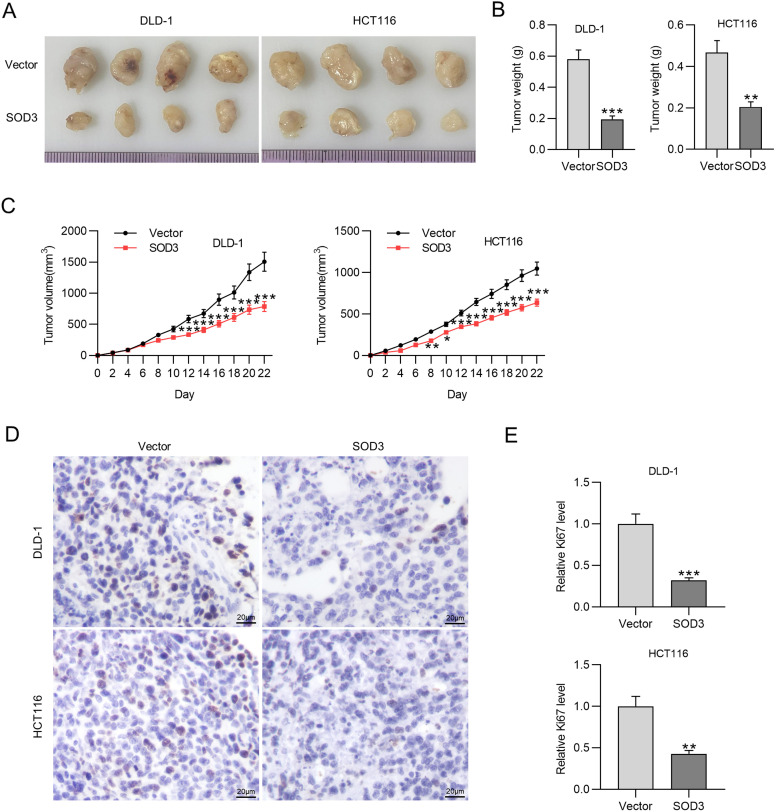
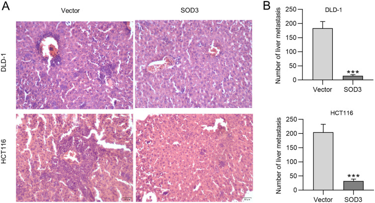
Materials and methods: Superoxide dismutase 3 expression was evaluated, and we analyzed clinical relevance of SOD3 expression in colorectal cancer. Subsequently, SOD3 roles in colorectal cancer progression were detected by gain of function experiments. Changes in subcutaneous tumor and liver nodule size after SOD3 overexpression were examined in nude mice. The expression of proliferation marker Ki67 was assessed by immunohistochemical staining.
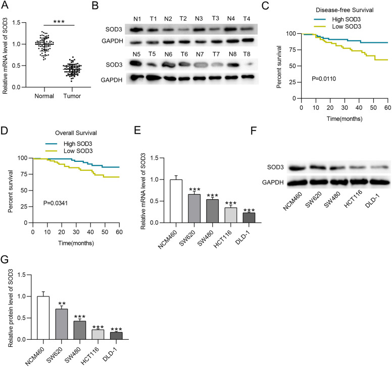
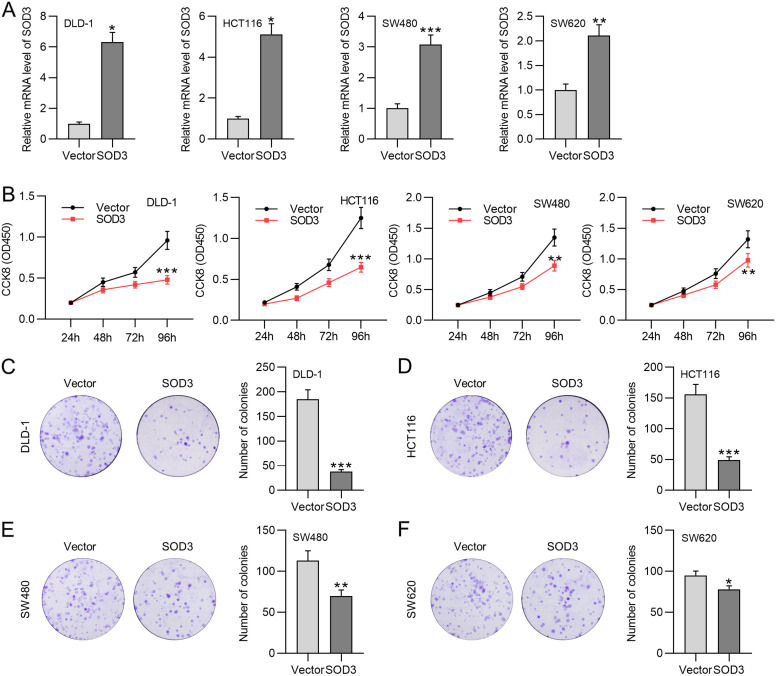
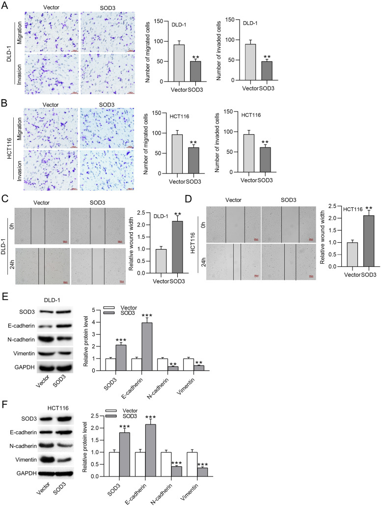
Results: Supperoxide dismutase 3 was downregulated in colorectal cancer (P <.01). Downregulation of SOD3 was correlated with unfavorable outcomes (P < .05). Superoxide dismutase 3 upregulation limited the proliferative (P <.05), migrative (P <.01) and invasive actions of colorectal cancer cells (P <.01) by suppressing epithelial-mesenchymal transition. Moreover, SOD3 overexpression reduced Ki67 expression (P <.01) and blocked tumor growth (P <01) and liver metastasis (P <.001) in mouse tumor model.
Conclusion: Superoxide dismutase 3 upregulation attenuates tumor growth and liver metastasis in colorectal cancer, suggesting that SOD3 has potential diagnostic and prognostic values regarding colorectal cancer treatment.
Conflict of interest statement
Figures
Similar articles
-
Genetic and epigenetic down-regulation of microRNA-212 promotes colorectal tumor metastasis via dysregulation of MnSOD.Gastroenterology. 2013 Aug;145(2):426-36.e1-6. doi: 10.1053/j.gastro.2013.04.004. Epub 2013 Apr 9. Gastroenterology. 2013. PMID: 23583431
-
cMET promotes metastasis and epithelial-mesenchymal transition in colorectal carcinoma by repressing RKIP.J Cell Physiol. 2021 May;236(5):3963-3978. doi: 10.1002/jcp.30142. Epub 2020 Nov 5. J Cell Physiol. 2021. PMID: 33151569
-
MCP-enhanced SOD3 activity inhibits gastric cancer and potentiate chemotherapy via modulating EGFR signaling.Life Sci. 2025 Feb 1;362:123358. doi: 10.1016/j.lfs.2024.123358. Epub 2024 Dec 31. Life Sci. 2025. PMID: 39746602
-
Extracellular Superoxide Dismutase: Growth Promoter or Tumor Suppressor?Oxid Med Cell Longev. 2016;2016:3612589. doi: 10.1155/2016/3612589. Epub 2016 May 12. Oxid Med Cell Longev. 2016. PMID: 27293512 Free PMC article. Review.
-
Extracellular superoxide dismutase and its role in cancer.Free Radic Biol Med. 2017 Nov;112:464-479. doi: 10.1016/j.freeradbiomed.2017.08.013. Epub 2017 Aug 24. Free Radic Biol Med. 2017. PMID: 28842347 Free PMC article. Review.
References
-
- Cancer international agency for research. Latest global cancer data: cancer burden rises to 19.3 million new cases and 10.0 million cancer deaths in 2020; 2020. https://www.iarc.fr/fr/news-events/latest-global-cancer-data-cancer-burd...
MeSH terms
Substances
LinkOut - more resources
Full Text Sources
Medical
Molecular Biology Databases
Miscellaneous
