

A giant pelvic infiltrating schwannoma misdiagnosed as an ovarian neoplasm that was resected using a laparoscopic approach: A case report
- PMID: 39128216
- PMCID: PMC11367093
- DOI: 10.1016/j.ijscr.2024.110138
A giant pelvic infiltrating schwannoma misdiagnosed as an ovarian neoplasm that was resected using a laparoscopic approach: A case report
Abstract
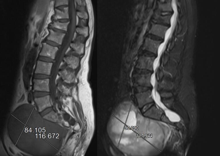
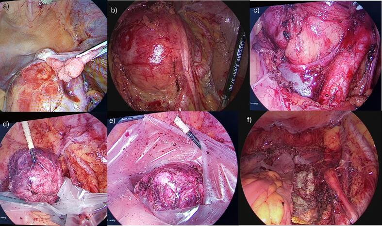
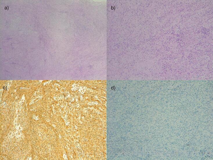
Introduction and importance: Large retroperitoneal schwannomas are rare and present significant challenges in surgical management, particularly when located in the pelvic region. Gynecologists can encounter rare problems when a pelvic schwannoma is mistaken for an adnexal pathology. Case Presentation: A 62-year-old woman presented with a giant retroperitoneal mass suspected of a potentially malignant ovarian tumor preoperatively. Computed tomography revealed a large mixed solid-cystic mass near the right adnexa measuring 118 × 100 × 80 mm. The cancer antigen 125 level was 196 U/mL. We performed a diagnostic-operative laparoscopy, which showed a retroperitoneal neoformation below the cava and aortic bifurcation adherent to the sacrum, right pelvic vessels, and hypogastric nerve up to the vagina. We carefully detached the mass from the nearby tissues using the most appropriate laparoscopic devices. The entire neoplasm was removed through the vagina into a surgical bag. The surgery lasted 180 min without complications. Histology revealed a grade I benign schwannoma. At the 12-month follow-up, the patient was asymptomatic without signs of recurrence. Clinical Discussion: Pelvic retroperitoneal schwannomas can mimic ovarian carcinomas; misdiagnosis may occur due to their rarity and the difficulty of interpreting preoperative imaging. In case of unexpected giant presacral schwannomas surgical management is challenging due to their peculiar location. Conclusion: This case underscores the need for a skilled, experienced team of gynecological oncologists to achieve favorable outcomes when performing laparoscopic surgery of giant pelvic retroperitoneal schwannoma. Adequate knowledge of the complex pelvic anatomy, careful surgical planning, and familiarity with the most appropriate surgical tools are critical points.
Keywords: Laparoscopy; Large retroperitoneal mass; Minimally invasive surgery; Presacral space; Schwannoma.
Copyright © 2024 The Authors. Published by Elsevier Ltd.. All rights reserved.
Conflict of interest statement
Declaration of competing interest The authors declare no conflicts of interest.
Figures
References
-
- Cury J., Coelho R.F., Srougi M. Retroperitoneal schwannoma: case series and literature review. Clinics (Sao Paulo) 2007;62:359–362. - PubMed
-
- Poojari V.G., Pai M.V., Nambiar J., Mathew M. Pelvic schwannoma mimicking as an adnexal mass. Int. J. Reprod. Contracept. Obstet. Gynecol. 2015;4:1206–1208.
-
- Sakalauskaite M., Stanaitis J., Cepkus S., Pleckaitis M., Lunevicius R. Retroperitoneal giant schwannoma eroding lumbal vertebra: a case report with a literature review. Open Med. 2008;3:233–244.
Publication types
LinkOut - more resources
Full Text Sources
