

Cosmetic retinoid use in photoaged skin: A review of the compounds, their use and mechanisms of action
- PMID: 39128883
- PMCID: PMC11788006
- DOI: 10.1111/ics.13013
Cosmetic retinoid use in photoaged skin: A review of the compounds, their use and mechanisms of action
Abstract
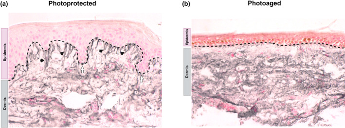
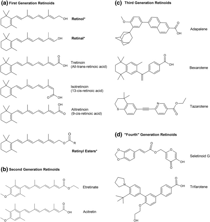
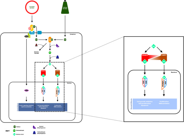
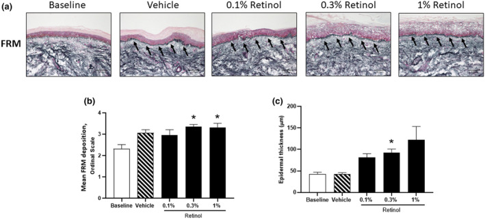
The inevitable attrition of skin due to ultraviolet radiation, termed photoaging, can be partially restored by treatment with retinoid compounds. Photoaged skin in lightly pigmented individuals, clinically presents with the appearance of wrinkles, increased laxity, and hyper- and hypopigmentation. Underlying these visible signs of ageing are histological features such as epidermal thinning, dermal-epidermal junction flattening, solar elastosis and loss of the dermal fibrillin microfibrillar network, fibrillar collagen and glycosaminoglycans. Retinoid compounds are comprised of three main generations with the first generation (all-trans retinoic acid, retinol, retinaldehyde and retinyl esters) primarily used for the clinical and cosmetic treatment of photoaging, with varying degrees of efficacy, tolerance and stability. All-trans retinoic acid is considered the 'gold standard' for skin rejuvenation; however, it is a prescription-only product largely confined to clinical use. Therefore, retinoid derivatives are readily incorporated into cosmeceutical formulations. The literature reported in this review suggests that retinol, retinyl esters and retinaldehyde that are used in many cosmeceutical products, are efficacious, safe and well-tolerated. Once in the skin, retinoids utilize a complex signalling pathway that promotes remodelling of photoaged epidermis and dermis and leads to the improvement of the cutaneous signs of photoaging.
L'altération inévitable de la peau due aux rayons ultraviolets, appelée photovieillissement, peut être partiellement restaurée par un traitement à base de composés rétinoïdes. Chez les personnes à la pigmentation claire, le photovieillissement de la peau se manifeste au plan clinique par l'apparition de rides, un relâchement accru et une hyperpigmentation ou hypopigmentation. Ces signes visibles du vieillissement sont sous‐tendus par des caractéristiques histologiques telles que l'amincissement de l'épiderme, l'aplatissement de la jonction dermo‐épidermique, l'élastose solaire et la perte du réseau microfibrillaire de fibrilline dermique, du collagène fibrillaire et des glycosaminoglycanes. Les composés rétinoïdes sont constitués de trois générations principales, la première génération (acide tout‐trans rétinoïque, rétinol, rétinaldéhyde et esters de rétinyle) étant principalement utilisée pour le traitement clinique et cosmétique du photovieillissement, avec des degrés variables d'efficacité, de tolérance et de stabilité. L'acide tout‐trans rétinoïque est considéré comme la référence en matière de rajeunissement de la peau; il s'agit toutefois d'un produit délivré uniquement sur ordonnance, dont l’utilisation est largement limitée au domaine clinique. Les dérivés rétinoïdes sont donc volontiers incorporés ds formulations cosméceutiques. La littérature citée dans cette synthèse bibliographique laisse penser que le rétinol, les esters de rétinyle et le rétinaldéhyde, utilisés dans de nombreux produits cosmétiques, sont efficaces, sûrs et bien tolérés. Une fois dans la peau, les rétinoïdes utilisent une voie de signalisation complexe qui favorise le remodelage de l'épiderme et du derme photovieillis, et conduit à l'amélioration des signes cutanés du photovieillissement.
Keywords: cosmeceuticals; formulation/stability; skin barrier; skin physiology/structure; topical retinoids.
© 2024 The Author(s). International Journal of Cosmetic Science published by John Wiley & Sons Ltd on behalf of Society of Cosmetic Scientists and Societe Francaise de Cosmetologie.
Conflict of interest statement
The authors declare no known conflicts of interest.
Figures
Similar articles
-
Retinoids in the treatment of photoageing: A histological study of topical retinoid efficacy in black skin.J Eur Acad Dermatol Venereol. 2024 Aug;38(8):1618-1627. doi: 10.1111/jdv.20043. Epub 2024 Apr 29. J Eur Acad Dermatol Venereol. 2024. PMID: 38682699
-
Retinoids in cosmeceuticals.Dermatol Ther. 2006 Sep-Oct;19(5):289-96. doi: 10.1111/j.1529-8019.2006.00086.x. Dermatol Ther. 2006. PMID: 17014484 Review.
-
Enhanced retinoid response by a combination of the vitamin A ester retinyl propionate with niacinamide and a flavonoid containing Ceratonia siliqua extract in retinoid responsive in vitro models.Int J Cosmet Sci. 2021 Feb;43(1):102-106. doi: 10.1111/ics.12669. Epub 2020 Nov 19. Int J Cosmet Sci. 2021. PMID: 33048363
-
Retinoids in the treatment of skin aging: an overview of clinical efficacy and safety.Clin Interv Aging. 2006;1(4):327-48. doi: 10.2147/ciia.2006.1.4.327. Clin Interv Aging. 2006. PMID: 18046911 Free PMC article. Review.
-
A short-term screening protocol, using fibrillin-1 as a reporter molecule, for photoaging repair agents.J Invest Dermatol. 2001 May;116(5):672-8. doi: 10.1046/j.1523-1747.2001.01322.x. J Invest Dermatol. 2001. PMID: 11348454 Clinical Trial.
Cited by
-
Photoaging: Current Concepts on Molecular Mechanisms, Prevention, and Treatment.Am J Clin Dermatol. 2025 May;26(3):321-344. doi: 10.1007/s40257-025-00933-z. Epub 2025 Mar 12. Am J Clin Dermatol. 2025. PMID: 40072791 Review.
-
Therapeutic Uses of Retinol and Retinoid-Related Antioxidants.Molecules. 2025 May 16;30(10):2191. doi: 10.3390/molecules30102191. Molecules. 2025. PMID: 40430363 Free PMC article. Review.
-
5'tiRNA-Glu-TTC targets TRPV3 and activates the PI3K/AKT signaling pathway to modulate skin photoaging.Noncoding RNA Res. 2025 Jul 14;15:29-43. doi: 10.1016/j.ncrna.2025.07.004. eCollection 2025 Dec. Noncoding RNA Res. 2025. PMID: 40741358 Free PMC article.
-
Usage Frequency and Ecotoxicity of Skin Depigmenting Agents.Pharmaceuticals (Basel). 2025 Mar 4;18(3):368. doi: 10.3390/ph18030368. Pharmaceuticals (Basel). 2025. PMID: 40143144 Free PMC article.
-
Novel Cyclized Hexapeptide-9 Outperforms Retinol Against Skin Aging: A Randomized, Double-Blinded, Active- and Vehicle-Controlled Clinical Trial.J Cosmet Dermatol. 2025 Jul;24(7):e70290. doi: 10.1111/jocd.70290. J Cosmet Dermatol. 2025. PMID: 40586182 Free PMC article. Clinical Trial.
References
-
- Madison KC. Barrier function of the skin: "la raison d'être" of the epidermis. J Invest Dermatol. 2003;121(2):231–241. - PubMed
-
- Arda O, Göksügür N, Tüzün Y. Basic histological structure and functions of facial skin. Clin Dermatol. 2014;32(1):3–13. - PubMed
-
- Fore J. A review of skin and the effects of aging on skin structure and function. Ostomy Wound Manage. 2006;52(9):24–35; quiz 6–7. - PubMed
-
- Svoboda M, Bílková Z, Muthný T. Could tight junctions regulate the barrier function of the aged skin? J Dermatol Sci. 2016;81(3):147–152. - PubMed
Publication types
MeSH terms
Substances
Grants and funding
LinkOut - more resources
Full Text Sources
Medical
