. 2024 Aug 11;7(1):975.
doi: 10.1038/s42003-024-06675-8.

Platelet extracellular vesicles preserve lymphatic endothelial cell integrity and enhance lymphatic vessel function

Affiliations

Platelet extracellular vesicles preserve lymphatic endothelial cell integrity and enhance lymphatic vessel function

Laurent Vachon et al. Commun Biol. .

Abstract

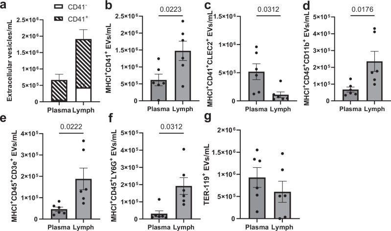
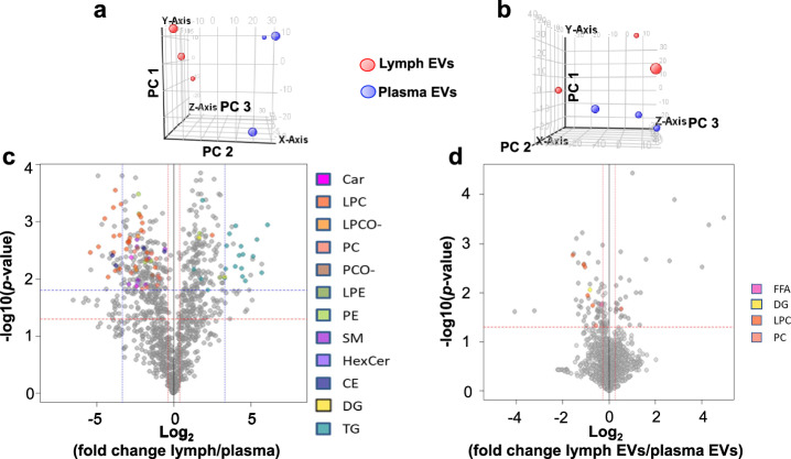
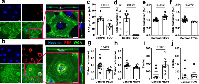
Lymphatic vessels are essential for preventing the accumulation of harmful components within peripheral tissues, including the artery wall. Various endogenous mechanisms maintain adequate lymphatic function throughout life, with platelets being essential for preserving lymphatic vessel integrity. However, since lymph lacks platelets, their impact on the lymphatic system has long been viewed as restricted to areas where lymphatics intersect with blood vessels. Nevertheless, platelets can also exert long range effects through the release of extracellular vesicles (EVs) upon activation. We observed that platelet EVs (PEVs) are present in lymph, a compartment to which they could transfer regulatory effects of platelets. Here, we report that PEVs in lymph exhibit a distinct signature enabling them to interact with lymphatic endothelial cells (LECs). In vitro experiments show that the internalization of PEVs by LECs maintains their functional integrity. Treatment with PEVs improves lymphatic contraction capacity in atherosclerosis-prone mice. We suggest that boosting lymphatic pumping with exogenous PEVs offers a novel therapeutic approach for chronic inflammatory diseases characterized by defective lymphatics.


Conflict of interest statement

The authors declare no competing interests.

Figures

Fig. 1
Fig. 1. Characterization of EVs in plasma and lymph by flow cytometry.
Plasma and lymph CFSE-positive EVs were identified by flow cytometry. a The proportion of EVs positive for CD41 amongst total MHCI+ EVs was identified in plasma and lymph. Different EV subsets were characterized using antibodies against a combination of anti-MHCI and (b) anti-CD41, (c) anti-CD41 and anti-CLEC-2, (d) anti-CD45 and anti-CD11b, (e) anti-CD45 and anti-CD3e and (f) anti-CD45 and anti-LY6G. g EVs derived from red blood cells were also identified using an anti-TER-119 antibody. n = 6 ± SEM. MHCI: major histocompatibility complex class I; EVs: extracellular vesicles.
Fig. 2
Fig. 2. Untargeted lipidomic analysis of lymph, plasma and their EVs.
Matched mice lymph and plasma, and EVs isolated from those two respective biological fluids, were analyzed by LC-QTOF-MS. Principal component analysis (PCA) plots for (a) plasma in blue and lymph in red and for (b) plasma EVs in blue and lymph EVs in red (PC 1, PC 2 and PC 3 refer to Principal Components 1, 2 and 3 on the Y, X and Z axes, respectively). c Volcano plot of untargeted lipidomics in lymph and plasma. The x axis represents the fold changes (in log2) of MS signal intensities values in lymph vs. plasma and the Y-axis represents the p-values (in –log10). Selection criteria were established with corrected p < 0.05 (horizontal blue line) and FC > 10 or < 0.1 (vertical blue lines), and with less stringent criteria of selection using FC > 1.3 or < 0.77 (vertical red lines). d Volcano plot of untargeted lipidomics in lymph EVs and plasma EVs. Selection criteria were established with corrected p < 0.05 (horizontal red line) and FC > 1.2 or < 0.83 (vertical red lines). Features that were discriminating lymph and plasma (or lymph EVs and plasma EVs) were annotated and validated using MS/MS analysis (color symbols). n = 3 (each n is a pool of 2–4 mice). PC: Principal Component; Car: acylcarnitines; CE: cholesteryl esters; FFA: free fatty acid; HexCer: hexosyl Ceramides; LPC: lysoglycerophosphatidylcholine; LPE: lysoglycerophosphatidylethanolamine; LPCO-: ether-linked lysoglycerophosphatidylcholine; PC: diacylglycerophosphatidylcholine; PE: diacylglycerophosphatidylethanolamine; PCO-: ether diacylglycerophosphatidylcholine; DG: diacylglycerols; SM: sphyngomyelins; TG: triacylglycerols.
Fig. 3
Fig. 3. Characterization of the effects of human platelet- and red blood cell-EVs on lymphatic endothelial cells in vitro.
PEVs stained with CellVue (red) were incubated on LEC in vitro for (a) 2 h and (b) 24 h and incubated with WGA (green) and Hoechst (blue). Z-stacks were acquired using a LSM 710 Confocal Microscope (Zeiss) equipped with a 63/1.4 oil DIC objective (white scale bar, 20 µm). (c) rbEVs, (d) PEVs and LECs treated with (e) rbEVs or (f) PEVs for 24 h were incubated with CM-H2DCFDA and ROS production was measured using flow cytometry. Superoxide dismutase (100 U/mL) was used as a negative control. The percentage of AnnexinV-Propidium iodide+ (PI+AnV-) LECs was determined after treatment with (g) PEVs or (h) rbEVs. The concentration of EVs produced from LECs in the culture supernatant was measured after treatment with (i) rbEVs or (j) PEVs. Each point (n) represents cells treated with PEVs produced from a single donor ± SEM. PEVs: platelet extracellular vesicles; rbEVs: red blood cell extracellular vesicles; WGA: Wheat Germ Agglutinin; ROS: reactive oxygen species; SOD: superoxide dismutase. AU: arbitrary unit.
Fig. 4
Fig. 4. Down- and upregulation of specific subsets of genes by lymphatic endothelial cells treated with PEVs assessed by RNA-sequencing and qPCR analysis.
Human LECs were treated for 24 h with PEVs. a Volcano plot of transcriptomic analysis was performed in human LEC. Y axis =  p-value (-LOG10) of the gene expression. X axis = fold change PEVs/Control (LOG2) of gene expression. Each gene is represented by a dot. Significant down- and upregulated genes in PEV-treated LECs are depicted in blue and red, respectively. mRNA expression in human LECs was also performed by qPCR analysis for (b) F11R, (c) PROX1, (d) FLT4, (e) TJP1 and (f) CDH5 mRNA. Human LECs were also treated for 24 h with rbEVs, and mRNA expression of (g) CDH5, (h) F11R, (i) PROX1 and (j) FLT4 was subsequently measured by qPCR analysis. Each point (n) represents cells treated with PEVs produced from a single donor ± SEM. PEVs: platelet extracellular vesicles; rbEVs: red blood cell extracellular vesicles; F11R: F11 Receptor; PROX1: Prospero homeobox protein 1; FLT4: Fms-related tyrosine kinase 4; TJP1: Tight junction protein-1; CDH5: cadherin 5.
Fig. 5
Fig. 5. Distribution of exogenous PEVs and outcomes on lymphatic function in vivo.
CellVue+ human PEVs (1 × 107) or vehicle control were injected in the dermis of the footpad in wild-type mice. Lymph nodes (LNs) draining the site of injection (green boxes) and LNs located at a distal site (beige boxes) were collected 15 min and 2 h post-injection. Then, they were (a) visualized with a fluorescence imaging scanner (IVIS Lumina II) or (b) digested with collagenase D to determine the percentage of cells containing EVs. c The concentration of free EVs within the LNs were assessed by flow cytometry using anti-mouse MHCII and anti-human CD62P antibodies. d Two hours post-PEV injection, popliteal lymphatic vessels draining the injection site were also collected, harvested and stained with anti-human CD41a antibody (red) and DAPI (blue) to be imaged with a confocal microscope (white scale bar, 20 µm). The lymphatic contraction capacity was measured in vivo 48 h after the injection of (e) PEVs or (f) rbEVs in Ldlr-/-, and (g) after injection of PEVs in wild-type mice. h The lymph nodes (N-D, beige; D, green) and (i) the aorta were also collected at 48 h and visualized with fluorescent imaging. Each point represents a mouse ± SEM. N-D: non-draining; D: draining; LN: lymph nodes; EVs: extracellular vesicles. PEVs: platelet extracellular vesicles. rbEVs: red blood cell extracellular vesicles. Created with Biorender.com.
Fig. 6
Fig. 6. Platelet extracellular vesicles preserve the integrity of lymphatic endothelial cells and enhance lymphatic vessel function.
CD41+ EVs travel from the blood circulation and enter the lymph mostly via a the lymphovenous junctions (crossing point between the lymphatic vessel and the subclavian vein) and b the lymphatic capillaries. CD41+ PEVs are also produced upon the adhesion and activation of platelets at the exterior of the lymphatic vessels before entering lymph. CD41+CLEC2+ EVs are mostly attached to the interior side of the lymphatic vessel. c Like platelets, CD41+ EVs bind to LECs via the interaction of their CLEC-2 receptor with podoplanin on LECs. d The specific signature of the EVs that bind LECs confers them the ability to enhance lymphatic contractions in mice known to display a defect in lymphatic pumping. Ultimately, enhancing lymphatic function with exogenous PEVs might become a new therapeutic option in the treatment of chronic inflammatory diseases where lymphatics are known to be defective, like atherosclerosis. Created with BioRender.com.

References

    1. Iqbal, J. & Hussain, M. M. Intestinal lipid absorption. Am. J. Physiol. Endocrinol. Metab.296, E1183–E1194 (2009). 10.1152/ajpendo.90899.2008 - DOI - PMC - PubMed
    1. Miteva, D. O. et al. Transmural flow modulates cell and fluid transport functions of lymphatic endothelium. Circ. Res.106, 920–931 (2010). 10.1161/CIRCRESAHA.109.207274 - DOI - PMC - PubMed
    1. Hoggan, G. & Hoggan, F. E. The lymphatics of the walls of the larger blood-vessels and lymphatics. J. Anat. Physiol.17, 1–23 (1882). - PMC - PubMed
    1. Lemole, G. M. The role of lymphstasis in atherogenesis. Ann. Thorac. Surg.31, 290–293 (1981). 10.1016/S0003-4975(10)60949-6 - DOI - PubMed
    1. Martel, C. et al. Lymphatic vasculature mediates macrophage reverse cholesterol transport in mice. J. Clin. Invest123, 1571–1579 (2013). 10.1172/JCI63685 - DOI - PMC - PubMed

Publication types

LinkOut - more resources