. 2024 Aug 11;21(1):200.
doi: 10.1186/s12974-024-03180-x.

The dopamine analogue CA140 alleviates AD pathology, neuroinflammation, and rescues synaptic/cognitive functions by modulating DRD1 signaling or directly binding to Abeta

Affiliations

The dopamine analogue CA140 alleviates AD pathology, neuroinflammation, and rescues synaptic/cognitive functions by modulating DRD1 signaling or directly binding to Abeta

Sehyun Chae et al. J Neuroinflammation. .

Abstract

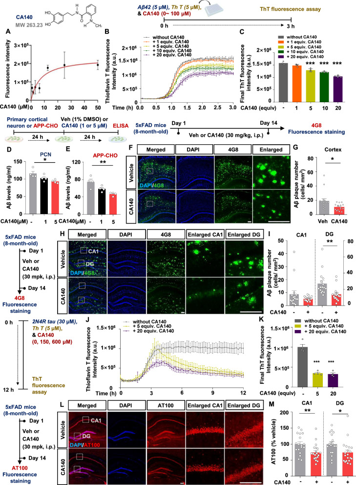
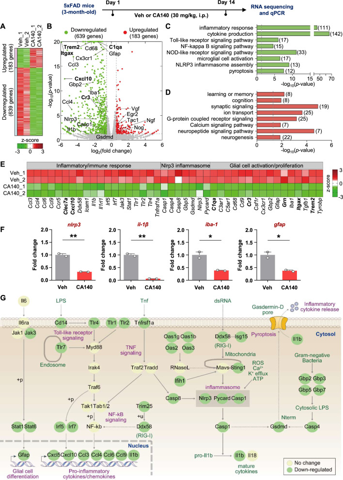
Background: We recently reported that the dopamine (DA) analogue CA140 modulates neuroinflammatory responses in lipopolysaccharide-injected wild-type (WT) mice and in 3-month-old 5xFAD mice, a model of Alzheimer's disease (AD). However, the effects of CA140 on Aβ/tau pathology and synaptic/cognitive function and its molecular mechanisms of action are unknown.

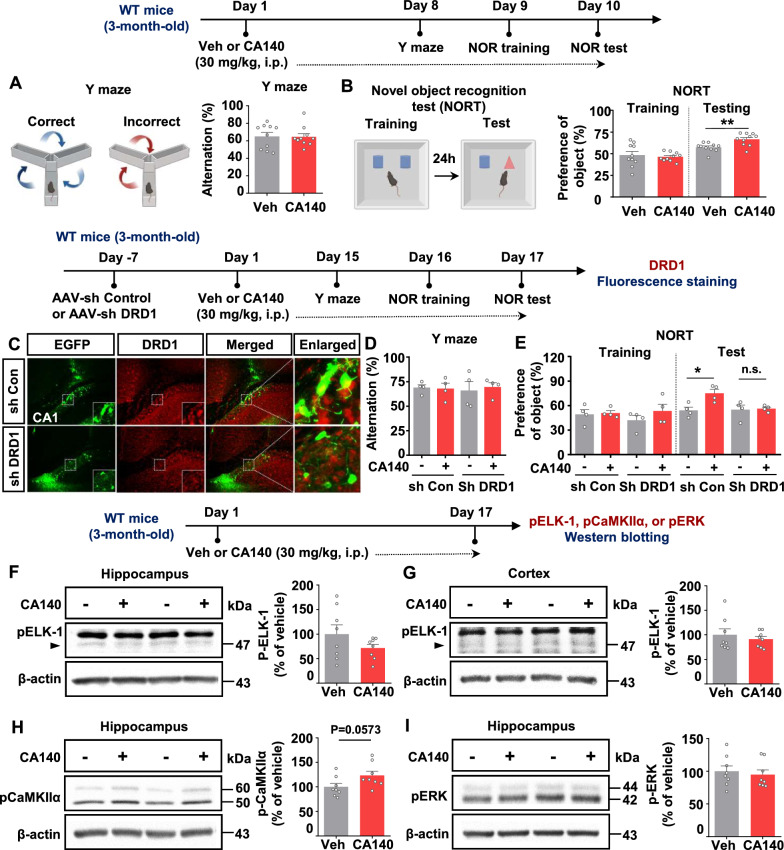
Methods: To investigate the effects of CA140 on cognitive and synaptic function and AD pathology, 3-month-old WT mice or 8-month-old (aged) 5xFAD mice were injected with vehicle (10% DMSO) or CA140 (30 mg/kg, i.p.) daily for 10, 14, or 17 days. Behavioral tests, ELISA, electrophysiology, RNA sequencing, real-time PCR, Golgi staining, immunofluorescence staining, and western blotting were conducted.

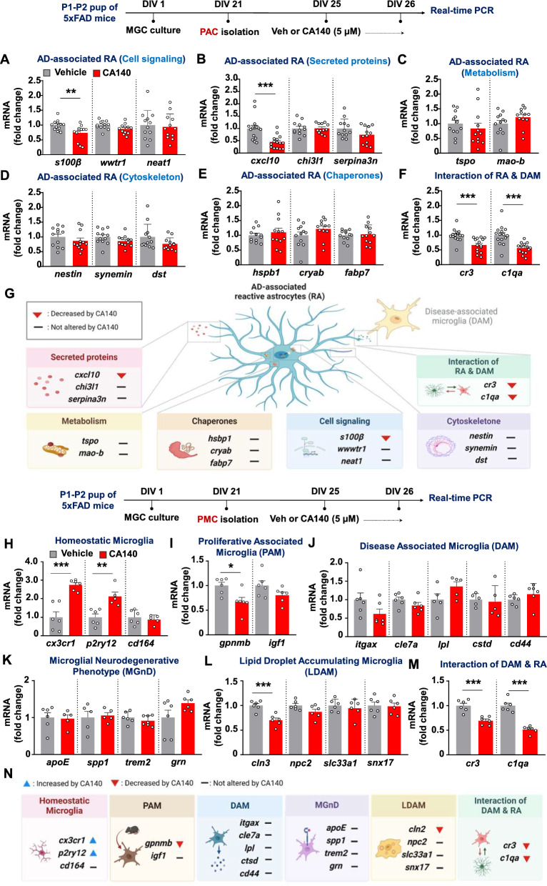
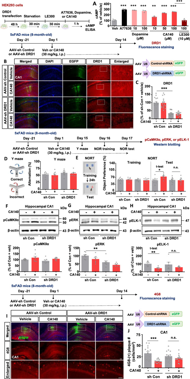
Results: In aged 5xFAD mice, a model of AD pathology, CA140 treatment significantly reduced Aβ/tau fibrillation, Aβ plaque number, tau hyperphosphorylation, and neuroinflammation by inhibiting NLRP3 activation. In addition, CA140 treatment downregulated the expression of cxcl10, a marker of AD-associated reactive astrocytes (RAs), and c1qa, a marker of the interaction of RAs with disease-associated microglia (DAMs) in 5xFAD mice. CA140 treatment also suppressed the mRNA levels of s100β and cxcl10, markers of AD-associated RAs, in primary astrocytes from 5xFAD mice. In primary microglial cells from 5xFAD mice, CA140 treatment increased the mRNA levels of markers of homeostatic microglia (cx3cr1 and p2ry12) and decreased the mRNA levels of a marker of proliferative region-associated microglia (gpnmb) and a marker of lipid-droplet-accumulating microglia (cln3). Importantly, CA140 treatment rescued scopolamine (SCO)-mediated deficits in long-term memory, dendritic spine number, and LTP impairment. In aged 5xFAD mice, these effects of CA140 treatment on cognitive/synaptic function and AD pathology were regulated by dopamine D1 receptor (DRD1)/Elk1 signaling. In primary hippocampal neurons and WT mice, CA140 treatment promoted long-term memory and dendritic spine formation via effects on DRD1/CaMKIIα and/or ERK signaling.

Conclusions: Our results indicate that CA140 improves neuronal/synaptic/cognitive function and ameliorates Aβ/tau pathology and neuroinflammation by modulating DRD1 signaling in primary hippocampal neurons, primary astrocytes/microglia, WT mice, and aged 5xFAD mice.

Keywords: Aβ; CA140; Dopamine D1 receptor; LTP; Learning and memory; Reactive gliosis; Tau.


Conflict of interest statement

The authors declare no competing interests.

Figures

Fig. 1
Fig. 1
CA140 reduces Aβ/tau aggregate formation, Aβ levels, and AD pathology in vitro and in vivo. A Graph of fluorescence intensity versus concentration of CA140 in the presence of aggregated Aβ42 (average fluorescence measurements from three independent experiments). B Real-time monitoring of the inhibitory effects of CA140 on amyloid formation. C Quantification of the ThT intensity of Aβ42 at the final time point. D, E Aβ levels in primary cortical neurons and APP-overexpressing CHO cells. F–I Eight-month-old 5xFAD mice were injected with vehicle (10% DMSO) or CA140 (30 mg/kg) daily for 14 days, and immunofluorescence (IF) staining of brain slices was conducted with an anti-Aβ17-24 (4G8) antibody (n = 16 brain slices from 4 mice/group). J, K Quantification of the ThT intensity of 2N4R full-length tau at the final time point. L, M 8-month-old 5xFAD mice were injected with vehicle (10% DMSO) or CA140 (30 mg/kg, i.p.) daily for 14 days, and IF staining of brain slices was conducted with an anti-TauThr212/Ser214 (AT100) antibody (Veh, n = 18 brain slices from 4 mice; CA140, n = 20 brain slices from 4 mice). Scale bar = 200 μm. *p < 0.05, **p < 0.01
Fig. 2
Fig. 2
Identification of genes affected by CA140 in 3-month-old 5xFAD mice. A Heatmap of 183 upregulated and 639 downregulated genes in CA140-injected 5xFAD mice compared with vehicle-injected 5xFAD mice. B Volcano plot showing differentially expressed genes (DEGs) in CA140-treated 5xFAD mice. The X- and Y-axes present the log2-fold-change and –log10 (p value), respectively. Red and green dots represent upregulated and downregulated genes, respectively. Gray dots represent genes without significant differences in expression. C, D Gene ontology biological processes (GOBPs) represented by the downregulated (C) and upregulated (D) genes. The dotted line indicates the p value cutoff used. The number of genes in each biological process is indicated in parentheses. E DEGs involved in the inflammatory response, glial cell activation/proliferation, and Nlrp3 inflammasome signaling. The color bar shows the z-score gradient. F Relative mRNA levels of the indicated genes in CA140- or vehicle-treated 5xFAD mice were analyzed by real-time PCR (n = 2 mice/group). G Network model describing the interactions among inflammatory response-related signaling pathways. The node colors represent downregulation (green) and no change (yellow) of the corresponding genes in CA140-injected 5xFAD mice. Nodes are arranged and connected according to the activation (arrows) information in the KEGG pathway and WikiPathways databases. Solid and dotted lines denote direct and indirect interactions, respectively. “+p”, phosphorylation. “+u”, ubiquitination. *p < 0.05, **p < 0.01
Fig. 3
Fig. 3
CA140 injection regulates microglial activation and astrocytic morphology in 8-month-old 5xFAD mice. A–H Eight-month-old 5xFAD mice were injected with vehicle (10% DMSO) or CA140 (30 mg/kg, i.p.) daily for 14 days, and immunofluorescence staining of brain slices was conducted with anti-Iba-1 and anti-GFAP antibodies (Iba-1: Veh, n = 23–24 brain slices from 4 mice; CA140, n = 17 brain slices from 4 mice; GFAP: Veh, n = 24 brain slices from 4 mice; CA140, n = 17 brain slices from 4 mice). Scale bar = 200 μm. *p < 0.05, **p < 0.01
Fig. 4
Fig. 4
CA140 administration reduces NLRP3, IL-1β levels and reactive gliosis in 8-month-old 5xFAD mice. A–H Eight-month-old 5xFAD mice were injected with vehicle (10% DMSO) or CA140 (30 mg/kg, i.p.) daily for 14 days, and immunofluorescence staining of brain slices was performed with anti-NLRP3 (A, B), anti-IL-1β (C, D), anti-CXCL10 (E, F) and anti-C1QA (G, H) antibodies (n = 16–23 brain slices from 4 mice). Scale bar = 200 μm. *p < 0.05, **p < 0.01, ***p < 0.001
Fig. 5
Fig. 5
CA140 treatment regulates reactive gliosis in primary astrocytes (PACs) and primary microglia (PMC) from 5xFAD mice. A–F PACs from 5xFAD mice were treated with vehicle (1% DMSO) or CA140 (5 μM) for 24 h, and the relative mRNA levels of the indicated genes were analyzed by real-time PCR (n = 12–15/group). G Summary illustration of the regulatory effect of CA140 on reactive astrogliosis in PACs from 5xFAD mice. HM PMCs from 5xFAD mice were treated with vehicle (1% DMSO) or CA140 (5 μM) for 24 h, and the relative mRNA levels of the indicated genes were analyzed by real-time PCR (n = 5–6/group). N Summary illustration of the regulatory effect of CA140 on microglial reactive state in PMCs from 5xFAD mice. *p < 0.05, **p < 0.01, ***p < 0.001
Fig. 6
Fig. 6
CA140 treatment reverses scopolamine (SCO)-induced impairments in long-term memory, dendritic spine number, and LTP in wild-type (WT) mice. A, B WT mice were injected daily with vehicle (10% DMSO) or CA140 (30 mg/kg, i.p.) for 14 days. On days 3–14, the mice were also injected daily with PBS or SCO (1 mg/kg, i.p.). Y-maze and NOR tests were performed on days 12 and 14, respectively (n = 8 mice/group). C–E Representative AO and BS dendrites from the hippocampal CA1 region and cortical layer V region of mice treated with CA140 and/or SCO (hippocampus AO: Veh, n = 77 neurons from 8 mice; SCO, n = 76 neurons from 8 mice; SCO + CA140, n = 76 neurons from 8 mice; hippocampus BS: Veh, n = 76 neurons from 8 mice; SCO, n = 77 neurons from 8 mice; SCO + CA140, n = 76 neurons from 8 mice; cortex AO and BS: n = 24 neurons from 4 mice/group). Scale bar = 10 μm. F WT mice were injected daily with CA140 (30 mg/kg, i.p.) or vehicle (10% DMSO) for 14 days and were also injected with SCO (1 mg/kg, i.p.) or PBS on days 3–14. G Representative excitatory postsynaptic current (EPSC) traces from the vehicle, SCO, and SCO + CA140 treatment groups. H Input–output curves from the vehicle, SCO, and SCO + CA140 treatment groups (Veh, n = 18 cells from 5 mice; SCO, n = 17 cells from 5 mice; SCO + CA140, n = 14 cells from 5 mice). I Representative EPSC traces before and after LTP induction in the vehicle, SCO, and SCO + CA140 treatment groups (1: before LTP; 2: after LTP). J, K Effects of SCO on LTP induction in the presence/absence of CA140. L Overlay of the two graphs in J, K. M Summary statistics for LTP induction (last 5 min: Veh, n = 20 cells from 8 mice; SCO, n = 22 cells from 8 mice; SCO + CA140, n = 22 cells from 7 mice). *p < 0.05, **p < 0.01, ***p < 0.001
Fig. 7
Fig. 7
CA140 improves long-term memory and dendritic spine number in 5xFAD mice. A, B Y-maze and NOR tests of 8-month-old 5xFAD mice injected with vehicle (10% DMSO) or CA140 (30 mg/kg, i.p.) daily for 17 days (n = 10 mice/group). C, D Representative hippocampal AO and BS dendrites of 8-month-old 5xFAD mice injected with vehicle (10% DMSO) or 30 mg/kg CA140 daily for 14 days (AO: Veh, n = 39 neurons from 5 mice; CA140, n = 35 neurons from 5 mice; BS: Veh, n = 35 neurons from 5 mice; CA140, n = 37 neurons from 5 mice). Scale bar = 10 μm. E Measurement of LTP in ex vivo hippocampal slices from 8-month-old 5xFAD mice injected with vehicle (10% DMSO) or CA140 (30 mg/kg, i.p.) daily for 14 days. Representative excitatory postsynaptic current (EPSC) traces before and after long-term potentiation (LTP) induction are shown (1: before LTP; 2: after LTP). F Effects of CA140 on LTP induction in 8-month-old 5xFAD mice. G Summary statistics for LTP induction (last 5 min: Veh, n = 26 cells from 7 mice; CA140, n = 23 cells from 7 mice). H Representative traces of mEPSCs of 6-month-old 5xFAD mice injected with vehicle (10% DMSO) or CA140 (30 mg/kg, i.p.) daily for 14 days. I, J Summary graphs of mEPSC amplitude and frequency in the vehicle and CA140-treated groups (Veh, n = 19 cells from 4 mice; CA140, n = 24 cells from 5 mice). KN Eight-month-old 5xFAD mice were injected with vehicle (10% DMSO) or CA140 (30 mg/kg, i.p.) daily for 14 days, and immunofluorescence staining was conducted with anti-synaptophysin (K, L) or anti-PSD-95 antibodies (M, N) (n = 16 brain slices from 4 mice/group). Scale bar = 200 μm. *p < 0.05, **p < 0.01, ***p < 0.001
Fig. 8
Fig. 8
CA140 alleviates cognitive function/Aβ pathology through DRD1 signaling in an aged AD mouse model. A HEK293 cells were transfected with DRD1 plasmid DNA, pretreated with a DRD1 antagonist (LE300, 10 μM) or vehicle (1% DMSO) for 30 min, and treated with A77636 (5 μM), dopamine (10 or 100 μM), or CA140 (10, 50, or 100 μM) for 1 h. cAMP levels were then measured by ELISA (n = 3/group). B–E Eight-month-old 5xFAD mice were injected with AAV-DRD1 shRNA or AAV-control shRNA in the bilateral hippocampal CA1 region. Three weeks after AAV injection, the mice were administered vehicle (10% DMSO) or CA140 (30 mg/kg, i.p.) daily for 14 days, and immunofluorescence staining of brain slices was performed with an anti-DRD1 antibody (n = 16 brain slices from 4 mice/group). Y-maze and NOR tests were performed on days 15–17 (n = 8–9 mice/group) F–H 8-month-old 5xFAD mice were injected with AAV-DRD1 shRNA or AAV-control shRNA in the bilateral hippocampal CA1 region. Three weeks after AAV injection, the mice were administered vehicle (10% DMSO) or CA140 (30 mg/kg, i.p.) daily for 17 days, and western blotting of brain lysates was performed with anti-p-Elk1, anti-p-ERK, anti-p-CaMKIIα, or β-actin antibodies (n = 6–8 mice/group). I, J Eight-month-old 5xFAD mice were injected with AAV-DRD1shRNA or AAV-control shRNA in the bilateral hippocampal CA1 region. Three weeks after AAV injection, the mice were administered vehicle (10% DMSO) or CA140 (30 mg/kg, i.p.) daily for 14 days, and immunofluorescence staining of brain slices was performed with anti-Aβ17-24 (4G8) antibody (n = 16 brain slices from 4 mice/group). Scale bar = 200 μm. *p < 0.05, **p < 0.01, ***p < 0.001
Fig. 9
Fig. 9
CA140 modulates DRD1 signaling to alter cognitive and synaptic function in wild-type (WT) mice. A, B WT mice were injected with vehicle (10% DMSO) or CA140 (30 mg/kg, i.p.) daily for 10 days, and Y-maze and novel object recognition (NOR) tests were conducted (n = 10 mice/group). C–E Y-maze and NOR tests of WT mice injected with AAV-Control shRNA or AAV-DRD1 shRNA, followed by daily injections of CA140 (30 mg/kg, i.p.) or vehicle (1% DMSO) for 17 days (n = 4 mice/group). F–I WT mice were injected with vehicle (10% DMSO) or CA140 (30 mg/kg, i.p.) daily for 17 days, and western blotting of brain lysates was conducted with anti-p-ELK-1, anti-p-CaMKIIα, or anti-p-ERK antibodies (n = 8 mice/group). *p < 0.05, **p < 0.01, ***p < 0.001
Fig. 10
Fig. 10
CA140 enhances dendritic spine formation by regulating functional synapses in primary hippocampal neurons (PHNs). A, C Dendritic spine density was measured in GFP-transfected PHNs treated with vehicle (1% DMSO) or CA140 (1 or 5 μM) for 24 h on DIV14 and DIV21. B, D Quantification of data from A and C (DIV14: Veh, n = 30; 1 μM CA140, n = 47; 5 μM CA140, n = 48; DIV21: Veh, n = 161; 1 μM CA140, n = 60; 5 μM CA140, n = 50). E–H PHNs were transfected with GFP for 24 h, treated with vehicle (1% DMSO) or CA140 (5 μM) for 24 h, and immunostained with anti-synaptophysin (E) or anti-PSD-95 (G) antibodies (synaptophysin intensity: Veh, n = 11; CA140, n = 12; synaptophysin puncta number: Veh, n = 55; CA140, n = 42; PSD-95 intensity: Veh, n = 16; CA140, n = 16; PSD-95 puncta number: Veh, n = 15; CA140, n = 14). Scale bar = 20 μm. **p < 0.01, ***p < 0.001
Fig. 11
Fig. 11
CA140 promotes dendritic spine formation through DRD1/CaMKII/ERK signaling in primary hippocampal neurons (PHNs). A, B DRD1 levels in GFP-transfected PHNs treated with vehicle (1% DMSO) or CA140 (5 μM) for 24 h were measured by immunostaining with anti-DRD1 antibodies (Veh, n = 152; CA140, n = 155). C, D Dendritic spine number in GFP-transfected PHNs pretreated with DRD1 inhibitor (LE300, 10 μM) or vehicle (1% DMSO) for 1 h and treated with CA140 (5 μM) or vehicle (1% DMSO) for 23 h (Veh, n = 79; CA140, n = 54; LE300, n = 72; LE300 + CA140, n = 62). E–H Immunostaining of p-CaMKIIα or p-ERK in GFP-transfected PHNs treated with CA140 (5 μM) or vehicle (1% DMSO) for 24 h (p-CaMKIIα: Veh, n = 93; CA140, n = 98; p-ERK: Veh, n = 29; CA140, n = 28). IL Immunostaining of p-CaMKIIα or p-ERK in GFP-transfected PHNs treated with LE300 (10 μM) or vehicle (1% DMSO) for 24 h (p-CaMKIIα: Veh, n = 99; LE300, n = 85; p-ERK: Veh, n = 78; LE300, n = 93). Scale bar = 10 μm. MP GFP-transfected PHNs were pretreated with vehicle (1% DMSO), CaMKIIα inhibitor (10 μM), or ERK inhibitor (PD98059) for 1 h and treated with CA140 (5 μM) or vehicle (1% DMSO) for 23 h. Then, dendritic spine number was measured (KN93: Veh, n = 37; CA140, n = 41; KN93 + Veh, n = 36; KN93 + CA140, n = 33; PD98059: Veh, n = 49; CA140, n = 45; PD98059 + Veh, n = 24; PD98059 + CA140, n = 34). Scale bar = 20 μm. *p < 0.05, **p < 0.01, ***p < 0.001

References

    1. DeTure MA, Dickson DW. The neuropathological diagnosis of Alzheimer’s disease. Mol Neurodegener. 2019;14(1):32. - PMC - PubMed
    1. DeKosky ST, Scheff SW. Synapse loss in frontal cortex biopsies in Alzheimer’s disease: correlation with cognitive severity. Ann Neurol. 1990;27(5):457–64. - PubMed
    1. Sobue A, Komine O, Yamanaka K. Neuroinflammation in Alzheimer’s disease: microglial signature and their relevance to disease. Inflamm Regen. 2023;43(1):26. - PMC - PubMed
    1. Muzio L, Viotti A, Martino G. Microglia in neuroinflammation and neurodegeneration: from understanding to therapy. Front Neurosci. 2021;15: 742065. - PMC - PubMed
    1. Perez-Nievas BG, Serrano-Pozo A. Deciphering the astrocyte reaction in Alzheimer’s disease. Front Aging Neurosci. 2018;10:114. - PMC - PubMed

MeSH terms