. 2025;23(2):209-231.
doi: 10.2174/1570159X22666240808124427.

MiRNA Dysregulation in Brain Injury: An In Silico Study to Clarify the Role of a MiRNA Set

Affiliations

MiRNA Dysregulation in Brain Injury: An In Silico Study to Clarify the Role of a MiRNA Set

Francesco Sessa et al. Curr Neuropharmacol. 2025.

Abstract

Background: The identification of specific circulating miRNAs has been proposed as a valuable tool for elucidating the pathophysiology of brain damage or injury and predicting patient outcomes.

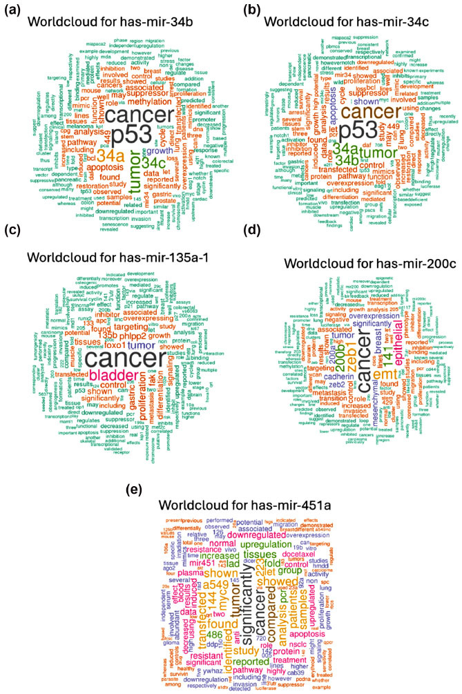
Objective: This study aims to apply several bioinformatic tools in order to clarify miRNA interactions with potential genes involved in brain injury, emphasizing the need of using a computational approach to determine the most likely correlations between miRNAs and target genes. Specifically, this study centers on elucidating the roles of miR-34b, miR-34c, miR-135a, miR-200c, and miR-451a.

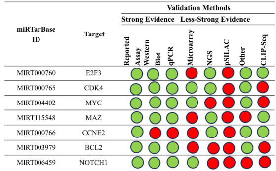
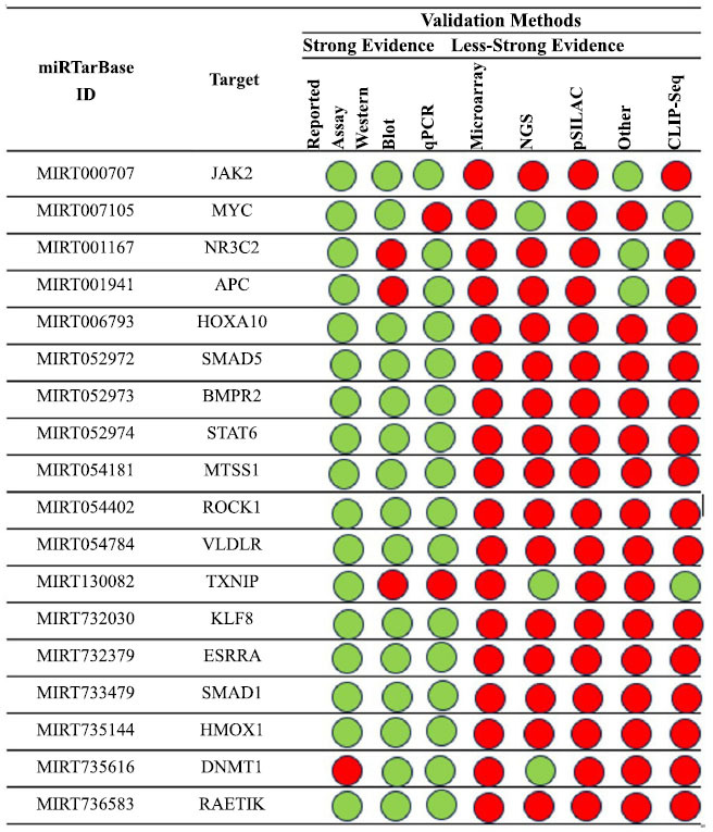
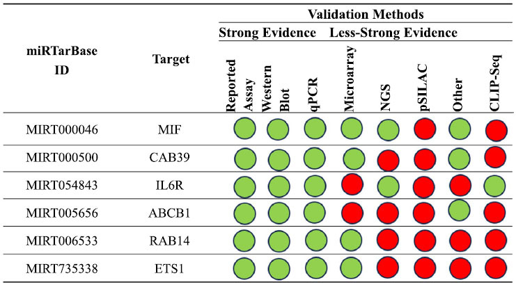
Methods: After a careful evaluation of different software available (analyzing the strengths and limitations), we applied three tools, one to perform an analysis of the validated targets (miRTarBase), and two to evaluate functional annotations (miRBase and TAM 2.0).

Results: Research findings indicate elevated levels of miR-135a and miR-34b in patients with traumatic brain injury (TBI) within the first day post-injury, while miR-200c and miR-34c were found to be upregulated after 7 days. Moreover, miR-451a and miR-135a were found overexpressed in the serum, while miRNAs 34b, 34c, and 200c, had lower serum levels at baseline post brain injury.

Conclusion: This study emphasizes the use of computational methods in determining the most likely relationships between miRNAs and target genes by investigating several bioinformatic techniques to elucidate miRNA interactions with potential genes. Specifically, this study focuses on the functions of miR-34b, miR-34c, miR-135a, miR-200c, and miR-451a, providing an up-to-date overview and suggesting future research directions for identifying theranomiRNAs related to brain injury, both at the tissue and serum levels.

Keywords: Brain injury; biomarkers; diagnosis; miRNA; prognosis; theranomiRNA..


Conflict of interest statement

The authors declare no conflict of interest, financial or otherwise.

Figures

Fig. (1)
Fig. (1)
Word cloud for hsa-mir-34b (a); hsa-miR-34c (b); hsa-miR-135a-1 (c); hsa-miR-200c (d); hsa-miR-451a (e).
Fig. (2)
Fig. (2)
The connection between cell functions and the uploaded miRNA set. These miRNAs are involved in a number of critical processes, including the development of the brain, nervous system, head, and animal organs.
Fig. (3)
Fig. (3)
Summary of the GO related to aging processes.
Fig. (4)
Fig. (4)
An overview of the primary illnesses associated with this miRNA dataset.
Fig. (5)
Fig. (5)
Summary of the miRNA interactions referred to hsa-mir-34b-5p, supported with at least 4 positive tools; green = validated methods, red = unvalidated methods. The methods are distinguished as strong or less strong tools.
Fig. (6)
Fig. (6)
The regulatory network of has-miR-34b and the MET gene (a and b) the regulatory network of interactions between the tested miRNA and MYC gene; (c) the regulatory network of interaction between miR-34b and NOTCH1 gene; the regulatory network of has-miR-34b and TGFBR2 gene (d) and YY1 gene (e).
Fig. (7)
Fig. (7)
An overview of the miRNA interactions associated with hsa-miR-34c, backed by a minimum of four positive tools; green indicates validated techniques, and red indicates unvalidated methods. The techniques have been classified as either powerful or weaker instruments.
Fig. (8)
Fig. (8)
The regulatory network of has-miR-34c and the E2F3 gene (a and b) shows the regulatory network of interaction between the tested miRNA and the CDK4 gene; (c) shows the network relative to the MAZ and (d) MYC genes.
Fig. (9)
Fig. (9)
An overview of the miRNA interactions associated with hsa-miR-135a, backed by a minimum of three positive tools; green denotes validated techniques, and red denotes unvalidated methods. The techniques have been classified as either powerful or weaker instruments.
Fig. (10)
Fig. (10)
The regulatory network of hsa-miR-135a and JAK2 and MYC gene.
Fig. (11)
Fig. (11)
A summary of the hsa-miR-200c interactions that are supported by at least four positive approaches; green indicates validated methods and red indicates unvalidated methods. The techniques have been classified as either powerful or weaker instruments.
Fig. (12)
Fig. (12)
The regulatory network of has-miR-200c and the ZEB1 gene (a and b) shows the regulatory network of the interaction between the tested miRNA and the TUBB3 gene; (c) shows the network relative to the BMI1 gene, ZEB2 gene (d) and FN1 gene (e).
Fig. (13)
Fig. (13)
The miRNA interactions with hsa-miR-451a are summarized, backed by a minimum of four positive tools. Green indicates validated techniques, while red indicates unvalidated methods. The techniques have been classified as either powerful or weaker instruments.
Fig. (14)
Fig. (14)
The regulatory network of has-miR-451a and the gene MIF (a and b) the regulatory network of interaction between the tested miRNA and the CAB39 gene and IL6R gene (c).

Similar articles

Cited by

References

    1. Shao F., Wang X., Wu H., Wu Q., Zhang J. Microglia and neuroinflammation: Crucial pathological mechanisms in traumatic brain injury-induced neurodegeneration. Front. Aging Neurosci. 2022;14:825086. doi: 10.3389/fnagi.2022.825086. - DOI - PMC - PubMed
    1. Ahluwalia M., Gaur P., Vaibhav K. Brain injury and neurodegeneration: molecular, functional, and translational approach. Biomedicines. 2023;11(7):1947. doi: 10.3390/biomedicines11071947. - DOI - PMC - PubMed
    1. Brett B.L., Gardner R.C., Godbout J., Dams-O’Connor K., Keene C.D. Traumatic brain injury and risk of neurodegenerative disorder. Biol. Psychiatry. 2022;91(5):498–507. doi: 10.1016/j.biopsych.2021.05.025. - DOI - PMC - PubMed
    1. Sessa F., Maglietta F., Bertozzi G., Salerno M., Di Mizio G., Messina G., Montana A., Ricci P., Pomara C. Human brain injury and mirnas: An experimental study. Int. J. Mol. Sci. 2019;20(7):1546. doi: 10.3390/ijms20071546. https://www.scopus.com/inward/record.uri?eid=2-s2.0-85064195391&doi=10.3... - DOI - PMC - PubMed
    1. Carvalho L.B., dos Santos Sanna P.L., dos Santos Afonso C.C., Bondan E.F., da Silva Feltran G., Ferreira M.R., Birbrair A., Andia D.C., Latini A., Foganholi da Silva R.A. MicroRNA biogenesis machinery activation and lncRNA and REST overexpression as neuroprotective responses to fight inflammation in the hippocampus. J. Neuroimmunol. 2023;382:578149. doi: 10.1016/j.jneuroim.2023.578149. - DOI - PubMed