. 2024 Sep;20(9):6045-6059.
doi: 10.1002/alz.14158. Epub 2024 Aug 11.

Association of cortical morphology, white matter hyperintensity, and glymphatic function in frontotemporal dementia variants

Affiliations

Association of cortical morphology, white matter hyperintensity, and glymphatic function in frontotemporal dementia variants

Die Xiao et al. Alzheimers Dement. 2024 Sep.

Abstract

Introduction: Frontotemporal dementia (FTD) can be phenotypically divided into behavioral variant FTD (bvFTD), nonfluent variant primary progressive aphasia (nfvPPA), and semantic variant PPA (svPPA). However, the neural underpinnings of this phenotypic heterogeneity remain elusive.

Methods: Cortical morphology, white matter hyperintensities (WMH), diffusion tensor image analysis along the perivascular space (DTI-ALPS), and their interrelationships were assessed in subtypes of FTD. Neuroimaging-transcriptional analyses on the regional cortical morphological deviances among subtypes were also performed.

Results: Changes in cortical thickness, surface area, gyrification, WMH, and DTI-ALPS were subtype-specific in FTD. The three morphologic indices are related to whole-brain WMH volume and cognitive performance, while cortical thickness is related to DTI-ALPS. Neuroimaging-transcriptional analyses identified key biological pathways linked to the formation and/or spread of TDP-43/tau pathologies.

Discussion: We found subtype-specific changes in cortical morphology, WMH, and glymphatic function in FTD. Our findings have the potential to contribute to the development of personalized predictions and treatment strategies for this disorder.

Highlights: Cortical morphologic changes, white matter hyperintensities (WMH), and glymphatic dysfunction are subtype-specific. Cortical morphologic changes, WMH, and glymphatic dysfunction are inter-correlated. Cortical morphologic changes and WMH burden contribute to cognitive impairments.

Keywords: MRI; cortical morphology; frontotemporal dementia; glymphatic function; neuroimaging‐transcriptional association; white matter hyperintensities.


Conflict of interest statement

Authors declare that they have no competing interests. Author disclosures are available in the supporting information.

Figures

FIGURE 1
FIGURE 1
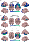
Cortical thickness reductions in variants of frontotemporal dementia (FTD). Brain regions showing significantly reduced cortical thickness in the bvFTD (A), nfvPPA (B), and svPPA (C) subtypes compared with matched healthy controls (HCs). The results were corrected for multiple comparisons using random field theory (RFT). The level of significance was set at RFT‐corrected p < 0.05. The color bar denotes the t‐value of the significant clusters of the corresponding contrast
FIGURE 2
FIGURE 2
Cortical surface area reductions in variants of frontotemporal dementia (FTD). Brain regions showing significantly reduced cortical surface area in the bvFTD (A), nfvPPA (B), and svPPA (C) subtypes compared with matched healthy controls (HCs). The results were corrected for multiple comparisons using random field theory (RFT). The level of significance was set at RFT‐corrected p < 0.05. The color bar denotes the t‐value of the significant clusters of the corresponding contrast
FIGURE 3
FIGURE 3
Local gyrification index (LGI) changes in variants of frontotemporal dementia (FTD). Brain regions showing significantly reduced LGI in the bvFTD (A), nfvPPA (B), and svPPA (C) subtypes compared with matched healthy controls (HCs). The results were corrected for multiple comparisons using random field theory (RFT). The level of significance was set at RFT‐corrected p < 0.05. The color bar denotes the t‐value of the significant clusters of the corresponding contrast
FIGURE 4
FIGURE 4
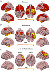
Brain regions with subtypes‐specific patterns in cortical thickness (A), surface area (B), and local gyrification index (LGI) (C) in frontotemporal dementia (FTD). The results were corrected for multiple comparisons using random field theory (RFT). The level of significance was set at RFT‐corrected p < 0.05. The color bar indicates the f‐value for the significant clusters of the vertex‐wise analysis of covariance (ANCOVA) among the three FTD subtypes
FIGURE 5
FIGURE 5
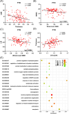
Correlates of imaging parameters in frontotemporal dementia (FTD). Negative correlations between cortical thickness and CDR score (A), between whole brain white matter hyperintensities (WMH) volume and Mini‐Mental State Examination (MMSE) score (B), and between whole brain WMH volume and local gyrification index (LGI) (C). (D) Positive correlation between diffusion tensor image analysis along the perivascular space (DTI‐ALPS) and cortical thickness. Functional enrichment analysis of the pivotal genes associated with greater intergroup regional deviances in cortical thickness, surface area, and LGI among the three FTD subtypes (E). Terms were retained with a threshold of false discovery rate (FDR) corrected p < 0.05

References

    1. Boeve BF, Boxer AL, Kumfor F, Pijnenburg Y, Rohrer JD. Advances and controversies in frontotemporal dementia: diagnosis, biomarkers, and therapeutic considerations. Lancet Neurol. 2022;21(3):258‐272. doi:10.1016/s1474-4422(21)00341-0 - DOI - PubMed
    1. Ulugut H, Pijnenburg YAL. Frontotemporal dementia: past, present, and future. Alzheimers Dement. 2023;19(11):5253‐5263. doi:10.1002/alz.13363 - DOI - PubMed
    1. Barker MS, Gottesman RT, Manoochehri M, et al. Proposed research criteria for prodromal behavioural variant frontotemporal dementia. Brain. 2022;145(3):1079‐1097. doi:10.1093/brain/awab365 - DOI - PMC - PubMed
    1. Piguet O, Kumfor F. Frontotemporal dementias: main syndromes and underlying brain changes. Curr Opin Neurol. 2020;33(2):215‐221. doi:10.1097/wco.0000000000000792 - DOI - PubMed
    1. Spinelli EG, Mandelli ML, Miller ZA, et al. Typical and atypical pathology in primary progressive aphasia variants. Ann Neurol. 2017;81(3):430‐443. doi:10.1002/ana.24885 - DOI - PMC - PubMed

LinkOut - more resources