. 2024 Jul 31;15(34):13841-13852.
doi: 10.1039/d4sc03642e. Online ahead of print.

More is different: progressive β-thiolation induced-porphyrin aggregation switches singlet oxygen photosensitization

Affiliations

More is different: progressive β-thiolation induced-porphyrin aggregation switches singlet oxygen photosensitization

Mengliang Zhu et al. Chem Sci. .

Abstract

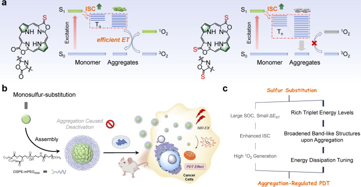
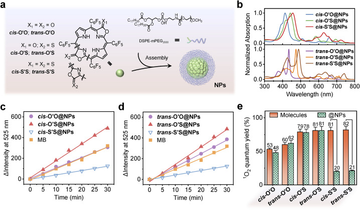
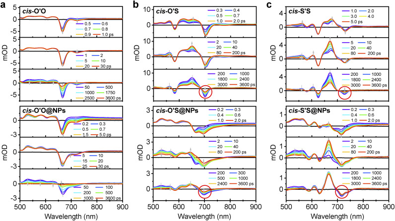
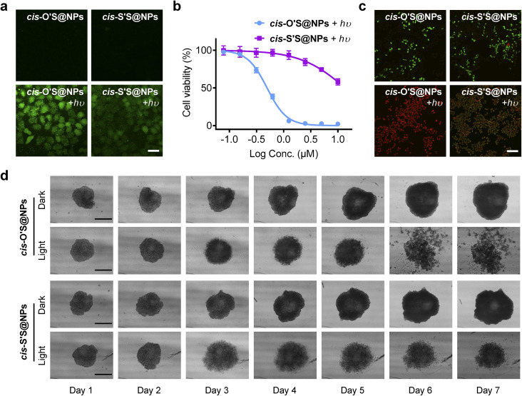
Incorporating sulfur atoms into photosensitizers (PSs) has been well-established to populate triplet states and increase singlet oxygen (1O2) production when exposed to light. In this work, we found that progressive thiolation of porphyrin β-periphery does promote intersystem crossing (ISC) between triplets and singlets, as seen in the excited state dynamics in dichloromethane or PS nanoparticles in water. However, in the latter case, more sulfur substitution deactivates 1O2 photosensitization, in contrast to the expected trend observed in dichloromethane. This observation was further supported by photocytotoxicity studies, where 1O2 photosensitization was switched off in living cells and multicellular spheroids despite being switched on in in vivo mice models. To understand the inconsistency, we performed molecular dynamics simulation and time-dependent density functional theory calculations to investigate possible aggregation and related excited states. We found that the extent of thiolation could regulate molecular packing inside nanoparticles, which gradually lowers the energy levels of triplet states even lower than that of 1O2 and, in turn, alters their energy dissipation pathways. Therefore, this study provides new insights into the design of metal-free PSs and sheds light on the excited state dynamics in aqueous media beyond the molecular level.


Conflict of interest statement

There are no conflicts to declare.

Figures

Scheme 1
Scheme 1. (a) Molecular structure of cis-O'S and cis-S'S and the excited state energy dissipation pathways in their aggregated states. (b) Schematic illustration of the cis-O'S self-assembly into nanospheres and subsequent PDT effect. (c) The proposed sulfur substitution strategy to obtain aggregation-regulated PDT effect by tuning excited state dynamics.
Fig. 1
Fig. 1. (a) One-pot synthesis of porphothiodilactone isomers. (b) X-ray crystal structures of cis-/trans-O'S, and cis-/trans-S'S. Hydrogen atoms and solvents are omitted for clarity. (c) Absorption spectra of cis-/trans-O'O, cis-/trans-O'S, and cis-/trans-S'S in CH2Cl2. (d) Emission spectra of cis-/trans-O'O, cis-/trans-O'S, and cis-/trans-S'S in CH2Cl2 with excitation at 405 nm (A405 = 0.10). (e) Phosphorescence spectra of 1O2 obtained from air-saturated CH2Cl2 solutions of cis-/trans-O'O, cis-/trans-O'S, and cis-/trans-S'S.
Fig. 2
Fig. 2. (a) Preparation of porphodilactone nanoparticles. (b) Absorption spectra of cis-O'O@NPs, cis-O'S@NPs, cis-S'S@NPs, trans-O'O@NPs, trans-O'S@NPs, and trans-S'S@NPs in aqueous solution. (c) Variation of SOSG emission intensity at 525 nm (λex = 488 nm) under light irradiation (700 nm, 5 mW cm−2, 0–30 min) in the presence of cis-O'O@NPs, cis-O'S@NPs, cis-S'S@NPs, and MB. (d) Variation of SOSG emission intensity at 525 nm (λex = 488 nm) under light irradiation (700 nm, 5 mW cm−2, 0–30 min) in the presence of trans-O'O@NPs, trans-O'S@NPs, trans-S'S@NPs, and MB. (e) Comparison of 1O2 quantum yields between porphodilactone molecules and nanoparticles.
Fig. 3
Fig. 3. (a) Femtosecond transient absorption (fs-TA) difference spectra for cis-O'O recorded in CH2Cl2 (0.5–1 ps, 1–30 ps, and 50–3600 ps) and cis-O'O@NPs recorded in H2O (0.2–5 ps, 5–30 ps, and 50–3600 ps) monitored over different delay regimes. (b) Femtosecond transient absorption (fs-TA) difference spectra for cis-O'S recorded in CH2Cl2 (0.3–2 ps, 2–200 ps, and 200–3600 ps) and cis-O'S@NPs recorded in H2O (0.2–2 ps, 2–200 ps, and 200–3600 ps) monitored over different delay regimes. (c) Femtosecond transient absorption (fs-TA) difference spectra for cis-S'S recorded in CH2Cl2 (1–5 ps, 5–200 ps, and 200–3600 ps) and cis-S'S@NPs recorded in H2O (0.2–2 ps, 2–200 ps, and 200–3600 ps) monitored over different delay regimes.
Fig. 4
Fig. 4. (a) Schematic illustration of the molecular aggregation of cis-S'S expected to persist in nanoparticles as deduced from equilibrated MD simulations in a water box (50 × 50 × 50 Å) and expected arrangements of the constituent aggregated dimers and trimers. (b) Normalized distribution of dihedral and transition dipole moment angle between cis-S'S dimers. (c) Depiction of the proposed excited state energy dissipation pathways and triplet energy levels of cis-O'O, cis-O'S, and cis-S'S dimers.
Fig. 5
Fig. 5. (a) Intracellular ROS level of HeLa cells treated with cis-O'S@NPs and cis-S'S@NPs under dark and light conditions (700 nm, 10 mW cm−2, 10 min), respectively. DCFH-DA was used as the fluorescent probe for ROS generation. Scale bar: 50 μm. (b) Photocytotoxicity of cis-O'S@NPs and cis-S'S@NPs (0–10 μM) on HeLa cells obtained by a CCK-8 assay. (c) Fluorescence images of Calcein AM/PI-stained HeLa cells treated with cis-O'S@NPs and cis-S'S@NPs under dark and light conditions, respectively. Scale bar: 200 μm. (d) Images of HeLa 3D MCSs treated with cis-O'S@NPs and cis-S'S@NPs (6 μM) under dark and light conditions, respectively. The images for day 1 were recorded before irradiation. Scale bar: 500 μm.
Fig. 6
Fig. 6. (a) Schematic illustration of the treatment regimen. (b) Tumor volume variation curves for the mice in control (PBS + ), cis-O'S@NPs, cis-S'S@NPs, cis-O'S@NPs + , and cis-S'S@NPs + groups during the treatment period (n = 4). ***p < 0.05. (c) Digital photographs of the tumors dissected from the mice in different groups. (d) Tumor weight of the mice in different groups at the end of treatment (mean ± SD, n = 4, **P < 0.05). (e) Body weight curves of the mice in different treatment groups (n = 4, mean ± SD).

References

    1. Li X. Lovell J. F. Yoon J. Chen X. Clinical development and potential of photothermal and photodynamic therapies for cancer. Nat. Rev. Clin. Oncol. 2020;17:657–674. doi: 10.1038/s41571-020-0410-2. - DOI - PubMed
    1. Zhao X. Liu J. Fan J. Chao H. Peng X. Recent progress in photosensitizers for overcoming the challenges of photodynamic therapy: from molecular design to application. Chem. Soc. Rev. 2021;50:4185–4219. doi: 10.1039/D0CS00173B. - DOI - PubMed
    1. Pham T. C. Nguyen V. N. Choi Y. Lee S. Yoon J. Recent strategies to develop innovative photosensitizers for enhanced photodynamic therapy. Chem. Rev. 2021;121:13454–13619. doi: 10.1021/acs.chemrev.1c00381. - DOI - PubMed
    1. Fan W. Huang P. Chen X. Overcoming the Achilles' heel of photodynamic therapy. Chem. Soc. Rev. 2016;45:6488–6519. doi: 10.1039/C6CS00616G. - DOI - PubMed
    1. Bastos E. L. Quina F. H. Baptista M. S. Endogenous photosensitizers in human skin. Chem. Rev. 2023;123:9720–9785. doi: 10.1021/acs.chemrev.2c00787. - DOI - PubMed

LinkOut - more resources