. 2024 Aug;76(4):3612-3616.
doi: 10.1007/s12070-024-04635-6. Epub 2024 Mar 18.

Hidradenocarcinoma of Nose: A Rare Case Report

Affiliations

Hidradenocarcinoma of Nose: A Rare Case Report

Krishna A Sahoo et al. Indian J Otolaryngol Head Neck Surg. 2024 Aug.

Abstract

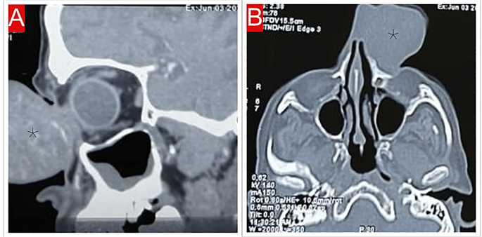
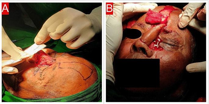
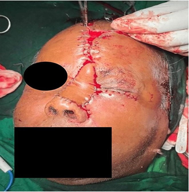
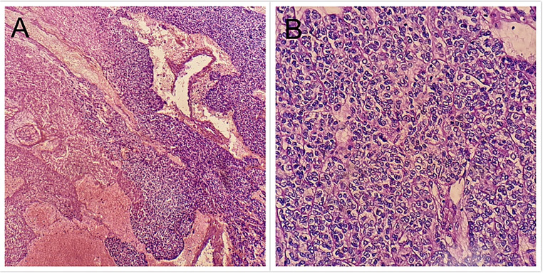
This report details the surgical management of a rare case of hidradenocarcinoma in a 70-year-old man presenting as a large multilobulated swelling on the dorsum of the nose. Following histopathological confirmation, the patient underwent wide complete excision of the tumour, coupled with sentinel lymph node dissection. Reconstruction involved the use of a paramedian forehead flap and cheek advancement flap. The successful outcome underscores the importance of early diagnosis and a comprehensive surgical approach for managing hidradenocarcinoma on the nasal dorsum.

Keywords: Cutaneous lesions on nose; Eccrine sweat gland tumour of nose; Hidradenocarcinoma nose; Malignant tumour of nose; Rare tumour of nose.


Conflict of interest statement

Conflict of InterestNot applicable.

Figures

Fig. 1
Fig. 1
A Image depicting a large, multilobulated mass situated on the dorsum of the nose. B Lateral view of face showing the nose mass causing obstruction of the left eye’s vision
Fig. 2
Fig. 2
A An image illustrating the sagittal section of the CT scan of the nose, revealing a heterogeneously enhancing mass over the nose. B Axial view CT scan displaying a mass over the nose without any evidence of cartilage or bone erosion
Fig. 3
Fig. 3
A An image depicting the measurement of the defect with skin markings for the paramedian forehead flap. B An image illustrating the rotation of the flap to the site of the defect
Fig. 4
Fig. 4
The image displays the paramedian forehead flap and cheek advancement flap, both of which were utilized to cover the defect, and subsequently sutured in place
Fig. 5
Fig. 5
A The microscopic image of the resected specimen, captured under lower magnification (40×), reveals clusters of squamous and clear cells arranged in a nest-like pattern within a fibro-vascular stroma distributed in the dermis. B The microscopic image of the resected specimen, taken under higher magnification (400×), demonstrates the presence of spindle cells with vesicular cytoplasm, as well as clear cells exhibiting atypical nuclei and hyperchromatism
Fig. 6
Fig. 6
The image above displays satisfactory results six months post-operation

References

    1. Romolo Fragola G, Tartaro GF, Nicoletti N, Zerbinati E, Nikolli GL, Giudice Raffaele Rauso: Hydro adenocarcinomaa rare tumor to be kept in mind. Oral Maxillofacial Surg Cases Volume 7:2021–100232. 10.1016/j.omsc.2021.100232
    1. Faulhaber D, Wörle B, Trautner B, Sander CA (2000) Clear cell hidradenoma in a young girl. J Am Acad Dermatol 42:693–695. 10.1067/mjd.2000.103274 10.1067/mjd.2000.103274 - DOI - PubMed
    1. de Lima AA, Santos M, de Morais PM, Rodrigues CAC (2021) Mar-Apr;96(2):251–253 Hidradenocarcinoma. An Bras Dermatol. 10.1016/j.abd.2020.03.023. Epub 2021 Feb 5. PMID: 33597101; PMCID: PMC8007541 - PMC - PubMed
    1. Hernández-Pérez E, Cestoni-Parducci R (1985) Nodular hidradenoma and hidradenocarcinoma. A 10-year review. J Am Acad Dermatol 12:15–20. 10.1016/S0190-9622(85)70002-3 10.1016/S0190-9622(85)70002-3 - DOI - PubMed
    1. Gauerke S, Driscoll JJ (2010) Hidradenocarcinomas: a brief review and future directions. Arch Pathol Lab Med 134:781–785. 10.5858/134.5.781 10.5858/134.5.781 - DOI - PubMed

LinkOut - more resources