. 2024 Aug;76(4):3570-3575.
doi: 10.1007/s12070-024-04615-w. Epub 2024 Apr 2.

A Rare Case of Lemierre's Syndrome due to Veillonella Parvula: A Dangerous and Forgotten Complication of a Septic Condition

Affiliations

A Rare Case of Lemierre's Syndrome due to Veillonella Parvula: A Dangerous and Forgotten Complication of a Septic Condition

Manuela Montatore et al. Indian J Otolaryngol Head Neck Surg. 2024 Aug.

Abstract

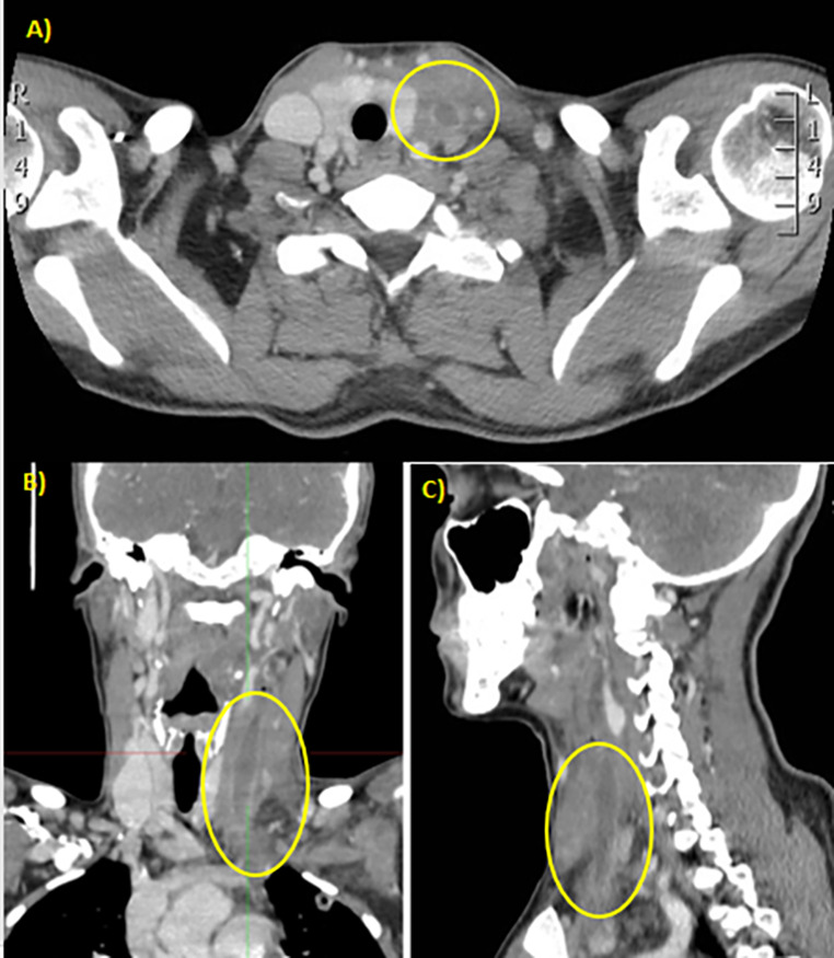
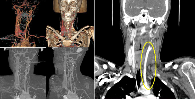
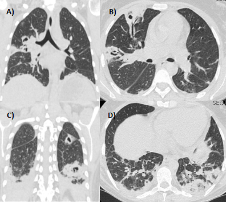
This clinical case presents an unusual case of Lemierre's syndrome (LS) in a young woman of 38-year-old. She arrived in the Emergency Department with a high fever and pharyngology resistant to antibiotic therapy with clarithromycin, ceftriaxone, and cortisone for two weeks. At the blood sampling, there is a marked leucocytosis, and the advice of the otolaryngologist is required given the strong pain in the throat. Due to the tonsillar abscess, a neck CT with a contrast medium is necessary for the otolaryngologist's opinion. The CT shows thrombosis of the jugular vein and left subclavian, with thickening of soft perivascular tissues; these findings suggest Lemierre's syndrome: a septic thrombophlebitis of the jugular vein that occurs as a complication of a peritonsillar abscess. The diagnostic process is then completed with a chest HR-CT, which reveals lung density and excavation areas suggesting tuberculosis. Blood culture reveals the presence of Veillonella Parvula (an anaerobic gram-negative coccus), sputum culture reveals the presence of some colonies of Enterobacter cloacae complex, real-time PCR examination on sputum reveals the presence of Streptococcus Pneumoniae and the borderline presence of rhinovirus. Microbiologists, after these results and neck and chest CT with a contrast agent, agree with the diagnosis of suspected LS at an early stage: a septic dissemination fortunately limited only to the neck and lungs region.

Keywords: Enterobacter cloacae; Leimerre’s syndrome; Oropharyngeal infection, thrombophlebitis; Septic emboli; Veillonella parvula; Vein thrombosis.


Conflict of interest statement

Conflict of InterestThe authors have no competing interests to disclose.

Figures

Fig. 1
Fig. 1
A CT of the neck, with contrast medium, shows that there is thrombosis of the jugular vein and subclavian left with thickening of soft perivascular tissues (A) in the axial section; (B) in the coronal section; (C) in the sagittal section
Fig. 2
Fig. 2
Angiotomography of the neck vessels. A filling defect is observed along the entire length of the left internal jugular vein
Fig. 3
Fig. 3
The HR-CT of the chest reveals pulmonary densities and areas of excavations at the posterior-basal segment of the LIS and anteroposterior of LSD probable infectious hypothesis, also caused by Mycobacterium tuberculosis. A) and B), a coronal section and an axial section, show the condition on the apex of the lungs. C) and D), a coronal section and an axial section, show the condition of the basal parenchyma of the lungs
Fig. 4
Fig. 4
The X-ray examination was performed in a single projection and showed a circumscribed parenchymal thickening at the level of the basal pulmonary field on the left. There is a dystelectasis stria at the peri-scissure site on the right, with traction relative to the pleura. Moderately thickened the peri-bronco vascular interstitial

References

    1. Fisher RG, Denison MR (1996) Veillonella parvula bacteremia without an underlying source. J Clin Microbiol 34(12):3235–3236. 10.1128/jcm.34.12.3235-3236.1996 10.1128/jcm.34.12.3235-3236.1996 - DOI - PMC - PubMed
    1. Salami A, Assouan C, Garba I, Konan E (2019) An unusual cause of Lemierre Syndrome. J Stomatol Oral Maxillofac Surg 120(4):358–360. 10.1016/j.jormas.2019.02.009 10.1016/j.jormas.2019.02.009 - DOI - PubMed
    1. Gowan RT, Mehran RJ, Cardinal P, Jones G (2000) Thoracic complications of Lemierre syndrome. Can Respir J 7(6):481–485. 10.1155/2000/451860 10.1155/2000/451860 - DOI - PubMed
    1. Bedan M, Gloksin W, Hagen TL (2023) Lemierre’s syndrome-A diagnostic challenge. Diagn Microbiol Infect Dis 107(2):116023. 10.1016/j.diagmicrobio.2023.116023 10.1016/j.diagmicrobio.2023.116023 - DOI - PubMed
    1. Amarnani S, Ranjan A (2022) Lemierre’s syndrome: a Lethal complication of Acute Tonsillitis. Cureus 14(10):e30072. 10.7759/cureus.30072 10.7759/cureus.30072 - DOI - PMC - PubMed

LinkOut - more resources