

Bronchopericardial Fistula: A Rare Complication of Necrotizing Pneumonia
- PMID: 39130980
- PMCID: PMC11316567
- DOI: 10.7759/cureus.64339
Bronchopericardial Fistula: A Rare Complication of Necrotizing Pneumonia
Abstract
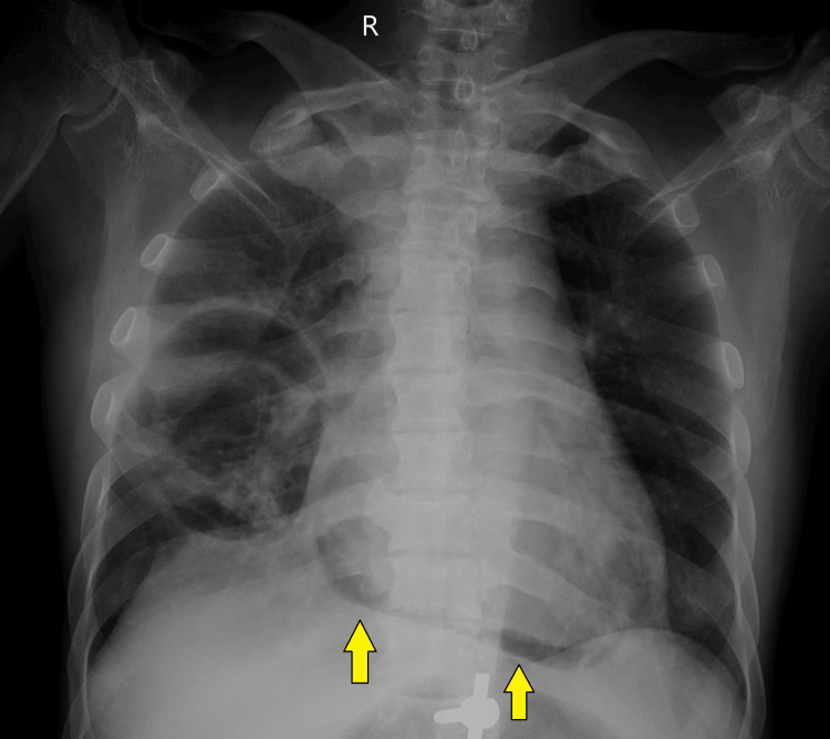
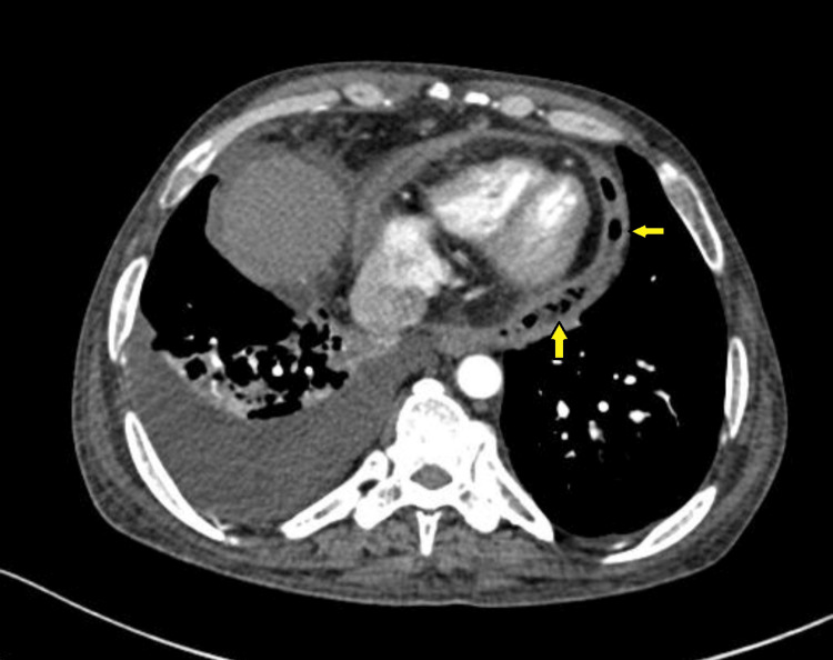
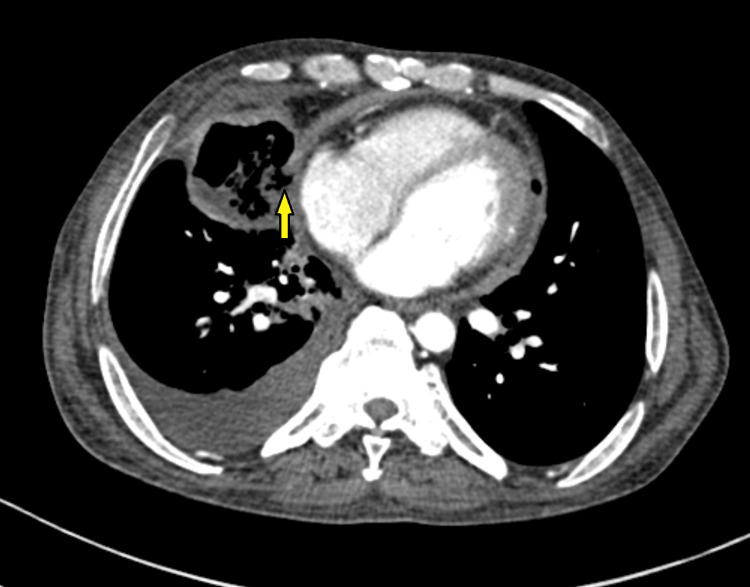
Pneumopericardium due to bronchopericardial fistula formation is a rare complication secondary to necrotizing pneumonia. Several such cases are reported due to different suppurative bacterial infections. Persistent fistulous communication has been reported to lead to tension pneumopericardium and hemodynamic instability, requiring urgent intervention such as pericardial drainage. A 41-year-old male patient, known to have chronic kidney disease and diabetes mellitus, presented with acute respiratory symptoms. Upon admission, the patient was febrile and required oxygen support via nasal prongs. A chest X-ray showed fibrocavitatory changes on the right side, with patchy air shadowing around the cardiac silhouette and a continuous diaphragm sign. A contrast-enhanced computed tomography (CECT) thorax revealed extensive areas of consolidation with necrotic areas within, forming a thin-walled cavity involving the right middle lobe. Also, suspicious communication of this cavity with the pericardial cavity along the right atrium was seen, with minimal pericardial collection and air foci within. The pleural fluid culture showed growth of Klebsiella pneumoniae. According to the antibiotic sensitivity report, the patient was started on IV meropenem and gentamicin for 21 days while monitoring kidney functions. The patient clinically improved on antibiotics, and follow-up radiological investigations showed resolution of pneumopericardium. In this patient, pneumopericardium was mild, and there was no evidence of tension pneumopericardium. Thus, conservative management with antibiotics was provided, with successful resolution. Unlike this case, if evidence of tension pneumopericardium had been present, emergency interventions for decompression would have been required, and these cases would have had a poor prognosis. This case demonstrates the importance of high suspicion and early diagnosis of pneumopericardium in patients with necrotizing pneumonia. Prompt treatment in these patients can prevent further life-threatening sequelae.
Keywords: broncho-pericardial fistula; cavity; klebsiella pneumoniae (kp); necrotising pneumonia; tension pneumopericardium.
Copyright © 2024, Bele et al.
Conflict of interest statement
Human subjects: Consent was obtained or waived by all participants in this study. Conflicts of interest: In compliance with the ICMJE uniform disclosure form, all authors declare the following: Payment/services info: All authors have declared that no financial support was received from any organization for the submitted work. Financial relationships: All authors have declared that they have no financial relationships at present or within the previous three years with any organizations that might have an interest in the submitted work. Other relationships: All authors have declared that there are no other relationships or activities that could appear to have influenced the submitted work.
Figures
Similar articles
-
A case of bronchopericardial fistula with tension pneumopericardium closed successfully by transpericardial intervention: A novel procedure.Catheter Cardiovasc Interv. 2017 Dec 1;90(7):1117-1120. doi: 10.1002/ccd.27376. Epub 2017 Oct 25. Catheter Cardiovasc Interv. 2017. PMID: 29068135
-
Tension pneumopericardium in chest trauma with gunshot wound.Ulus Travma Acil Cerrahi Derg. 2003 Oct;9(4):304-6. Ulus Travma Acil Cerrahi Derg. 2003. PMID: 14569491
-
Pneumopericardium in a patient with AIDS.Tex Heart Inst J. 2002;29(1):51-3. Tex Heart Inst J. 2002. PMID: 11995852 Free PMC article.
-
A fatal complication of an incarcerated diaphragmatic hernia: pyo-pneumopericardium due to a gastro-pericardial fistula Case report and literature review.Ann Ital Chir. 2016;87:75-8. Ann Ital Chir. 2016. PMID: 27026068 Review.
-
[2 cases of the tension pneumopericardium following blunt chest trauma resulting in the cardiac tamponade].Nihon Kyobu Geka Gakkai Zasshi. 1994 Aug;42(8):1242-6. Nihon Kyobu Geka Gakkai Zasshi. 1994. PMID: 7963843 Review. Japanese.
References
-
- A case of bronchopericardial fistula with tension pneumopericardium closed successfully by transpericardial intervention: a novel procedure. Shelke AB, Kawade R, Gandhi S. Catheter Cardiovasc Interv. 2017;90:1117–1120. - PubMed
-
- An unusual case of bronchopericardial fistula secondary to necrotizing pneumonia. Bharti P, Agarwal V, Kuruvilla J, Singla S. Egypt J Radiol Nucl Med. 2022;53:159.
-
- CT of bronchopericardial fistula: an unusual complication of multidrug-resistant tuberculosis in HIV infection. Bennett JA, Haramati LB. AJR Am J Roentgenol. 2000;175:819–820. - PubMed
Publication types
LinkOut - more resources
Full Text Sources