Review
. 2024 Mar 26;3(3Part B):101292.
doi: 10.1016/j.jscai.2023.101292. eCollection 2024 Mar.

Computed Tomography in Infectious Endocarditis

Affiliations
Review

Computed Tomography in Infectious Endocarditis

Eefje M Dalebout et al. J Soc Cardiovasc Angiogr Interv. .

Abstract

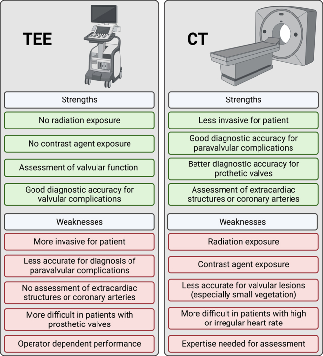
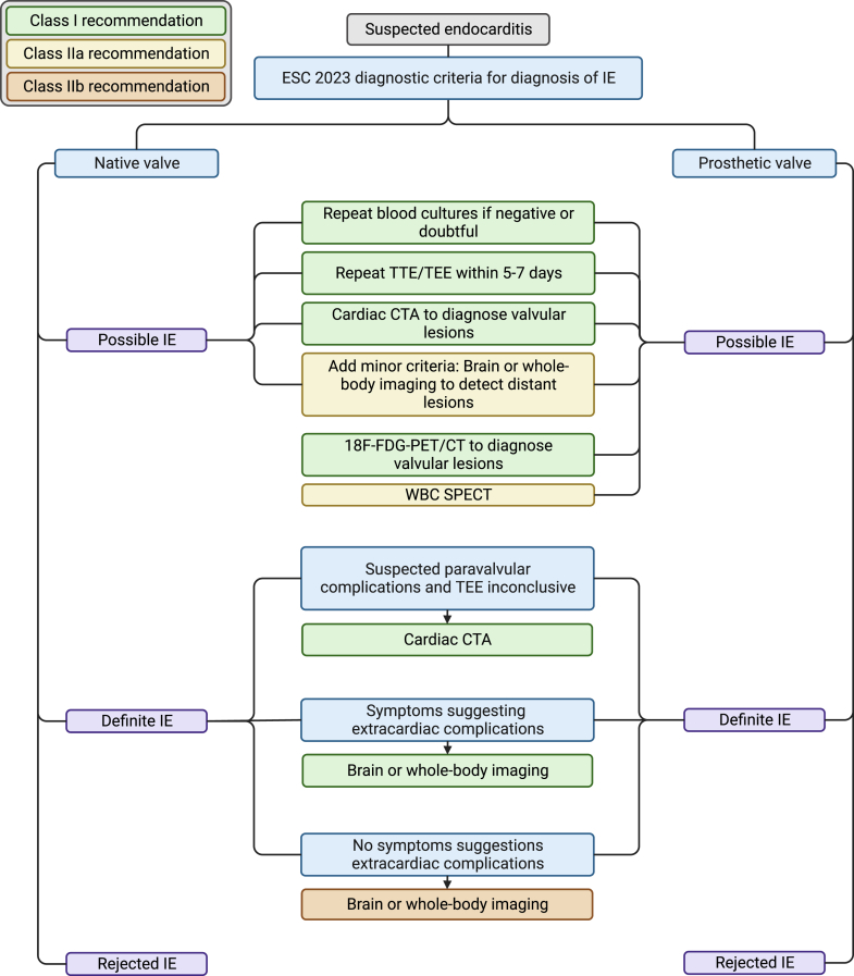
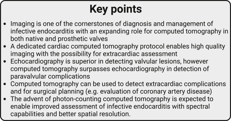
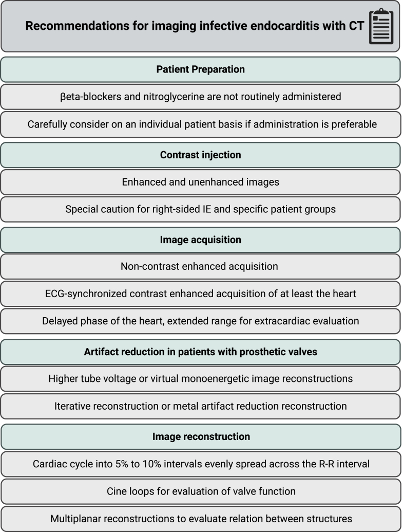
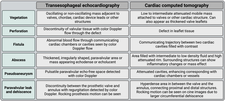
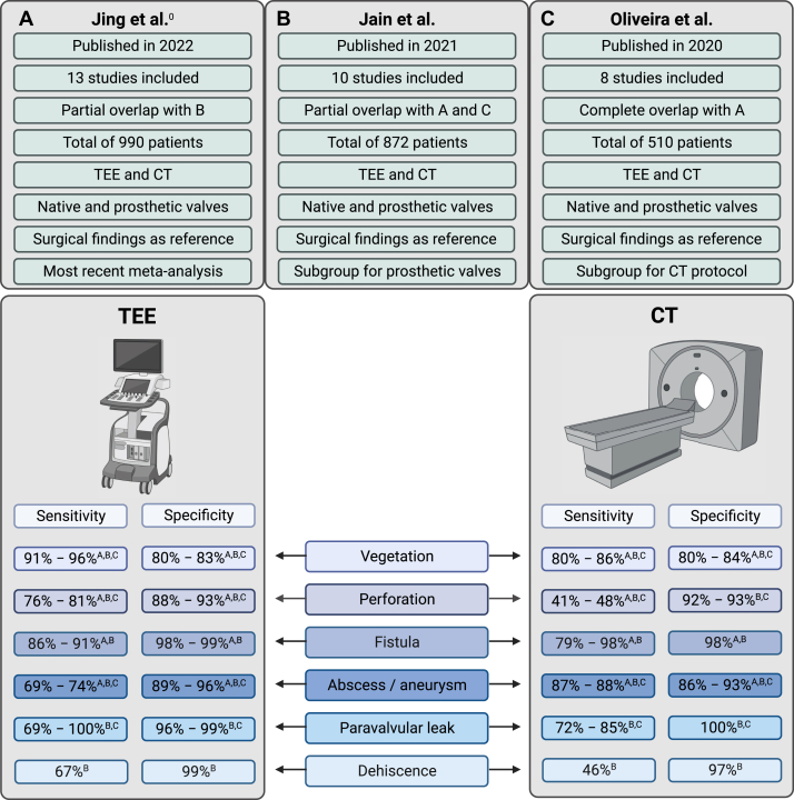
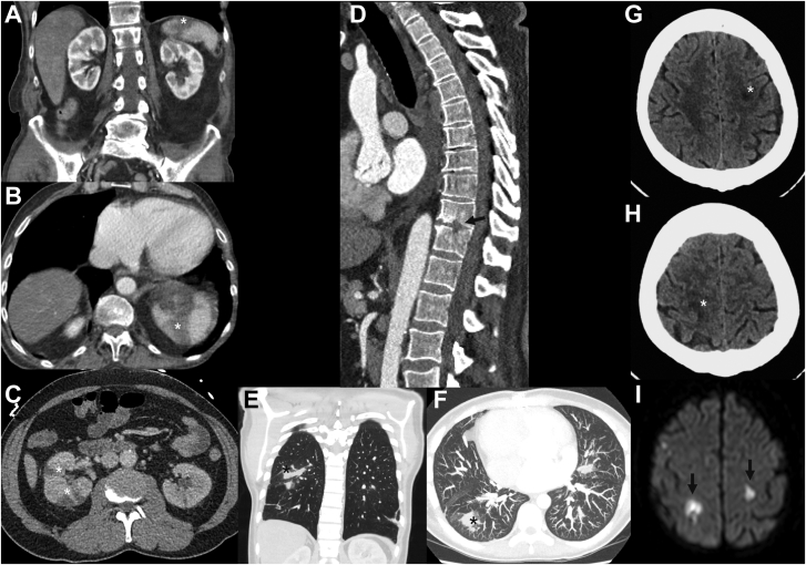
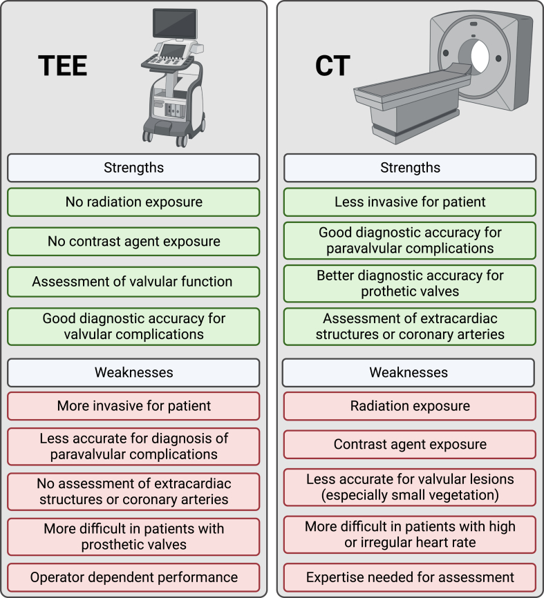
Imaging is one of the cornerstones in diagnosis and management of infective endocarditis, underlined by recent guidelines. Echocardiography is the first-line imaging technique, however, computed tomography (CT) has a class I recommendation in native and prosthetic valve endocarditis to detect valvular lesions in case of possible endocarditis and to detect paravalvular and periprosthetic complications in case of inconclusive echocardiography. Echocardiography has a higher diagnostic accuracy than CT in detecting valvular lesions, but not for diagnosing paravalvular lesions where CT is superior. Additionally, CT is useful and recommended by guidelines to detect extracardiac manifestations of endocarditis and in planning surgical treatment including assessment of the coronary arteries. The advent of photon-counting CT and its improved spatial resolution and spectral imaging is expected to expand the role of CT in the diagnosis of infective endocarditis. In this review, we provide an overview of the current role of CT in infective endocarditis focusing on image acquisition, image reconstruction, interpretation, and diagnostic accuracy.

Keywords: cardiac imaging; computed tomography; infective endocarditis.


Figures

None
Graphical abstract
Figure 1
Figure 1
The diagnostic pathway for patients with suspected infective endocarditis (IE) and the role of computed tomography based on the 2023 European Society of Cardiology (ESC) guidelines for the management of IE. The color of the boxes indicates the class of recommendation. CTA, computed tomography angiography; TEE, transesophageal echocardiography; TTE, transthoracic echocardiography; WBC SPECT, white blood cell single photon emission computed tomography; 18F-FDG-PET/CT, 18F-fluorodeoxyglucose positron emission tomography and computed tomography.
Figure 2
Figure 2
Key points of this review.
Figure 3
Figure 3
Pledgets used in surgical aortic valve replacement shown on computed tomography in contrast-enhanced, true non-contrast, and virtual non-contrast images. (A) Contrast-enhanced image, (B) true non-contrast image, (C) virtual non-contrast image.
Figure 4
Figure 4
Summary of computed tomography (CT) protocol recommendations for imaging infective endocarditis (IE). ECG, electrocardiography.
Figure 5
Figure 5
Overview of signs of endocarditis that can be seen on echocardiography and computed tomography.,,, ,
Figure 6
Figure 6
Summaryof3 meta-analyses considering the diagnostic accuracy of transesophageal echocardiography(TEE)and computed tomography(CT)for infective endocarditis. The most important study characteristics are provided and the range of pooled sensitivity and specificity for different signs of endocarditis. (A) Meta-analysis by Jing et al; (B) meta-analysis by Jain et al; (C) meta-analysis by Oliveira et al.
Figure 7
Figure 7
Patient with a native aortic valve endocarditis and vegetations. Vegetations (arrows) on the aortic valve and ascending aorta are seen on transesophageal echocardiography (A) and computed tomography (B and C).
Figure 8
Figure 8
Thickened valve leaflets and vegetations in a patient with aortic biological prosthetic heart valve endocarditis. Thickened valve leaflets and vegetations on the valve are seen on both transesophageal echocardiography (A and B) and computed tomography (C and D). Also, notice the thickening of the aortic root (asterisks) indicating aortic root abscess formation.
Figure 9
Figure 9
Perforation of the anterior mitral valve leaflet in a patient with native mitral valve endocarditis. The transesophageal echocardiography (A and B) images show the Doppler jet traversing the anterior mitral valve leaflet (arrow) indicating a perforation. The computed tomography images (C and D) show a focal discontinuity (arrow) in the valve leaflet compatible with a perforation.
Figure 10
Figure 10
Fistula between the aortic root and right atrium in a patient with aortic biological prosthetic heart valve endocarditis. Fistula between the aortic root and right atrium (asterisks) shown on transesophageal echocardiography with a Doppler jet traversing from the aortic root to the right atrium (A and B) and computed tomography with a contrast-enhanced trajectory between the aortic root and right atrium (arrows) (C).
Figure 11
Figure 11
Aneurysm between the aortic root and left atrium. Aneurysm between the aortic root and left atrium filling with blood in the systolic phase and collapsing in the diastolic phase (asterisks) in a patient with endocarditis of a mechanical aortic valve shown on transesophageal echocardiography (A and B) and computed tomography (C and D).
Figure 12
Figure 12
Extracardiac complications shown on computed tomography. Wedge-shaped hypoattenuating regions in the spleen and kidney (A, B, and C) are compatible with infarct due to embolized vegetations. Destruction of part of the vertebral bodies due to spondylodiscitis (D) and pulmonary septic emboli (E and F). Cerebral ischemia due to septic emboli seen on computed tomography (G and H) and magnetic resonance imaging (I).
Central Illustration
Overview of strengths and weaknesses of transesophageal echocardiography (TEE) and computed tomography (CT).

References

    1. Momtazmanesh S., Saeedi Moghaddam S., Malakan Rad E., et al. Global, regional, and national burden and quality of care index of endocarditis: the global burden of disease study 1990-2019. Eur J Prev Cardiol. 2022;29(8):1287–1297. doi: 10.1093/eurjpc/zwab211. - DOI - PubMed
    1. Delgado V., Ajmone Marsan N., de Waha S., et al. 2023 ESC Guidelines for the management of endocarditis. Eur Heart J. 2023;44(39):3948–4042. doi: 10.1093/eurheartj/ehad193. - DOI - PubMed
    1. Writing Committee Members. Otto C.M., Nishimura R.A., et al. 2020 ACC/AHA guideline for the management of patients with valvular heart disease: a report of the American College of Cardiology/American Heart Association joint committee on clinical practice guidelines. J Am Coll Cardiol. 2021;77(4):e25–e197. doi: 10.1016/j.jacc.2020.11.018. - DOI - PubMed
    1. Habets J., Tanis W., Reitsma J.B., et al. Are novel non-invasive imaging techniques needed in patients with suspected prosthetic heart valve endocarditis? A systematic review and meta-analysis. Eur Radiol. 2015;25(7):2125–2133. doi: 10.1007/s00330-015-3605-7. - DOI - PMC - PubMed
    1. Parra J.A., Hernández L., Muñoz P., et al. Detection of spleen, kidney and liver infarcts by abdominal computed tomography does not affect the outcome in patients with left-side infective endocarditis. Med (Baltim) 2018;97(33) doi: 10.1097/MD.0000000000011952. - DOI - PMC - PubMed

LinkOut - more resources