[Preprint]. 2024 Aug 17:2024.07.28.605513.
doi: 10.1101/2024.07.28.605513.

Monkey lateral prefrontal cortex subregions differentiate between perceptual exposure to visual stimuli

Affiliations

Monkey lateral prefrontal cortex subregions differentiate between perceptual exposure to visual stimuli

Kyoko Leaman et al. bioRxiv. .

Update in

Abstract

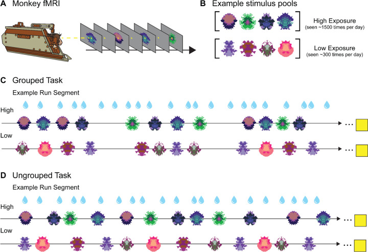
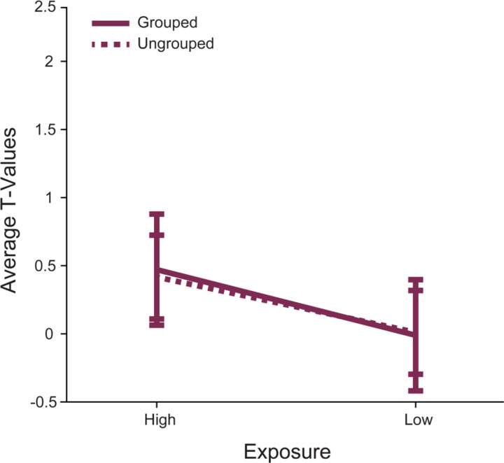
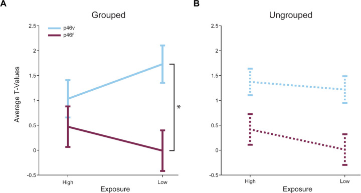
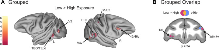
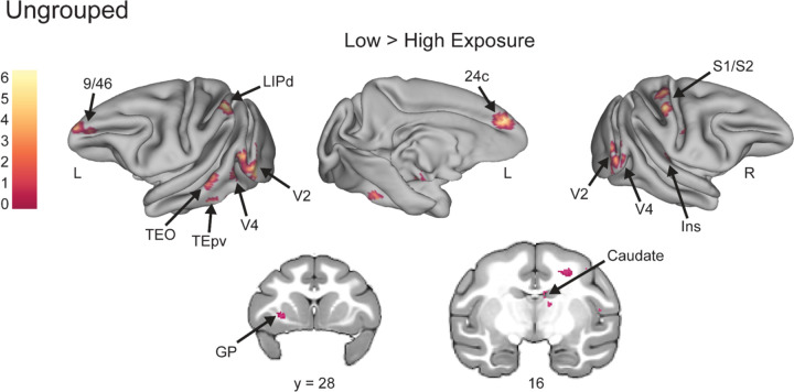
Each day, humans must parse visual stimuli with varying amounts of perceptual experience, ranging from incredibly familiar to entirely new. Even when choosing a novel to buy at a bookstore, one sees covers they have repeatedly experienced intermixed with recently released titles. Visual exposure to stimuli has distinct neural correlates in the lateral prefrontal cortex (LPFC) of nonhuman primates. However, it is currently unknown if this function may be localized to specific subregions within LPFC. Specifically, we aimed to determine whether the posterior fundus of area 46 (p46f), an area that responds to deviations from learned sequences, also responds to less frequently presented stimuli outside of the sequential context. We compare responses in p46f to the adjacent subregion, posterior ventral area 46 (p46v), which we propose may be more likely to show exposure-dependent responses due to its proximity to novelty responsive regions. To test whether p46f or p46v represent perceptual exposure, we performed awake functional magnetic resonance imaging (fMRI) on three male monkeys as they observed visual stimuli that varied in their number of daily presentations. Here we show that p46v, but not p46f, shows preferential activation to stimuli with low perceptual exposure, further localizing exposure-dependent effects in monkey LPFC. These results align with previous research that has found novelty responses in ventral LPFC and are consistent with the proposal that p46f performs a sequence-specific function. Further, they expand on our knowledge of the specific role of LPFC subregions and localize perceptual exposure processing within this broader brain region.


Figures

Figure 1.
Figure 1.. No-report Grouped and Ungrouped viewing tasks.
A. Monkeys in “sphynx” position fixate on central fixation square during fMRI scanning for both tasks. B. Example High and Low Exposure stimulus pools for both Ungrouped and Grouped tasks depict images that would be used for a single scanning session. Over the course of a standard scanning session, High Exposure images will be presented ~1500 times on average while Low Exposure images will be presented ~300 times on average. C. Example partial Grouped task run showing two of three possible block types (snippets of High and Low Exposure blocks are shown, the third deviant block type is not reported here). Three pseudorandomized four-item example groupings are shown for a High Exposure block (top row) and Low Exposure block (bottom row). Four-item image groups appear in six possible timing templates. All blocks contained 30 four-item groupings (120 total image presentations). The yellow square indicates the 14 second fixation period between each block. D. Example partial Ungrouped task run showing snippets of High and Low block types (the third is not shown). Twelve pseudorandomized image presentations with jittered timing are shown for a High Exposure block (top row) and Low Exposure block (bottom row). All blocks contained 120 image presentations. The yellow square indicates the 14 second fixation period between each block. Blue water droplets schematize reward delivery, which is decoupled from image events and delivered on a graduated schedule based on the duration the monkey has maintained fixation.
Figure 2.
Figure 2.. Right area p46f does not respond differently to Low Exposure versus High Exposure images.
The mean T-values for each condition > baseline are shown. The error bars represent the 95% confidence intervals (1.96 × standard error of the mean). Responses to stimuli in the Grouped task are shown in a solid line, while responses in the Ungrouped task are shown with a dashed line. Responses to High and Low Exposure stimuli were different such that there was a marginal main effect of stimulus exposure.
Figure 3.
Figure 3.. Right areas p46v and p46f respond differently across conditions in both tasks.
The mean T-values for each condition > baseline are shown. The error bars represent the 95% confidence intervals (1.96 × standard error of the within-bin mean). Responses in p46v are shown in blue and p46f is shown in purple. A. High Exposure compared to Low Exposure images in the Grouped task show a significant interaction effect with ROI (indicated by asterisk, p = 0.05) and a highly significant main effect of ROI. B. High Exposure compared to Low Exposure images in the Ungrouped task show a significant main effect of ROI and a marginal main effect of exposure.
Figure 4.
Figure 4.. Whole brain contrasts show known novelty-responsive regions, including area IT, respond more to Low Exposure images in the Grouped task.
Voxel-wise contrast of Low Exposure Images > High Exposure Images in the Grouped task false discovery rate (FDR) error cluster corrected for multiple comparisons (FDRc < 0.05, height p < 0.005 unc., extent = 116) is shown. A. Significant clusters in inferotemporal cortex (TEO/TEpd), visual area 2 (V2), ventral visual area 4 (V4v), somatosensory areas 1 and 2 (S1/S2), and ventral lateral prefrontal cortex (45/46v) are shown on inflated brains. Color bar indicates t value. B. Significant clusters in lateral orbitofrontal cortex (13l) and ventral lateral prefrontal cortex (45/46v) are shown in pink. The p46v ROI used in previous analyses is shown in blue and overlap between the contrast and ROI is shown in yellow.
Figure 5.
Figure 5.. Novelty-sensitive temporal regions are active for Low Exposure stimuli in the Ungrouped task.
Voxel-wise contrast of Low Exposure Images > High Exposure Images in Ungrouped task false discovery rate (FDR) error cluster corrected for multiple comparisons (FDRc < 0.05, height p < 0.005 unc., extent = 91) is shown. Significant clusters in prefrontal area 9/46, dorsal lateral intraparietal area (LIPd), inferotemporal cortex (TEO/TEpv), visual area 2 (V2), visual area 4 (V4), anterior midcingulate cortex (A24c), somatosensory areas 1 and 2 (S1/S2), Insula (Ins), globus pallidus (GP), and caudate are shown on inflated brains and coronal slices. Color bar indicates t value for inflated brain clusters.

References

    1. Anderson B., Mruczek R. E. B., Kawasaki K., & Sheinberg D. (2008). Effects of Familiarity on Neural Activity in Monkey Inferior Temporal Lobe. Cerebral Cortex, 18(11), 2540–2552. 10.1093/cercor/bhn015 - DOI - PMC - PubMed
    1. Balan P. F., Zhu Q., Li X., Niu M., Rapan L., Funck T., Wang H., Bakker R., Palomero-Gallagher N., & Vanduffel W. (2024). MEBRAINS 1.0: A new population-based macaque atlas. Imaging Neuroscience, 2, 1–26. 10.1162/imag_a_00077 - DOI - PMC - PubMed
    1. Barbas H., & Mesulam M.-M. (1985). Cortical afferent input to the principals region of the rhesus monkey. Neuroscience, 15(3), 619–637. 10.1016/0306-4522(85)90064-8 - DOI - PubMed
    1. Chavis D. A., & Pandya D. N. (1976). Further observations on corticofrontal connections in the rhesus monkey. Brain Research, 117(3), 369–386. 10.1016/0006-8993(76)90089-5 - DOI - PubMed
    1. Ghazizadeh A., Fakharian M. A., Amini A., Griggs W., Leopold D. A., & Hikosaka O. (2020). Brain Networks Sensitive to Object Novelty, Value, and Their Combination. Cerebral Cortex Communications, 1(1), tgaa034. 10.1093/texcom/tgaa034 - DOI - PMC - PubMed

Publication types

LinkOut - more resources