[Preprint]. 2024 Aug 4:2024.07.08.602591.
doi: 10.1101/2024.07.08.602591.

Single-nanoparticle electrophoretic mobility determination and trapping using active-feedback 3D tracking

Affiliations

Single-nanoparticle electrophoretic mobility determination and trapping using active-feedback 3D tracking

Alexis Johnson et al. bioRxiv. .

Abstract

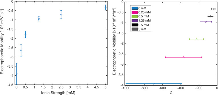
Nanoparticles (NP) are versatile materials with widespread applications across medicine and engineering. Despite rapid incorporation into drug delivery, therapeutics, and many more areas of research and development, there is a lack of robust characterization methods. Light scattering techniques such as dynamic light scattering (DLS) and electrophoretic light scattering (ELS) use an ensemble-averaged approach to the characterization of nanoparticle size and electrophoretic mobility (EM), leading to inaccuracies when applied to polydisperse or heterogeneous populations. To address this lack of single-nanoparticle characterization, this work applies 3D Single-Molecule Active Real-time Tracking (3D-SMART) to simultaneously determine NP size and EM on a per-particle basis. Single-nanoparticle EM is determined by using active feedback to "lock on" to a single particle and apply an oscillating electric field along one axis. A maximum likelihood approach is applied to extract the single-particle EM from the oscillating nanoparticle position along the field-actuated axis, while mean squared displacement is used along the non-actuated axes to determine size. Unfunctionalized and carboxyl-functionalized polystyrene NPs are found to have unique EM based on their individual size and surface characteristics, and it is demonstrated that single-nanoparticle EM is a more precise tool for distinguishing unique NP preparations than diffusion alone, able to determine the charge number of individual NPs to an uncertainty of less than 30. This method also explored individual nanoparticle EM in various ionic strengths (0.25-5 mM) and found decreased EM as a function of increasing ionic strength, in agreement with results determined via bulk characterization methods. Finally, it is demonstrated that the electric field can be manipulated in real time in response to particle position, resulting in one-dimensional electrokinetic trapping. Critically, this new single-nanoparticle EM determination and trapping method does not require microfluidics, opening the possibility for the exploration of single-nanoparticle EM in live tissue and more comprehensive characterization of nanoparticles in biologically relevant environments.


Figures

Figure 1:
Figure 1:. Experimental scheme for single-particle EM measurements.
(A) Field-stimulation open-bath chamber. Inset: diffusing nanoparticle under applied electric field. (B) 3D-SMART microscope illustration. (C) Example 3D trajectory of a freely diffusing nanoparticle in water, captured by 3D-SMART.
Figure 2:
Figure 2:. Three-dimensional motion of nanoparticles in solution under various applied voltage conditions.
(A-C) Illustration depicting the voltage conditions applied along the X direction: 0V, constant application of +2 V, and sinusoidal oscillation between +2 V and −2 V at a frequency of 1 Hz. (D-F) Representative 3D trajectories of 196 nm carboxyl-functionalized PS NPs diffusing under the field conditions specified in (A-C). (G-I) X position of nanoparticles versus time under the field conditions specified in (A-C). (J-L) Mean squared displacement versus lag time of particles under the field conditions specified in (A-C).
Figure 3:
Figure 3:. Directed particle motion in the X direction due to electric field stimulation.
Nanoparticle X position with (A) zero applied voltage, (B) constant applied voltage, and (C) oscillating applied voltage.
Figure 4:
Figure 4:. Relative EM uncertainty versus trajectory duration.
(A) Relative uncertainty of electrophoretic mobility for 196 nm carboxyl-functionalized PS NPs calculated at each second of continuous tracking. The dotted red line denotes the desired cutoff criteria: 20 seconds of single particle tracking (vertical), and 10% relative uncertainty of measurement (horizontal). (B) Histogram of relative uncertainty values reached for individual trajectories at 20 seconds of tracking (N = 102).
Figure 5:
Figure 5:. Electrophoretic Mobility and Diffusion Coefficients of various sizes and surface functionalities of PS NPs.
(A) Real-time single-nanoparticle determined diffusion coefficient of polystyrene nanoparticles. (B) Real-time single-nanoparticle determined EM of polystyrene nanoparticles. * = p<0.05, ** = p<0.01, ***=p<0.001, ****=p<0.0001
Figure 6:
Figure 6:. Ionic Strength dependence of single-particle EM.
(A) EM of 196 nm carboxyl functionalized polystyrene nanoparticles vs ionic strength for 0–5 mM NaCl. (B) EM versus calculated charge number in NaCl solution. For both plots, error bars represent the standard deviation of the population.
Figure 7:
Figure 7:. One-dimensional nanoparticle confinement via electrokinetic feedback.
(A) 3D trajectory of 110 nm nanoparticle with electrokinetic feedback. (B) XY view of particle confinement trajectory using ±2 V feedback. (C) X position distribution of unconfined and confined particles. (D) Particle confinement of X position (blue) due to electrokinetic feedback (orange) with 40 μm boundary (black). (E) 1D MSD of directed motion along X-axis (blue) and free diffusion along the Y axis (blue).

Similar articles

References

    1. Allen T. M.; Cullis P. R. Drug Delivery Systems: Entering the Mainstream. Science 2004, 303 (5665), 1818–1822. DOI: doi:10.1126/science.1095833. - DOI - PubMed
    1. Lee D.-E.; Koo H.; Sun I.-C.; Ryu J. H.; Kim K.; Kwon I. C. Multifunctional nanoparticles for multimodal imaging and theragnosis. Chemical Society Reviews 2012, 41 (7), 2656–2672, 10.1039/C2CS15261D. DOI: 10.1039/C2CS15261D. - DOI - PubMed
    1. Attia M. A.; Essa E. A.; Elebyary T. T.; Faheem A. M.; Elkordy A. A. Brief on recent application of liposomal vaccines for lower respiratory tract viral infections: From influenza to COVID-19 vaccines. Pharmaceuticals 2021, 14 (11), 1173. - PMC - PubMed
    1. Lim S. H.; Wong T. W.; Xian T. W. Overcoming colloidal nanoparticle aggregation in biological milieu for cancer therapeutic delivery: Perspectives of materials and particle design. Advances in Colloid and Interface Science 2024, 103094. DOI: 10.1016/j.cis.2024.103094. - DOI - PubMed
    1. Bhattacharjee S. DLS and zeta potential – What they are and what they are not? Journal of Controlled Release 2016, 235, 337–351. DOI: 10.1016/j.jconrel.2016.06.017. - DOI - PubMed

Publication types

LinkOut - more resources