[Preprint]. 2024 Jul 29:2024.07.28.605514.
doi: 10.1101/2024.07.28.605514.

Degradability tunes ECM stress relaxation and cellular mechanics

Affiliations

Degradability tunes ECM stress relaxation and cellular mechanics

Badri Narayanan Narasimhan et al. bioRxiv. .

Abstract

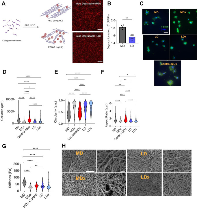
In native extracellular matrices (ECM), cells can use matrix metalloproteinases (MMPs) to degrade and remodel their surroundings. Likewise, synthetic matrices have been engineered to facilitate MMP-mediated cleavage that enables cell spreading, migration, and interactions. However, the intersection of matrix degradability and mechanical properties has not been fully considered. We hypothesized that immediate mechanical changes result from the action of MMPs on the ECM and that these changes are sensed by cells. Using atomic force microscopy (AFM) to measure cell-scale mechanical properties, we find that both fibrillar collagen and synthetic degradable matrices exhibit enhanced stress relaxation after MMP exposure. Cells respond to these relaxation differences by altering their spreading and focal adhesions. We demonstrate that stress relaxation can be tuned through the rational design of matrix degradability. These findings establish a fundamental link between matrix degradability and stress relaxation, which may impact a range of biological applications.


Conflict of interest statement

Declaration of interests The authors declare no competing interests.

Figures

Figure 1.
Figure 1.. Cells sense changes in collagen architecture and degradability independent of stiffness.
MD - More degradable, MDx - More degradable gels with collagenase treatment, LD - Less degradable, LDx - Less degradable gels with collagenase treatment, MDx-Control - same stiffness as that of MDx gels. (A) Scheme for producing collagen gels of varying degradability through macromolecular crowding with PEG (created with BioRender.com). Scale bar represents 20 μm. (B) Degradation rate quantified by monitoring change in absorbance at 313 nm after adding collagenase, Student’s t-test was performed between samples. (C) Representative immunofluorescent images of HFFs spreading on MD, MDx, LD and LDx gels. F-actin in green, nucleus in blue. Scale bar represents 50 μm. Quantification of HFF spreading: (D) Area, (E) Circularity, and (F) Aspect ratio. Cells were analyzed after 4 hr culture, n=183 cells across N=3 biological replicates. (G) Stiffness of gels measured using AFM. (H) SEM images of MD, MDx, LD and LDx gels, imaged at 15,000x and 30,000x magnification. Scale bar represents 2 μm for the left panel and 500 nm for the corresponding right panel. Statistical significance was determined using ANOVA, *p <0.05, **p < 0.01, and ****p < 0.0001.
Figure 2.
Figure 2.. Differences in degradability of collagen gels correspond to differences in stress relaxation.
MD - More degradable, MDx - More degradable gels with collagenase treatment, LD - Less degradable, LDx - Less degradable with collagenase treatment, MDx-Control - same stiffness as that of MDx gels. (A) Average of relaxation curves. (B) Standard linear solid parameter F∞. (C) Parameter Τ relaxation time. (D) Power law rheology fit parameter β. The data are obtained from n=3 independent samples, N = 15 measurements per sample. For all graphs, statistical significance was determined by ANOVA, *P <0.05, **P < 0.01, ***P < 0.001, and ****P < 0.0001.
Figure 3.
Figure 3.. Rational design of synthetic stress relaxing gels as a function of degradability.
(A) PVA gels of tunable degradability are generated by varying the ratio of non-degradable to degradable cross-links (created with BioRender.com). (B) Stiffness of hydrogels, N=45 across three independent samples, ND - non-degradable, NDx - non-degradable treated with collagenase, CD - collagenase degradable, and CDx - collagenase degradable treated with collagenase. (C) Stress relaxation of hydrogels assessed using AFM. (D) Final normalized force, n=15 measurements per gel. For all graphs, statistical significance was determined using ANOVA, *P <0.05 and ****P < 0.0001.
Figure 4.
Figure 4.. The fibroblast response to degradation of synthetic degradable gels is similar to their response to degradation of collagen gels.
NDx - Non-degradable gels with collagenase treatment, CDx - Collagenase degradable gels with collagenase treatment. Human foreskin fibroblasts (HFF) spreading on gels (A) Phalloidin (green) and DAPI (blue) stained images. Scale bar = 50 μm. (B) Vinculin focal adhesions and corresponding filtered images showing adhesions. Scale bar = 20 μm. Quantification of cell spreading: (C) Area, (D) Circularity, and (E) Aspect ratio, n=247 cells measured across N=3 biological replicates. (F) Number of focal adhesions per cell with area between 1–10 μm2 and (G) Percentage of area occupied by focal adhesions per cell quantified using vinculin staining, n=17 and n=21 cells analyzed for NDx and CDx conditions across N=3 biological replicates. For all graphs, statistical significance was determined using student’s t-test, **P < 0.01 and ****P < 0.0001.
Figure 5.
Figure 5.. Degradation induced stress relaxation effects on cell spreading are cell type dependent.
(A) C2C12 myoblasts spreading on NDx and CDx gels. Assessment of C2C12 spreading: (B) Area, (C) Circularity, and (D) Aspect ratio after 24 hr of culture, n=193 cells across N=3 biological replicates.(E) MCF10A spreading on NDx and CDx gels. Quantification of MCF10A spreading: (F) Area, (G) Circularity, and (H) Aspect ratio after 24 hr culture, n=143 cells across N=3 biological replicates. For all graphs, student’s t-test was performed, ****P < 0.0001.

References

    1. Akhmanova M., Osidak E., Domogatsky S., Rodin S., Domogatskaya A., Stem Cells Int. 2015, 2015, 167025. - PMC - PubMed
    1. Pelham R. J. Jr, Wang Y. l., Proc. Natl. Acad. Sci. U. S. A. 1997, 94, 13661. - PMC - PubMed
    1. Mason B. N., Starchenko A., Williams R. M., Bonassar L. J., Reinhart-King C. A., Acta Biomater. 2013, 9, 4635. - PMC - PubMed
    1. Carvalho E. M., Kumar S., Acta Biomater. 2023, 163, 146. - PubMed
    1. Adebowale K., Gong Z., Hou J. C., Wisdom K. M., Garbett D., Lee H.-P., Nam S., Meyer T., Odde D. J., Shenoy V. B., Chaudhuri O., Nat. Mater. 2021, 20, 1290. - PMC - PubMed

Publication types

LinkOut - more resources