Review
. 2022 Jul 13;1(5):100405.
doi: 10.1016/j.jscai.2022.100405. eCollection 2022 Sep-Oct.

Left Atrial Appendage Occlusion: Current Stroke Prevention Strategies and a Shift Toward Data-Driven, Patient-Specific Approaches

Affiliations
Review

Left Atrial Appendage Occlusion: Current Stroke Prevention Strategies and a Shift Toward Data-Driven, Patient-Specific Approaches

Keegan Mendez et al. J Soc Cardiovasc Angiogr Interv. .

Abstract

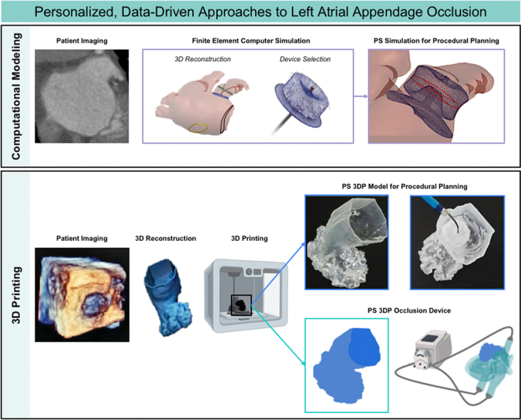
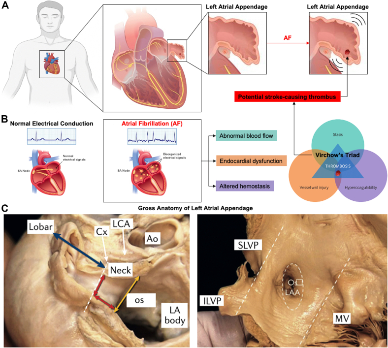
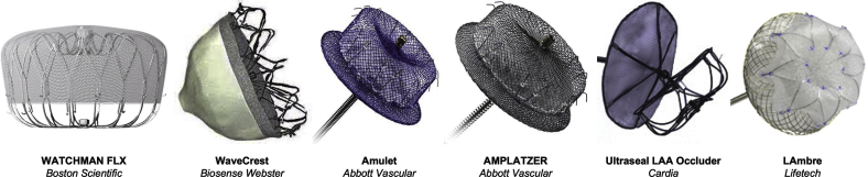
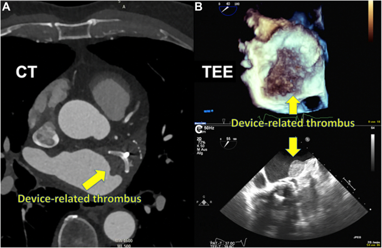
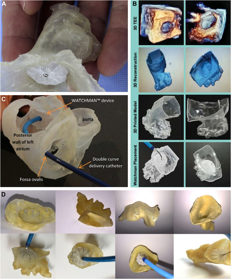
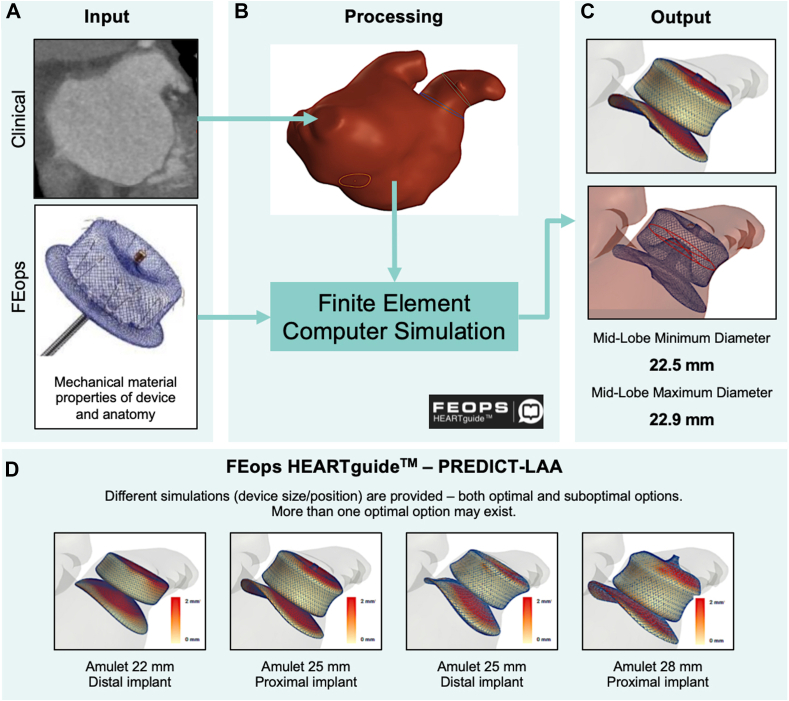
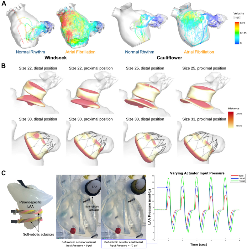
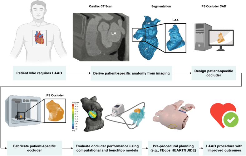
The left atrial appendage (LAA) is a complex structure with unknown physiologic function protruding from the main body of the left atrium. In patients with atrial fibrillation, the left atrium does not contract effectively. Insufficient atrial and LAA contractility predisposes the LAA morphology to hemostasis and thrombus formation, leading to an increased risk of cardioembolic events. Oral anticoagulation therapies are the mainstay of stroke prevention options for patients; however, not all patients are candidates for long-term oral anticoagulation. Percutaneous occlusion devices are an attractive alternative to long-term anticoagulation therapy, although they are not without limitations, such as peri-implant leakage and device-related thrombosis. Although efforts have been made to reduce these risks, significant interpatient heterogeneity inevitably yields some degree of device-anatomy mismatch that is difficult to resolve using current devices and can ultimately lead to insufficient occlusion and poor patient outcomes. In this state-of-the-art review, we evaluated the anatomy of the LAA as well as the current pathophysiologic understanding and stroke prevention strategies used in the management of the risk of stroke associated with atrial fibrillation. We highlighted recent advances in computed tomography imaging, preprocedural planning, computational modeling, and novel additive manufacturing techniques, which represent the tools needed for a paradigm shift toward patient-centric LAA occlusion. Together, we envisage that these techniques will facilitate a pipeline from the imaging of patient anatomy to patient-specific computational and bench-top models that enable customized, data-driven approaches for LAA occlusion that are engineered specifically to meet each patient's unique needs.

Keywords: computed tomography; imaging; left atrial appendage; left atrial appendage occlusion; stroke.


Conflict of interest statement

Dr Wang is a consultant or advisory to Edwards Lifesciences Corporation, Boston Scientific Corp, Abbott, and Neochord Inc and receives funding grants from Boston Scientific Corp. Dr O’Neill is a consultant or advisory to Edwards Lifesciences Corporation and Abbott. Prof. Roche is a consultant to Holistick Medical and Surmodics and is on the board of directors for Affluent Medical and the scientific advisory board for Helios Cardiovascular. Ms Mendez and Mr Kennedy reported no financial interests.

Figures

None
Graphical abstract
Figure. 1
Figure. 1
Location, anatomy, and morphology of the left atrial appendage (LAA). (A) Location of the LAA within the heart and the representation of clot formation during atrial fibrillation. (B) Atrial fibrillation (AF) is caused by abnormal electrical activity. The central feature of AF is rapid and uncoordinated atrial activity. Disordered electrical propagation causes disorganized stimulation of the myocardium and subsequent arrhythmic contractions. AF decreases contractility, resulting in blood stasis and diminished peak flow velocities. AF is also associated with endothelial damage, fibrosis, and inflammation, especially within the LAA, which leads to a prothrombotic and hypercoagulable state. This association is consistent with the Virchow triad, which synthesizes the pathogenesis of clot formation in the LAA in patients with AF: abnormal blood flow, endocardial dysfunction, and altered hemostasis. (C) The LAA is located near the atrioventricular groove between the left ventricle and the pulmonary artery trunk, with the base close to the proximal left circumflex artery. The LAA is divided into 3 anatomic regions: the opening or ostium, neck, and lobar region. The ostium can be teardrop-shaped, round, elliptical, foot-like, or triangular, and its diameter can range from 10 to 40 mm. The lobar region usually consists of 2 lobes and is the most spatially complex region of the LAA, with heavy trabeculations and pectinate muscles. The length and width of the neck, as well as the number of lobes, vary considerably. The LA body is smooth-walled. AF, atrial fibrillation; Ao, aorta; ILPV, inferior left pulmonary vein; Cx, circumflex artery; LA, left atrium; LCA, left coronary artery; MV, mitral valve; os, ostium; SLPV, superior left pulmonary vein. Reproduced from Nishimuraet al, 2019, and Caliskan et al, 2017.
Figure. 2
Figure. 2
Decision tree for left atrial appendage closure in patients with an indication for stroke prevention due to atrial fibrillation. CAD, coronary artery disease; HAS-BLED, scoring system developed to assess 1-year risk of major bleeding in people taking anticoagulants for atrial fibrillation (Hypertension, Abnormal renal and liver function, Stroke, Bleeding, Labile INR, Elderly, Drugs or alcohol); LAA, left atrial appendage; NOAC, novel oral anticoagulant; OAC, oral anticoagulant; Vit-K, vitamin K. Reproduced from Glikson et al, 2020.
Figure. 3
Figure. 3
Commercially available, CE-mark approved percutaneous left atrial appendage occlusion devices. Adapted from Glikson et al, 2020. CE, Conformité Européenne.
Figure. 4
Figure. 4
Device-related thrombosis (DRT) detected during imaging follow-up. (A) DRT detected using cardiac computer tomography in a 75-year-old patient 590 days after left atrial appendage closure. (B, C) DRT in 3- and 2-dimensional transesophageal echocardiography in an 87-year-old patient 226 days after left atrial appendage closure. CT, computed tomography; TEE, transesophageal echocardiography. Reproduced from Sedaghat et al, 2021.
Figure. 5
Figure. 5
The use of 3-dimensional (3D) printed models in left atrial appendage occlusion. (A) The Watchman device placed within a flexible 3D-printed model demonstrates the clinical utility of 3D printing for device sizing and avoiding procedural complications. (B) 3D-printed models of the left atrial appendage using real-time 3D transesophageal echocardiographic data for assistance with physician planning and decision making. (C) 3D-printed model of a patient’s specific left atrial and left atrial appendage anatomy for assistance in the bench-test selection of catheter curvature for device implantation. (D) Computed tomography-based 3D-printed models for preprocedural planning in left atrial appendage device closure. Adapted from Otton et al, 2015; Liu et al, 2016; Wang et al, 2016; and Obasare et al, 2017.
Figure. 6
Figure. 6
FEops HEARTguide workflow and simulations. Example of computed tomography-based virtual 3-dimensional planning software for patient-specific procedural planning. (A) Preprocedural computed tomography scan is uploaded on a web-based platform. (B) Image processing to extract 3-dimensional patient-specific anatomical reconstruction and landmarks for the procedure. Patient-specific reconstruction, in combination with the device model, serves as input for computational finite element analysis. (C) Model outputs several options in terms of device size and position, including left atrial appendage wall apposition plots, deformation visualization, and measurements. (D) Use of simulation output in clinical practice: for a single patient, different simulations in terms of device size and position are provided to the operator to help guide decision making before the procedure. Scale bar: white color indicates perfect apposition, and red color indicates gaps of ≥2 mm between the device and the walls. PREDICT-LAA, Value of FEops HEARTguide Patient-Specific Computational Simulation in the Planning of Percutaneous Left Atrial Appendage Closure With the Amplatzer Amulet Device. Reproduced from Garot et al, 2020.
Figure. 7
Figure. 7
Computational and bench-top models for left atrial appendage occlusion procedural planning and device testing and validation. (A) Computational fluid dynamic analysis of the left atrial appendage (LAA) to predict the risk of thrombosis. Instantaneous velocity streamlines passing through the LAA orifice for both normal and pathologic atrial fibrillation condition. (B) Computational modeling to optimize preprocedural planning in left atrial appendage occlusion. Apposition plots for patients implanted with Amulet (top) and Watchman (bottom) devices. Different sizes and positions are virtually simulated, with white color indicating perfect apposition and red color indicating gaps of ≥2 mm between the device and the walls. (C) Design and development of bench-top circulatory model with interchangeable, patient-derived LAA geometries. Patient-derived silicone casting of LAA geometry that is incorporated into a cardiac simulator. Soft robotic actuators are used as artificial muscles to make the LAA contract cyclically. LAA pressure waveforms can be modulated by varying the actuation pressure and regime of the soft robotic actuators. LAA, left atrial appendage occlusion. Adapted from Bosi et al, 2018; Bavo et al, 2020; and Mendez et al, 2021.
Figure. 8
Figure. 8
Vision flowchart of personalized engineering approach for left atrial appendage occlusion. Design, fabrication, validation, and implantation of a patient-specific left atrial appendage occlusion device. Adapted from Robinson et al, 2018; Gu et al, 2018; Senadeera et al, 2020; and FEops HEARTguide. CAD, computer-aided design; CT, computed tomography; LAAO, left atrial appendage occlusion; PS, patient-specific.
Central Illustration
Personalized, data-driven approaches for left atrial appendage occlusion. Computational modeling technologies enable simulation of LAA occlusion device placement before procedures (top). Patient imaging can be coupled with 3D printing to create patient-specific LAA occlusion devices (bottom).

Similar articles

Cited by

References

    1. Tabata T., Oki T., Yamada H., et al. Role of left atrial appendage in left atrial reservoir function as evaluated by left atrial appendage clamping during cardiac surgery. Am J Cardiol. 1998;81(3):327–332. - PubMed
    1. Al-Saady N.M., Obel O.A., Camm A.J. Left atrial appendage: structure, function, and role in thromboembolism. Heart. 1999;82(5):547–554. doi: 10.1136/hrt.82.5.547. - DOI - PMC - PubMed
    1. Regazzoli D., Ancona F., Trevisi N., et al. Left atrial appendage: physiology, pathology, and role as a therapeutic target. BioMed Res Int. 2015;2015 doi: 10.1155/2015/205013. - DOI - PMC - PubMed
    1. Caliskan E., Cox J.L., Holmes D.R., et al. Interventional and surgical occlusion of the left atrial appendage. Nat Rev Cardiol. 2017;14(12):727–743. doi: 10.1038/nrcardio.2017.107. - DOI - PubMed
    1. Beigel R., Wunderlich N.C., Ho S.Y., Arsanjani R., Siegel R.J. The left atrial appendage: anatomy, function, and noninvasive evaluation. J Am Coll Cardiol Imaging. 2014;7(12):1251–1265. - PubMed

LinkOut - more resources