Review
. 2022 Jun 30;1(5):100391.
doi: 10.1016/j.jscai.2022.100391. eCollection 2022 Sep-Oct.

Management of Pediatric Pulmonary Vein Stenosis

Affiliations
Review

Management of Pediatric Pulmonary Vein Stenosis

Ryan Callahan et al. J Soc Cardiovasc Angiogr Interv. .

Abstract

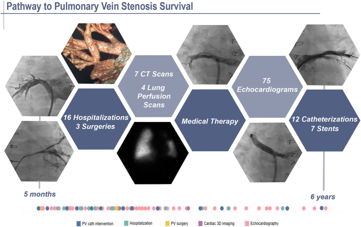
Pediatric intraluminal pulmonary vein stenosis has evolved into a chronic illness, with improving survival. Although significant knowledge gaps remain, medical providers have found success in the management of patients with pulmonary vein stenosis using a comprehensive multimodality treatment strategy. This review discusses the core principles employed by 4 centers dedicated to improving pulmonary vein stenosis outcomes, including how to make the diagnosis, educating the family, treatment strategy, the importance of surveillance, and the management of symptoms and comorbidities.

Keywords: congenital heart disease; management; pulmonary vein stenosis; therapy.


Figures

None
Graphical abstract
Figure 1
Figure 1
Tree diagram of pediatric pulmonary venous obstructions. The tree diagram demonstrates the mechanisms of pulmonary venous obstruction, including intraluminal PVS. The various types of PVS are outlined. PVS, pulmonary vein stenosis; Qp, pulmonary blood flow; TAPVC, total anomalous pulmonary venous connection.
Figure 2
Figure 2
Chest radiograph of a 3-month-old child presenting with respiratory failure secondary to multivessel pulmonary vein stenosis. The image demonstrates a bilateral pattern of interstitial opacities as a result of pulmonary venous hypertension and interstitial edema. R, right.
Figure 3
Figure 3
Nuclear medicine lung perfusion scan obtained before and after transcatheter intervention. The lung perfusion scan demonstrates decreased flow to the left lung secondary to severe left common pulmonary vein stenosis (left). There is improved flow to the left lung following transcatheter intervention (right).
Figure 4
Figure 4
Optical coherence tomography scan of the pulmonary vein. Image demonstrates pulmonary vein with a history of stent implantation and current evidence of ingrowth, also known as in-stent restenosis (left, center). There are several areas of wall injury following cutting balloon venoplasty (right).
Figure 5
Figure 5
Comparison of pulmonary artery wedge angiography versus retrograde venography in the right upper pulmonary vein. Wedge angiography (left) demonstrating possible ostial narrowing of the right upper pulmonary vein. Retrograde venography (right) via long sheath with opacification of the entire right upper and right middle pulmonary veins and better delineation of stenoses distal to the venoatrial junction.
Figure 6
Figure 6
Left common pulmonary vein venography. Retrograde venography via guide catheter demonstrates in-stent restenosis, opacification of the entire left lower vein, and atresia of the left upper segment with interlobar collaterals.
Central Illustration
The pathway to pulmonary vein stenosis (PVS) survival. The health care interaction timeline demonstrates the clinical path of a 5-month-old infant who initially presented with cardiogenic shock, severe coarctation, left ventricular dysfunction and multivessel PVS. Over the course of 6 years, the patient required numerous transcatheter and surgical pulmonary vein interventions, systemic sirolimus medical therapy, surveillance testing, and frequent hospitalizations. The timeline demonstrates a decreasing frequency of tests and interventions with increasing age, illustrating the impact of therapy on PVS stabilization. 3D, 3-dimensional; cath, catheter; CT, computed tomography; PV, pulmonary vein.

References

    1. Jadcherla A.V., Backes C.H., Cua C.L., Smith C.V., Levy P.T., Ball M.K. Primary pulmonary vein stenosis: a new look at a rare but challenging disease. Neoreviews. 2021;22(5):e296–e308. doi: 10.1542/neo.22-5-e296. - DOI - PubMed
    1. Vanderlaan R.D., Rome J., Hirsch R., Ivy D., Caldarone C.A. Pulmonary vein stenosis: treatment and challenges. J Thorac Cardiovasc Surg. 2021;161(6):2169–2176. doi: 10.1016/j.jtcvs.2020.05.117. - DOI - PubMed
    1. Humpl T., Fineman J., Qureshi A.M. The many faces and outcomes of pulmonary vein stenosis in early childhood. Pediatr Pulmonol. 2021;56(3):649–655. doi: 10.1002/ppul.24848. - DOI - PubMed
    1. Nasr V.G., Callahan R., Wichner Z., Odegard K.C., Dinardo J.A. Intraluminal pulmonary vein stenosis in children: a “new” lesion. Anesth Analg. 2019;129(1):27–40. doi: 10.1213/ANE.0000000000003924. - DOI - PubMed
    1. McLennan D.I., Solano E.C.R., Handler S.S., Lincoln J., Mitchell M.E., Kirkpatrick E.C. Pulmonary vein stenosis: moving from past pessimism to future optimism. Front Pediatr. 2021;9:747812. doi: 10.3389/FPED.2021.747812. - DOI - PMC - PubMed

LinkOut - more resources